Cellular Injury, Adaptations, Maladaptive Changes (PATHO #1)
 Call Kai
 Learn
Learn Practice Test
 Spaced Repetition
 Match
Match Flashcards
 Knowt Play
1/54

There's no tags or description
Looks like no tags are added yet.
Name | Mastery | Learn | Test | Matching | Spaced | Call with Kai | Chat |
|---|
No analytics yet
Send a link to your students to track their progress
55 Terms
prokaryotes
a cell structure
Cyanobacteria(blue-green algae), bacteria, rickettsia
have no organelles or nuclear membrane
eukaryotes
cells of higher animals and plants
Protein synthesis, transport outer cell membrane, enzyme conent
three components of a cell
plasma membrane (outer membrane of cell)
cytoplasm (fluid filling)
nucleus (organs of cell)
nucleus
lg membrane- bound organelle
cell division, genetic info (controls cell activity and stores DNA) deoxyribonuceluic acid)
cytoplasm organelles
cytoplasmic matrix
fills space btwn nucelus and plasma membrane, aqueous mass
plasma membrane
protect cell
cell to cel recognition
gates, channels and pump
consists of lipids and proteins
lipids
bilayer made of up phosoplipids (phosphate bound to lipid) and glycolipids (carbohydrate bound to lipid)
trasnferring of ions and moleculues into and out of cell
proteins
some proteins can pass thry the membrane and some cant
trasnportation of cell
passive vs active
passive transport
no energy required
osmosis
diffusion
active transport
requires energy when trasnporting particles across the plasma membrane
etiology
og cause of cell alteration or diease
etiologic agents
causes of cell alteration or diease e.g infection, trauma
disease etiology
characterstic changes w specific etiologic agents
e.g cold temp=frostbite
in response: cell may devolop adaptive compensatory or maladaptive changes
cell size change
atrophy, hypertrophy (physiological vs pathological)
atrophy
decrease in cell size
hypertrophy
increase in cell size
cell number/apperance change
hyperplasia, metapasia dysplasia
hyperplasia
increase in cell number
metaplasia
reversiable replacement of one mature cell type by another less mature cell type or chnage in phenotype
dysplasia
derranged cellular growth
atrophy explained
decrease in cell size, decrease organ size if enough cells shrink
physiologic (normal in normal in early development)
pathlogic (adverse from disease/injury)results from decrease in workload, pressure, use, blood supply, nurtrion e,g paralysis shrinks skeletal muscle
hypertrophy explained
increase in cell size, increase organ size
physiologic (results from increased demand, stimularion by hormones, growth factors)
pathologic (results from chronic hemodynamic overload) e.g cardiac hypertropgy or strength training
physiological hypertropphy
cell enlargemnt w adequate supporting tissues
e.g enlargemnet of cardiac cells w exersise traning
pathological hypertrophy
increase in cell size, w/o increase in support structures
e.g enlargemnt of heart tissue due to hypertension
hyperplasia explained
increase in number of cells
only in cells capable of mitosis
can result from hormonal stimulation
e.g estrogen stimulates growth of breast cellls in pregency
may evolce inro maladaptive compensation (cell number increases to much, e.g keloid formation)
hyperplsia keloid formation
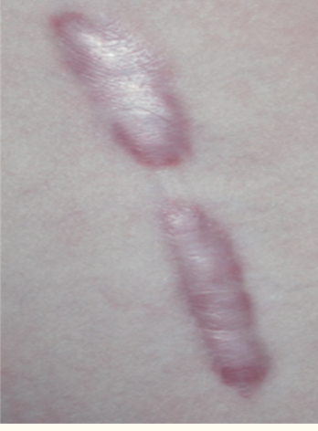
metaplasia explained
reversible replacement of one mature cell type by another
associated w tissue damage, repair, regenration
reprogramming of stem cells or undifferentaied mesenchymal cells
metaplasia explained
replacemet of one cell type w another r
genetic reprogramming (ensure cell survival e.g GERD)
metaplasia in GERD
lower esophageal cells, squamous epithliam transition to comulmnar stomack like
dysplasia explained
abnormal changes in size, shape, orgninization of mature cells
may b reversible if triggering stimulis is removed
tissue appears disorderly but is not cancer
(if changes penetrate basemnt membrane: invasive neoplasm) deranged cellular growth (often a result of chronic inflammation or precancerous condition.)
cells vary in size, shape, orgnization comparted w normal e.g cervical dysplasia detetced by PAP
neoplasia
new growth (disorganized, uncoordinated, uncontrolled)
cancerous
often interchanged with term tumor
neoplasms may b bengign or malignant
benign neoplasm
cells resemble normal cells
well differentated cells
do not metastasize
well defined boarders
maligant neoplasm
cells appear differet from healthy cells
poorly differentiated cells
increased likelihood of metastatiss
poorly defined boarders
cellular injury
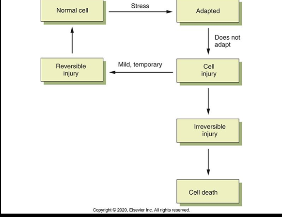
oxidative stress and free radicals
an imbalence of free radicals and antioxidants in your body that leads to cell damage
exposure of cells to reactive oxygen specisis (ROS)— free radicals—cause cellular damage and targets DNA
clincal e.g heart disease, diabetes, cancer
antioxidants—-vitamin E—-reduce oxidative stress fror many diease processes
TIPS
T- toxins (endogenous (within body) exogenous (external enviorment e.g all)
I-infections
P-phsyical injury
S-serum deficit injury
toxins: cellular injury mechanisms
chemical or toxic injury (xeobiotics-toxic, mutagentic, carcinogenic)
lead, carbon monoxide, ethonal, mercury, social street drugs
chemical agenets of toxins cellular injury mech
over the counter and prescrived drugs, opiod abuse, leading cause of child posing is medications
toxins cellular injury mech (enviormental, heavy metals, ethanol)
emviormental- air pollution
heavy metals-lead cadmium arsenic mercury
ethonal-fetal alcohol syndrome, detal alcohol specturm disorders
infectious injury
pathogenecity of microorganism
bacterial or viral
disease prodcuing potential (invasion and destructuion, toxin production, production of hypersensitivry reactions)
physical unitential and intential injuries
blunt force injuries (result of application of mechanical force to body) results in tearinf shearing crushing of tissues e.g motor vechiles accidents and falls)
contusions, lacerations, fractures
unintetial and intentional injuries
sharp force injuries (stab wounds)
gunshot wounds
asphyxial injuries (suffocation, drowning)
s-serum deficit injury: cellular injury mechanism: hypoxia
hypoxic injury (single most common cause of cellualr injury)
results from ischemia (reduced supply of blood)
reduced oxygen conent in ambient air
loss of hemoglobin
decrease production of rbc
diseases of the respiratory and cadiovascular systems
posiinning of the oxidative enzymes (cytochromes) within the cells)
anozia0total lack of o2 caused by obstruction
ischemia
ischemia reperfusion injury
cell injury and death caused by rstoration of blood flow and oxygen
mechanism: oxidative stress, increase intracellualr calcium concentration, inflammation
apoptosis
programmed cellular death
normal part of aging
necrosis
associated w inflmmation and related to cellular injury
cell degeneration: necrosis
cell death due to injury
irreversaible process (membrane disintregrates, lysomal activation and autolysis
initatieates inflmmatory reaction
infarction
ischemic nercosis: death of tossue results from prolonged ischemia e.g MI cell contents cardiac proteins released into circulation
gangrene
prolonged ischemia, infaraction and necrosis
closttridum perfrigens (emits indefitable gas as it destory tissues, gas gangrene)
necrosis
gangeronous necrosis (death of tossue from severe hypoxic injury
dry: skin becomes dry shrivled brown/black
wet: arease become cold, swollen, black (Gas gangrene caused by clostridium
barrettes esophogus
metaplastic changes in GERD
may lead to cancer
endothilium could b consider body larget organ, active tissue is bulnarable to injury
ceullular injury
wellness and disease are reflected in cells, injury to a cell or its componements can lead to illness (biochemical lesion froms on the cell at the point of injury, a. chnages the chemistry of metabolic reaactions within cell e.g chronic alcoholism)
aging and altered cellular and tissue biology
aging—lose structure adn function—decrease—decrease in size or waste away
Aging and Altered Cellular and Tissue Biology- FRATLITY
weakness, decrease staminia, functional decline in older adults
increase vulnarability to falls, disability, disease, death
somatic death= death of entire body