Renal System
 Call Kai
 Learn
 Practice Test
 Spaced Repetition
 Match
 Flashcards
 Knowt Play
1/182

There's no tags or description
Looks like no tags are added yet.
183 Terms
Functions of the kidney
removal of metabolic waste and foreign chemicals through urine excretion
regulates water and ion balance
acid-base balance
gluconeogenesis
production of hormones/enzymes
Hormones that kidney secretes
EPO, 25-(OH)2D, renin
Macro structure of urinary system
kidneys→ureters→bladder→urethra
Ureter function
urine transport
Bladder function
urine storage
Urethra function
urine excretion
Macro structure of kidney
renal cortex
renal medulla
renal pelvis
ureter beginning

Nephron
structural and functional unit of the kidney
Two component of the nephron
renal corpuscle and renal tubule
Structure of nephron
glomerulus
proximal convoluted tubule
loop of Henle
distal convoluted tubule
cortical collecting duct
medullary collecting duct
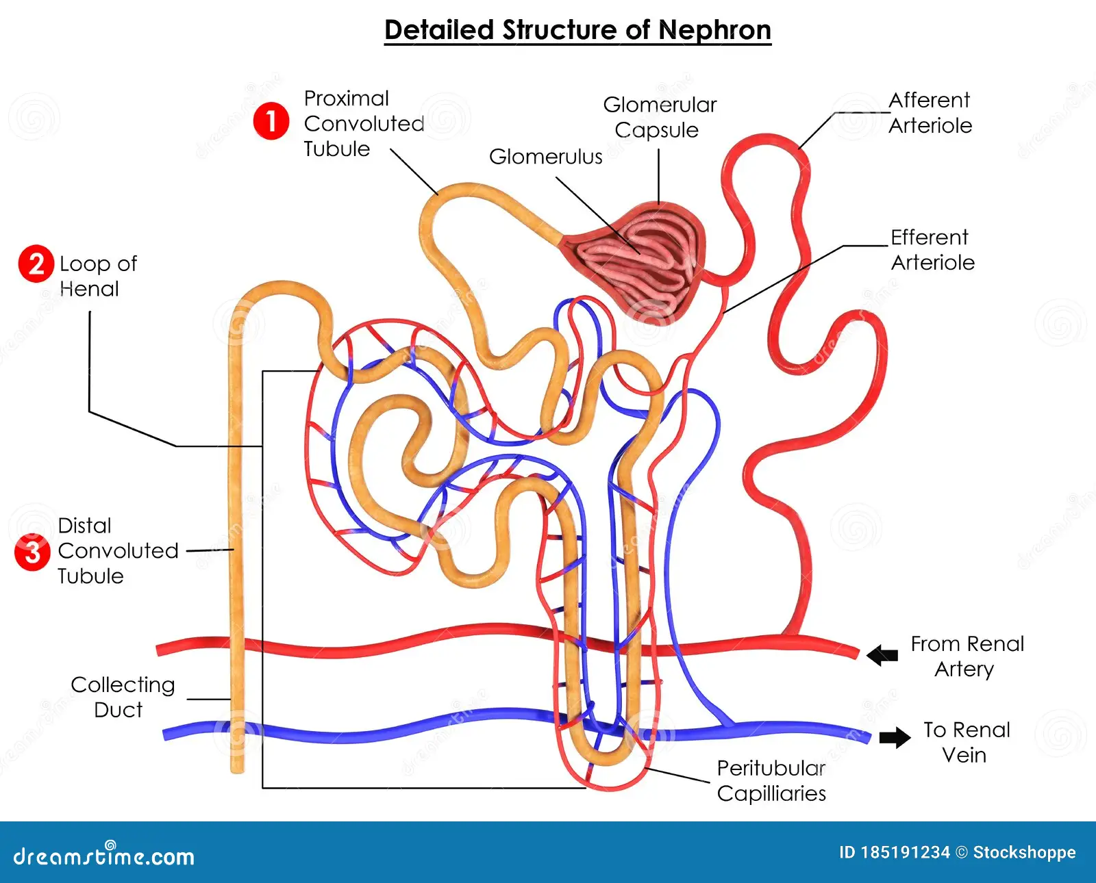
Blood flow to and from nephron
blood brought to glomerulus through afferent arteriole
blood brought away from glomerulus through efferent arteriole
peritubular capillaries surround loop of henle
Glomerular filter
3 ply filter
leaky capillary wall fenestrations
basement membrane
filter slits between podocytes

Makup of basement membrane
full of proteins and anions
Mesangial cells
cell type contained in glomerulus. smooth-muscle like cells that help regulate blood flow in glomerulus via contraction. Also engulf macromolecules that get hung up during filtration
Two type of nephron
juxtamedullary nephrons
cortical nephrons
Juxtamedullary nephrons
located mostly in the medulla
have long loops of Henle
concentrate urine
Cortical nephrons
located mostly in the cortex
short loop of Henle
3 functions of the kidney
glomerular filtration
tubular secretion
tubular reabsorption
Glomerular filtration
move material from blood to glomerulus
Tubular secretion
move material from blood to tubule directly
Tubular reabsorption
move material from tubule back to peritubular capillaries
What drives glomerular filtration?
Starling forces
Permeability of glomerular capillaries to water and solute
incredibly permeable
glomerular pressure compared to systemic blood pressure
Much higher than would normally be found in capillaries. This is because blood is carried to and from by arterioles
Surface area of glomerular capillaries
Huge surface area→good for efficient filtration
What CANNOT cross glomerular filter
large anions, proteins RBCs, anything bound to proteins
Purpose of inability of proteins to cross glomerular filter
Must maintain plasma oncotic pressure!
Hydrostatic pressure of glomerular capillaries vs bowman’s capsule
Much higher in glomerular capillaries
Oncotic pressure in glomerular capillaries vs Bowman’s capsule
Oncotic pressure in bowman’s capsule virtually nonexistent
Effect of constricting afferent arteriole on glomerular filtration
decreases hydrostatic pressure and thus decreases filtration
Effect of constricting efferent arteriole on glomerular filtration
backs up pressure, which increases filtration
Effect of dilating afferent arteriole on glomerular filtration
increases hydrostatic pressure, which increases filtration
Effect of dilating efferent arteriole on glomerular filtration
loosens pressure, causing hydrostatic pressure to drop and thus decrease filtration
Where does tubular reabsorption begin?
begins as soon as filtrate enters tubule
Main type of transport that tubular reabsorption occurs by
Usually through transepithelial transport (either active or passive). Few paracellular transport can occur
Is tubular reabsorption active or passive?
Can be either!
Materials that are reabsorbed
water, glucose, urea, sodium, amino acids, vitamins, important ions
Type of transport that tubular secretion usually occurs by
transepithelial transport. Not much paracellular transport
Is tubular secretion active or passive?
Can be either!
Glucose reabsorption graph based on different BGLs
usually all glucose is reabsorbed
however, if BGL gets too high, kidney stops glucose reabsorption and starts excretion

Why does glucose reabsorption stop at a certain point?
Glucose transporters are saturated
Calculating glucose lost
filter load—reabsorption
Most abundant cation in filtrates
sodium!
Importance of sodium
helps do water balance
movement of other substaces depends on sodium moving down its gradient
Type of transport involved in sodium reabsorption
Almost always active
Importance of tubular secretion
remove drug and drug metabolites from blood
eliminate urea/uric acid
remove excess potassium
regulate pH
Metabolic role of kidneys
can do gluconeogenesis to replenish BGL during fasting
break down organic compounds in preparation for secretion
How do you regulate reabsorption and secretion?
regulate amount of transport proteins
based on autocrine, paracrine, and hormonal signals
Where does majority of reabsorption occur?
PCT and loop of Henle
How is sodium reabsorbed in PCT?
Sodium transferred to epithelial cells via cotransport and antitransport
Sodium returned to interstitial fluid of capillaries via sodium potassium ATPase on basolateral membrane
Potassium leak channels on the basolateral membrane allow potassium to flow down concentration gradient, allows Na/K ATPase to keep functioning
How is sodium reabsorbed in DCT?
moves down gradient using sodium channels from tubule to epithelial cells
moves into interstitial fluid of capillaries via Na/K ATPase
K is secreted through K leak channels located on the luminal membrane, allowing Na/K ATPase to keep functioning
Reabsorption of water
Water follows sodium! This is because as sodium decreases, osmolarity decreases and water flows down the osmolarity gradient
Type of transporter used by water
aquaporin
Renal clearance
quantitative metric of filtration function that describes the volume of plasma cleared of a substance in one minute
Renal clearance equation
C=UV/P
C→clearance
U→concentration of substance in urine
V→flow rate
P→concentration of substance in plasma
What type of substance should you use to measure renal clearance?
Use a substance that is ONLY filtered (not reabsorbed or secreted)
What is urine composed of?
Filtrate that isn’t reabsorbed
How is micturition reflex triggered?
Via the stretch of smooth muscles in bladder
Types of nerves that innervate bladder
parasympathetic
sympathetic
somatic
Three muscles of bladder
detrusor
internal sphincter
external sphincter
Nervous system input to detrusor muscle during bladder filling
Parasympathetic innervation of detrusor muscle is inhibited
Nervous system input to internal sphincter during bladder filling
Sympathetic input to internal sphincter is stimulated
Nervous system input to external sphincter during bladder filling
Somatic input of external sphincter is stimulated
Nervous system input to detrusor muscle during bladder emptying
Parasympathetic innervation of detrusor muscle is promoted
Nervous system input to internal sphincter during bladder emptying
Sympathetic input to internal sphincter is inhibited
Nervous system input to external sphincter during bladder emptying
Somatic input of external sphincter is inhibited
Total daily intake and loss of water
Intake: about 3 L
Loss: about 3 L
they’re equal!
Sources of fluid loss
urine (major)
insensible loss in skin and lungs
sweat and feces
What type of process is sodium reabsorption: passive or active?
Active
Places sodium is reabsorbed
All tubular segments (70% in PCT) except in descending loop of Henle and the medullary collecting duct
How does water reabsorption occur?
Through diffusion thanks to aquaporins. Dependent on sodium
Places of water reabsorption
Everywhere except in the ascending loop of henle and the cortical+medullary collecting ducts→unless hormones act upon collecting
Hormone influence on cortical collecting duct
Aldosterone acts on cortical collecting duct to increase sodium and water reabsorption
Vasopressin acts to increase ONLY water reabsorption
PTH acts to increase Ca2+ reabsorption
How does aldosterone increase sodium reabsorption in the cortical collecting duct?
upregulated sodium channels and sodium potassium ATPase
induces obligatory potassium secretion by upregulating K+ channels in luminal membrane
Water absoprtion in the cortical collecting duct under the influence of aldosterone
water follows sodium through aquaporins
What happens when sodium is reabsorbed but not water?
Changes in osmolarity, which induces water reabsorption
Vasopressin (ADH) effect
increases aquaporin proteins in luminal membrane of medullary collecting duct
increases urea transporters
directly acts on arterioles to increase vasoconstriction and therefore TPR
Aquaporin regulation
vasopressin (ADH) binds to receptor on basolateral membrane of collecting duct epithelial cells
adenylyl cyclase activated through GPCR mechanism, causing a rise in cAMP and subsequent PKA activation
PKA phosphorylates AQP2, which is contained within vesicles
Phosphorylation causes vesicles to fuse with luminal membrane
Aquaporins added to luminal membrane
Are other aquaporins always present in the basolateral membrane in collecting duct?
Yes!
Importance of urea
sets up osmotic gradient for H2O reabsorption in medullary collecting duct. Without urea, you can’t achieve a hyperosmolar environment that favors reabsorption
Glomerular filtration rate
volume of filtrate formed each minute
What affects glomerular filtration rate?
volume of surface area available
filtration membrane permeability (can’t regulate this though)
net filtration pressure/starling forces
blood pressure/blood flow
Relationship between glomerular filtration rate and net filtration pressure
directly proportional
Cascade of sodium loss effects on GFR and blood pressure
Decreased sodium leads to decreased MAP, which stimulates increased renal sympathetic output, leading renal arteriolar constriction and decreased GFR, which decreases the amount of sodium and water excreted

Where is aldosterone synthesized?
zona glomerulosa of adrenal cortex
Renin angiotensin system
angiotensinogen→angiotensin I→angiotensin II→aldosterone→Na+ and H2O retention; vasoconstriction→increased blood pressure
Production of angiotensinogen
Produced in the liver all the time
Activation of angiotensinogen into angiotensin I
Converted into angiotensin I by renin ONLY when blood pressure fall
Conversion of angiotensin I into angiotensin II
Angiotensin converting enzyme in the lungs
Effects of angiotensin II
binds zona glomerulosa cells in adrenal cortex
directly increases vasoconstriction
3 components of the juxtaglomerular apparatus
afferent arteriole
macula densa in the DCT
juxtaglomerular cells wrapped around afferent arteriole
Purpose of JG apparatus
control secretion of renin by sensing changes in blood pressure
What does the macula densa sense?
has chemoreceptors that sense NaCl content of filtrate
What do the JG cells sense?
mechanoreceptors that sense arteriolar wall stretch, and therefore BP. Also the cells that directly secrete renin
Three stimuli that stimulate renin secretion from JG cells
arterial stretch
Na content reported from macula densa
sympathetic innervation of JG cells
Effects of decreased plasma volume on renin secretion diagram
All of this leads to increased secretion of renin by JG cells which decreases sodium and water loss and promotes their reabsorption

Where is atrial natriuretic peptide (ANP) produced?
cardiac atrial cells
When is ANP produced?
When atria are more distended→greater plasma volume
What kind of signal is ANP and what is the consequence on signaling?
Water soluble hormone; therefore, it must act through a cell surface receptor
ANP and sodium excretion diagram
ANP secretion leads to renal afferent arteriole dilation and efferent arteriole constriction, increasing GFR and therefore sodium excretion
