Cardiovascular and Respiratory Anatomy and Physiology
 Call Kai
 Learn
 Practice Test
 Spaced Repetition
 Match
 Flashcards
 Knowt Play
1/250

Earn XP
Description and Tags
Comprehensive practice flashcards covering cardiovascular and respiratory anatomy, comparative physiology, blood cell biology, hemodynamics, acid-base regulation, and pathophysiology across animal species.
251 Terms
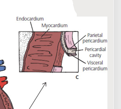
Visceral pericardium
The serous membrane on the immediate surface of the heart, composed of simple squamous epithelium; also known as the epicardium.

Parietal pericardium
The outer pericardial sac surrounding the heart, lined by simple squamous epithelium with loose connective tissue.

Pericardial space
The potential space between the visceral pericardium and parietal pericardium that contains pericardial fluid.

Epicardium
The outermost tissue layer of the heart wall, consistent with the visceral pericardium.

Myocardium
The thick middle muscular layer forming the wall of the heart, composed of striated cardiac muscle and connective tissue.
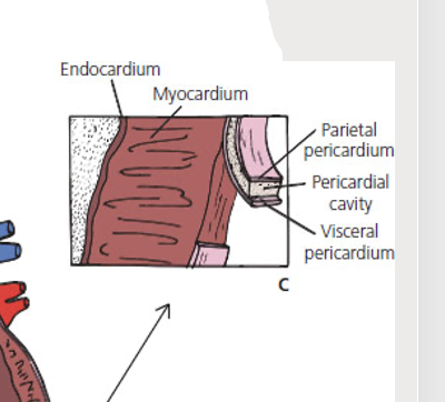
Endocardium
The smooth innermost lining of the heart chambers and valves, composed of simple squamous epithelium.
Base of heart
Higher flat part
Apex of heart
Lower pointed part on sternum
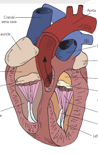
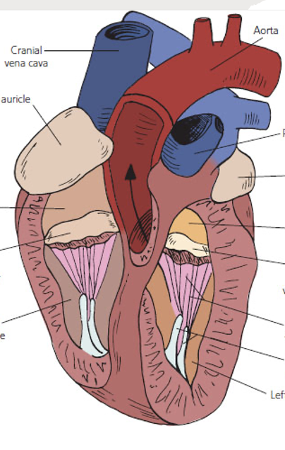
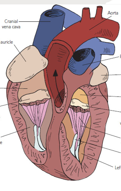
4 chambers of heart
Right and Left Aria (s. atrium) and Right and left Ventrical
Right atrium
The thin-walled, irregularly shaped craniodorsal heart chamber that receives deoxygenated systemic venous return from the cranial and caudal venae cavae; goes to tricuspid valve

Left atrium
The thin-walled craniodorsal heart chamber that receives oxygenated blood returning from the lungs via the pulmonary veins; goes to bicuspid valve
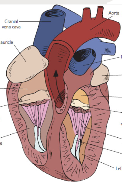
Auricle (Left and right)
Outer pouch of atria, when atrium swell up with blood, blood goes there as well

Interatrial septum
The internal muscular and membranous wall that divides the right atrium from the left atrium;
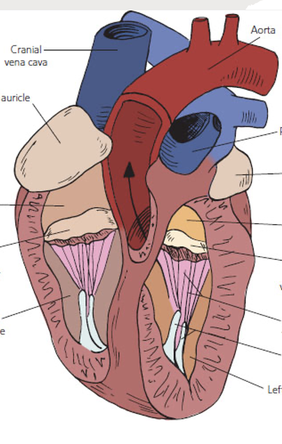
Right ventricle
The thick-walled, angled caudoventral chamber that pumps deoxygenated blood through the pulmonary valve into the pulmonary artery; goes to pulmonary semilunar valve

Left ventricle
The thickest-walled caudoventral chamber that ejects oxygenated blood under high pressure through the aortic valve into the systemic aorta; goes to aorta semilunar valve
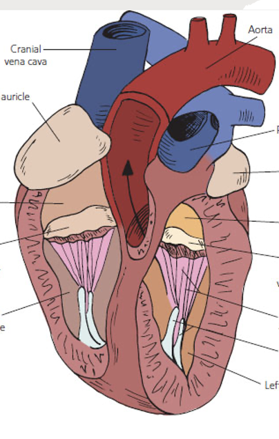
Interventricular septum
The thick muscular wall separating the right and left ventricles of the heart.

Atrioventricular (AV) valves
Connective tissue flap valves (cusps) situated between the atria and the ventricles that prevent backflow of blood during ventricular systole; right is tricuspid and left is mitral; if flow backwards forms clot
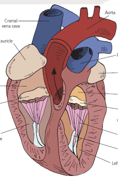
Tricuspid valve
The right atrioventricular valve located between the right atrium and the right ventricle.
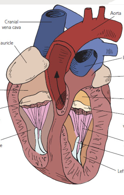
Mitral valve
The left atrioventricular valve (bicuspid valve) located between the left atrium and the left ventricle.

Chordae tendineae
Fibrous cord-like 'heart strings' connecting the free margins of the AV valve cusps to the papillary muscles.

Papillary muscles
Muscular projections extending from the interior ventricular walls that anchor the chordae tendineae to prevent valve prolapse during systole; muscle contracts as it pulls on heart strings and open av vavles

Semilunar (SL) valves
Crescent-shaped pocket valves located at the exits of the ventricles into the great arterial trunks; separates ventricles from blood vessel so blood stays in vessel

Aortic semilunar valve
The semilunar valve positioned between the left ventricle and the systemic aorta.
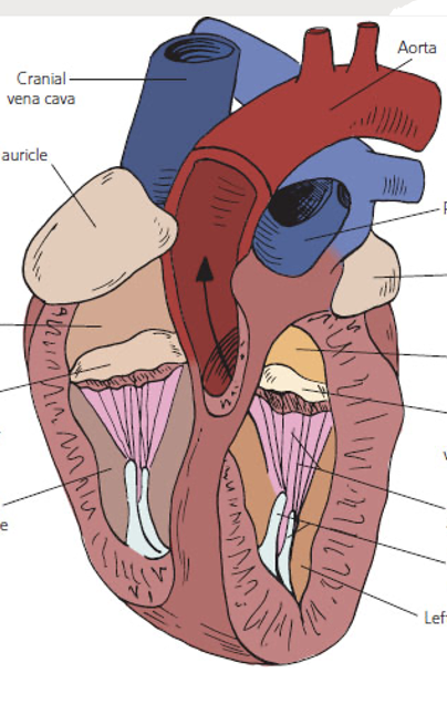
Pulmonary semilunar valve
The semilunar valve positioned between the right ventricle and the pulmonary artery trunk.

Canine thoracic heart anatomy (left lateral view)
Anatomical landmarks showing: 1. Flattened base, 2. Apex, 3. Right ventricle, 4. Left ventricle, 5. Right ventricular margin, and 6. Left ventricular margin.
Dual circulatory system
A circulatory arrangement characteristic of mammals and birds comprising two distinct loops: the pulmonary circuit and the systemic circuit.
Pulmonary circuit
The circulatory pathway through the right side of the heart, directing deoxygenated blood to the lungs for gas exchange and returning oxygenated blood to the left atrium.
Systemic circuit
The circulatory pathway powered by the left side of the heart, delivering oxygenated blood to body tissues and returning deoxygenated blood to the right atrium.
Oxygenated blood path (capillaries)
Aorta (Left) → artery → arterioles → Capillaries (deox) → venules → veins → Cranial and caudal vena cava (right)
Mammal and bird circulatory
Duel, 4 chambered

Cardiac action potential
The characteristic cyclical electrical sequence across myocardial cells comprising fluctuating resting potential (Phase 4), rapid Na+ depolarization (Phase 0), early repolarization (Phase 1), a Ca++ influx plateau (Phase 2), and K+ efflux repolarization (Phase 3).
What does action potential need
Na, K, CALCIUM
Leaky sodium channels
Cardiac pacemaker channels in nodal tissue that allow a gradual influx of Na+ to slowly elevate resting membrane potential toward threshold.
Plateau phase (Phase 2)
A prolonged period of sustained depolarization in cardiac muscle fibers resulting from the inward movement of Ca2+.
Repolarization (phase 3)
K+ leaves cell
Intercalated disks
Specialized cell junctions connecting adjacent cardiac muscle cells that facilitate rapid ion flow so the myocardium contracts synchronously as a functional syncytium; beat in unison
Purkinje fibers
Specialized, modified myocardial cells organized in the ventricular walls that conduct action potentials rapidly throughout the ventricles; sends signals through heart
Intrinsic cardiac conduction
The ability of heart tissue to initiate its own action potentials and contractions without requiring external nerve impulses.
Sinoatrial (SA) node
The primary pacemaker of the heart situated in the right atrium that sets the base rate of electrical firing.
Atrioventricular (AV) node
The specialized conduction node that receives impulses from the atria and introduces a brief pause to permit complete atrial emptying before ventricular contraction.
Atrioventricular (AV) bundle
The conducting bundle of His that exits the AV node and divides into right and left bundle branches descending along the interventricular septum.
Conduction network pathway (AP flow)
SA node (makes action potential) → AV node (pause at atria contract) → AV bundle (distributes impulse, branches) → Purkinje fibers (ventricles contract)

Electrocardiogram (ECG)
A diagnostic recording that measures electrical potential changes generated by myocardial depolarization and repolarization across the body surface; basically electricity in the heart
Sinus rhythm
The normal, regular cardiac rhythm initiated and controlled by the sinoatrial node.
Arrhythmia
Any abnormality, irregularity, or deviation from the normal sinus rhythm of the heart.
Leads
Points of electrode attachment, has P wave, PR interval, QRS complex, and T wave
P wave
The initial deflection on an electrocardiogram tracing representing the electrical depolarization of both atria.

PR interval
The ECG segment representing the conduction time of the action potential through the atria and the atrioventricular node.

QRS complex
The rapid, high-amplitude ECG waveform produced by the depolarization of the ventricles.

T wave
The ECG waveform corresponding to the electrical repolarization of the ventricles.

Lub-dub
1 beat caused by contraction; Lub from the closure of the atrioventricular valves and Dub from the closure of the semilunar valves.
Systole
The phase of the cardiac cycle during which ventricular myocardium contracts, creating high intracardiac pressure to eject blood.
Diastole
The phase of the cardiac cycle during which ventricular muscle relaxes and ventricles fill with blood under lower pressure.
First heart sound (S1)
The 'lub' sound produced by the closure of the atrioventricular (mitral and tricuspid) valves and resulting turbulent blood flow as ventricles contract.
Second heart sound (S2)
The 'dub' sound generated by the closure of the aortic and pulmonary semilunar valves at the conclusion of ventricular systole.
Third heart sound (S3)
An occasional low-frequency sound occurring during rapid ventricular filling, observed particularly in animals with large, slow hearts.
Heart murmur
An abnormal cardiac acoustic vibration caused by turbulent blood flow, commonly resulting from incomplete valve closure or stenotic openings.
Holosystolic murmur
A pathologic cardiac murmur that lasts throughout the entire duration of ventricular systole.
Pathway of blood in heart (underlined are oxygenated)
Left atrium → left AV valve (mitral) → left ventricle → aortic SL valve → aorta → arteries to various body parts → smaller arteries → arterioles→ capillaries → venules → small veins → larger veins → cranial and caudal vena cava → right atrium → right AV valve (tricuspid) → right ventricle → pulmonary SL valve → pulmonary artery → right and left pulmonary artery to each lung → smaller arteries → arterioles→ capillaries of alveoli → alveoli venules → small veins → larger veins → pulmonary vein → left atrium
Umbilical vein
A single fetal blood vessel that carries relatively oxygen-rich and nutrient-rich blood from the placenta to the fetus.
Umbilical arteries
A pair of fetal vessels that transport relatively deoxygenated, nutrient-depleted blood from the fetus back to the placenta.
Mammal fetal circuit
Fetas not breathing and liver not working but relies on the mother's placenta for oxygen and nutrients. Blood is shunted through the umbilical cord directly to the fetal circulation; since fetus blood consistent with mom blood, must be shunted from liver
3 shunts (bypass fetas liver/lung)
Ductus venosus
A fetal vascular shunt that connects the umbilical vein directly to the caudal vena cava, bypassing the fetal liver.
Foramen ovale
An interatrial fetal aperture that allows blood to pass directly from the right atrium to the left atrium, bypassing the unexpanded lungs.
Ductus arteriosus
A fetal vascular connection diverting blood directly from the pulmonary artery into the descending aorta, bypassing the pulmonary circulation.
Portal system
A vascular arrangement wherein blood flows from one capillary bed to another capillary bed through a vein, bypassing general systemic circulation; hepatic or remal
Hepatic portal system
The portal circuit that detoxify blood, vein collects blood from gastrointestinal tract to liver first and then heart, blood enters caudal vena cava

Renal portal system
A vascular shunt network found in non-mammalian vertebrates that directs venous blood from the hind limbs and tail directly through the kidneys to maintain perfusion and clear toxins; done during brumation/hibernation to maintain kidney function
Single circuit system
2 chambers, in fish
Operculum
The bony protective covering situated over the gills of fish, active in respiratory ventilatory movement.
Fish heart
A two-chambered cardiovascular organ consisting of one atrium and one ventricle through which deoxygenated blood passes only once per complete circuit.
Amphibian heart
A three-chambered organ consisting of two atria and a single shared ventricle, resulting in partial mixing of oxygenated and deoxygenated blood.
Reptilian heart
A three-chambered organ containing two atria and an incompletely divided ventricle whose internal trabeculae minimize the mixing of arterial and venous blood.
Crocodilian heart
A unique reptilian anatomical exception featuring a fully partitioned four-chambered heart with two separate atria and two separate ventricles.
3 layers in arteries in vein
Tunica externa (adventitia), tunica media, tunica intima
Tunica adventitia (tunica externa)
The outermost fibrous layer of a blood vessel or lymphatic vessel, formed primarily of loose connective tissue.
Tunica media
The middle layer of a blood vessel composed of smooth muscle cells and elastic or loose connective tissue fibers that controls vascular diameter, thick in arterioles due to elasticity and contraction for regulating blood flow.
Tunica intima (tunica interna)
The innermost lining of all blood and lymph vessels, consisting of an endothelial layer of simple squamous epithelium supported by a basement membrane, nutrients and electrolytes pass through
Arteries
Away from heart, carry oxygenated blood except pulmonary arteries.
Veins
Blood vessels that carry deoxygenated blood towards the heart, except pulmonary veins which carry oxygenated blood from the lungs.
Arteries as pressure reservoirs
The physiologic capacity of large elastic arterial walls to distend during systole and recoil during diastole, maintaining continuous forward hydrostatic pressure.
Veins as volume reservoirs
The capacitance property of thin-walled, compliant systemic veins that permits them to expand and hold large fractions of total blood volume at low pressures.
Endothelium
The simple squamous epithelial monolayer lining the interior surface of blood vessels, heart chambers, and lymphatics; the only cellular layer comprising capillaries.
Capillaries
¬Found all over body
¬Endothelium (tunica intima) only
¬Entered by arterioles
¬Exited by venules
¬Precapillary sphincter
-cell does not have direct access to blood; site of nutrient and gas exchange, and communicates with it when in fluid (O2, electrolyte, nutrient)
Precapillary sphincter
A ring of smooth muscle encircling the origin of a capillary from an arteriole or metarteriole that regulates blood flow into capillary beds; if something needs oxygen they open and let blood through. If not they close
Capillary hydrostatic pressure
The outward filtration force generated by fluid pressure within the capillary lumen that pushes plasma water and dissolved solutes into the interstitium; as water increase, pressure increase; blood cells and proteins do not leave, but if sphincter is too big, will leave and cause bleeding

Oncotic pressure
The inward osmotic pull generated primarily by non-diffusible plasma proteins (such as albumin) that draws fluid from the interstitial space back into capillaries; more proteins in space, more pressure

Starling forces
The net physical balance between hydrostatic pressure gradients and oncotic (colloid osmotic) pressure gradients determining fluid transudation across capillary walls.
Lymph vessel
A conduit for lymph that transports lymph fluid, proteins, and immune cells from tissues back to the circulatory system, playing a crucial role in fluid balance and immune response; recovers lost fluid and plasma
Venous valves
Semilunar folds of the tunica intima positioned within peripheral veins that facilitate unidirectional return of blood toward the heart and prevent backflow.
Skeletal muscle pump
The mechanical squeezing action of contracting skeletal muscles against deep veins that propels venous blood upward past one-way valves.
Thoracic respiratory pump
The generation of negative intrathoracic pressure during inhalation that creates a vacuum effect, sucking venous blood into the thoracic cavity toward the right atrium.
Exercise
Do this regularly to have better cardiovascular health
Lymphatic system
An accessory vascular system composed of thin-walled vessels lined by three tunics that recovers extravasated interstitial fluid, proteins, and lymphocytes; returns fluid to cranial vena cava and maintains fluid balance.
Lymphatic system layers
Same as vein
Lymph
The typically clear, colorless interstitial fluid collected and transported by lymphatic vessels, containing water, proteins, and leukocytes.
Thoracic duct
The major lymphatic trunk collecting lymph from the caudal body, abdomen, and thorax, emptying into the cranial vena cava near the heart.
Lacteals
Specialized lymphatic capillaries situated in the intestinal villi that absorb dietary lipids and fat-soluble substances (milky white); does not contain lymph but contains chyle
Chyle
The milky-white lymphatic fluid collected from lacteals within the gastrointestinal tract due to a high concentration of absorbed emulsified fats.