Anatomy Test 2
 Call Kai
 Learn
Learn Practice Test
 Spaced Repetition
 Match
Match Flashcards
 Knowt Play
1/91

There's no tags or description
Looks like no tags are added yet.
Name | Mastery | Learn | Test | Matching | Spaced | Call with Kai | Chat |
|---|
No analytics yet
Send a link to your students to track their progress
92 Terms
5 primary functions of skeletal system
1. Support
2. Storage of minerals and lipids
3. Blood cell production
4. Protection
5. leverage
Long bone
Longer than it is wide; example: humerus
short bone
Roughly cube-shaped; example: carpal
Flat bone
Thin, flattened bone; example: parietal bone
Irregular bone
Complex shape; example: vertebra
Sesamoid bone
Bone that develops within a tendon; example: patella
Sutural bone
Small bone located within a skull suture
fossa
bowl/depression. Example: intercondyle fossa on femur
Tubercle
Small knob or protrusion exaple greater and lesser tubercle on humerus. none on femur
Tuberosity
Rough area. Each bone has only one. Example ulna tuberosity, deltoid tuberosity(humerus), radial tuberosity, ischial tuberosity, gluteal (femur), tibial tuberosity (front)
Condyle
Rounded end that articulates with another bone, usually distal. Example: Femur has medial/lateral epicondyle and condyle. Humerous is has medial/lateral epicondyle and condyle in middle with capitiium and trochea.
Sinus
cavity within bone to reduce bone density and weight
Diaphysis vs Metaphysis vs Epiphysis
Diaphysis → Shaft of a long bone
Epiphysis → End of a long bone
Metaphysis → Region between the diaphysis and epiphysis
Osteon
Basic
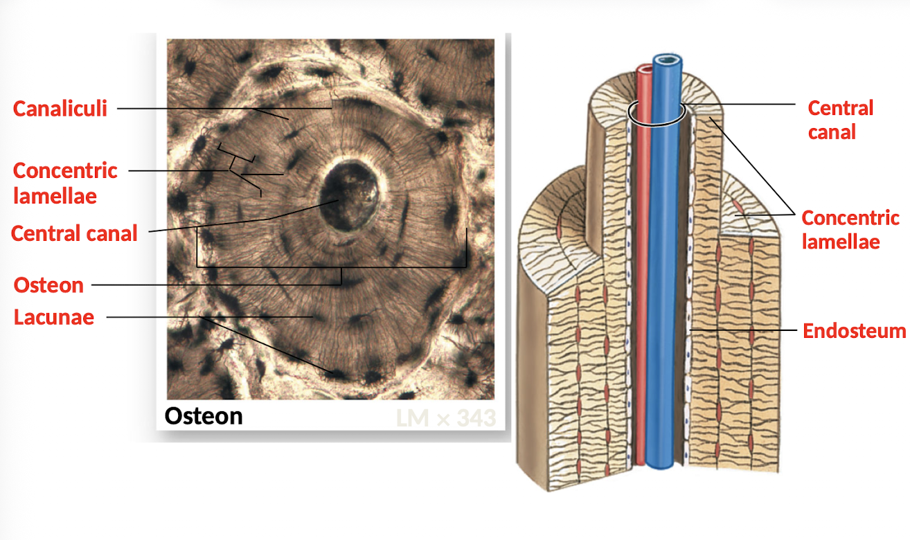
structural unit of compact bone
Importance of canaliculi
Allow osteocytes embedded in hard matrix to exchange nutrients/wastes and communicate through their extensions
Two major components of bone matrix
Mineral crystals (2/3) + protein fibers (1/3)
whats in mineral crystas of bone matrix and what does it do
Hydroxyapatite → Mineral crystals that contribute hardness/strength to bone
whats in protein fiber of matrix and what does it do
Collagen → Protein fibers that provide flexibility/tensile strength
spongey bone structure
Trabeculae (not osteons) with no blood vessals
spongey bone structure function
Reducses bone mass making bones light weight, space for bone marrow and distributes forces
yellow vs red bone marrow
Red bone marrow → Contains blood vessels; associated with blood cell production
Yellow bone marrow → Found in some bones and stores lipids
Periosteum. What is it and what are the functions
Covers and lines the inside of bones (outer membrane)
Isolates bone
Route for circulation and innervation
Aids bone growth and repair
Endosteum. What is it and what are the functions
Inner membrane in central canal
Lines medullary cavity
Covers trabeculae
Lines central canals
Contains bone cells
Active in growth and repair
Endochdonral Osstification
Bone builds from cartilage. Babies are mostly cartilage; then blood vessels develop nearby, which hollow out the cartilage to form bone. Hyaline cartilage remains at the ends of bones. Can be primary (diaphysis) and secondary (epiphysis)
Intramembranous ossification
Dermal bones such as mandible and clavicle.
Appositional Growth
Compact bone thickens and strengthens long bones with layers of circumferential lamellae. Bones dont get longer they get thicker
Bone remodeling
Ongoing replacement/recycling of bone matrix involving many bone cells which requires a blood supply
Why is remodeling important?
Daily maintenance, mineral recycling, adaptation to stress and fracture repair
Name 4 bone-cell types (make up 2% of bone mass, 98% matrix)
Osteogenic cells, osteoblasts, osteocytes, osteoclasts
Osteoprogenitor/Osteogenic cell
can transform into osteoblast or osteoclast, stem cell
Osteoblast
Bulids bone
Osteoclast
Destorys/ Reabsorbs bone. Breaks matrix down to maintain bone mass
Osteocyte
Mature bone cell that maintains bone matrix
Bone homeostasis
Balance between osteoblast bone building and osteoclast bone resorption
How do osteoclasts breakdown bone
Protease breaks down proteins. Cathepsin breaks down collagen. Low pH makes it acidic, which breaks down hydroxyapatite
explain chemical aspect of osteoclasts
inside osteroclast carbonic anahydrase enzyme speeds up process to make carbonic acid.
Why is calcium homeostasis so important
bone strength
muscle contracton
nerve signaling
blood clotting
heart function
2nd messanger
horomone realese
heart function
Factors that effect bone remodeling
Lifting weights, diet, movement level, racket sports (using one arm more)
Calcitriol (thyroid gland)
hormone made in the kidneys
helps absorb calcium and phosphorous from the digestive tract
its synthesis requires Vitamin D3
Goal is to lower blood calcium levels
Bone → osteoclasts inhibited while osteoblasts continue locking calcium into matrix
Intestine → decreased calcium absorption
Kidney → allows more calcium loss
Parathyroid hormone
Goal is to raise blood calcium levels
Bone → increased calcium release
Intestine → increased calcium absorption
Kidney → retains more calcium → less lost in urine
What is the axial skeleton
forms the central axis of your body. It has the skull throacic cage and vertebrae
axial skeleton functions
Supports and protects organs in body cavities
Attaches muscles of the head, neck, and trunk
Facilitates respiratory movements
Stabilizes parts of the appendicular skeleton
Coronal suture
Frontal + parietal bones
Sagittal suture
Right parietal + left parietal
Lambdoid suture
Parietal bones + occipital bone
Squamous suture
Parietal + temporal
Metopic/frontal suture
Associated with the frontal bone; your lecture labels the frontal/metopic suture.
Sinuses. what are they lined with?
cavities that decrease the weight of the skull. Lined with mucous membranes to protect the entrances of the respiratory system, cleans air.
Functions of the temporal bones
form lateral walls of cranium
contribute to the zygomatic arches
articulate with the mandible
surround/protect the inner ear
provide muscle attachment
Bony nasal septum
Perpendicular plate of ethmoid + vomer
jaw articulation
Temporal bone + mandible
zygomatic arch
Temporal process of zygomatic bone + zygomatic process of temporal bone
Functions of the sphenoid:
1. Forms part of the floor of the cranium
2. Unites the cranial and facial bones
3. Strengthen sides of the skull
4. Contains sphenoidal sinuse
• Functions of the ethmoid:
1. Forms anteromedial floor of cranium
2. Forms roof of the nasal cavity
3. Forms part of the nasal septum and medial orbital wall
4. Contains ethmoid air cells (small sinuses)
Nasal conchae function
The conchae increase the surface area/contact of air moving through the nasal cavity and create turbulence, helping condition inhaled air. Conchae help prepare incoming air before it travels deeper into the respiratory tract.
Vertebral arch
Pedicles + laminae
Hyoid bone
does NOT articulate with the skeleton.
Instead, it is held in place by muscles.
Its functions in your lecture include:
supporting the larynx
attaching muscles of the tongue
Vertebral column parts
Cervical → 7
Thoracic → 12
Lumbar → 5
Sacrum
Coccyx
what are c1 and c2 and their special features
atlas: occipital condyle
axis: dens
Cervical Vertebrae features
large foremen (thick spinal cord) , transverse foramen
Thoracic vertebrae features
Costal facets for rib connection, heart shaped body
Lumbar vertebrae
small foramen and big body
Rib classification
True ribs
Ribs 1–7
False ribs
Ribs 8–12
Floating ribs
Ribs 11–12
Important relationship:
All floating ribs are false ribs, but not all false ribs are floating ribs.
Rib articulations
Ribs articulate with thoracic vertebrae. Anteriorly, they articulatewitho the sternum through costal cartilage,
What is the appendicular skeleton
Think limbs + the structures attaching the limbs to the axial skeleton.
Pectoral Girdle and functions
Clavicle + scapula
functions:
Connects the arms to the body
Positions the shoulders
Provides a base for arm movement
Connects with the axial skeleton at the manubrium Bone Tissue (Chapter 6)
clavicle ends and Articulations
Sternal end (medial) → manubrium of sternum
Acromial end (lateral) → acromion of scapula
is radius or ulna lateral? do they articulate w eachother
Radius = lateral/thumb side
Ulna = medial/pinky side
Radius and ulna articulate with each other proximally AND distally.
Pelvic girdle and function
2 coxal bones
Each coxal bone is actually made from 3 fused bones:
Ilium + ischium + pubis
The pelvic girdle is strong enough to bear the weight of the body.
Pelvis
2 coxal bones + sacrum + coccyx
Patella
kneecap sesamoid bone (bone devloping in tendon)
Two important tarsal bones
Talus → involved in ankle articulation
Calcaneus → heel bone
Variations in the Skeleton (tells ppl abt how they lived)
muscle strength and mass
medical history
sex and age
body size
nutrition/environment
cause of death
types joints (3)
synarthrosis (immovable), amphiarthrosis (slightly movable), diarthrosis (freely movable)
suture
synarthrosis, coronal, saggital
gamphosis
synarthrosis tooth-maxillia/mandible
synchondrosis
synarthrosis, costal cartillage, epiphyseal cartillage/plates
synostosis
synarthrosis, metopic suture, ephiphyseal lines, coxal bone, transverse lines
syndesmosis
amphiarthrosis, interosseous membrane and distal tibia-fibula joint
synphysis
amphiarthrosis: pubic symphysis and intervertbreal discs
Gliding
Diarthrosis: intercarpal/intertarsal TMJ
Hinge
Diarthrosis: elblow, knee, ankle
Pivot
Diarthrosis: atlas-axis and radio-ulnar (proximal)
Condyler
Diarthrosis, sit in depression: radius-scaphoid, occipital-atlas, metacarpal/tarsal w/proximal phalange
saddle
Diarthrosis: thumb (metacarpal 1+trapezium)
Ball + Socket
Diarthrosis: hip (coxal/femur) and shoulder (humerous/scapula)
The RANK pathway
OPG blocks RANKL → less osteoclast activation → less bone breakdown
RANK = receptor involved in osteoclast activation
RANKL = binds RANK → promotes osteoclast formation/activation
OPG = binds/blocks RANKL so it cannot activate RANK → decreases osteoclast activity
Estrogen role in rank pathway
Estrogen↑ → OPG ↑ → RANKL blocked → osteoclast activity ↓
what does menopause cause
decrease in estrogen which causes RANKL to not be blocked and increases osteoclast activity and increases osteoporosis risk
Denosumab
binds RANKL.
Therefore:
Denosumab → RANKL blocked → can't activate RANK → osteoclast activation ↓ → bone resorption ↓
what does prednisone do
↓ OPG
AND
↑ RANKL
Therefore:
↑ osteoclast formation/activity → ↑ bone resorption
But it ALSO causes:
osteoblast apoptosis → ↓ bone formation
So prednisone hurts bone from both directions:
More destruction + less rebuilding = bone loss