Exam 2: PHOTOS ONLY (with desc)
 Call Kai
 Learn
 Practice Test
 Spaced Repetition
 Match
 Flashcards
 Knowt Play
1/88

There's no tags or description
Looks like no tags are added yet.
89 Terms
caput succedaneum
soft tissue swelling that crosses the suture lines caused by pressure on the head during birth
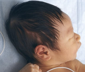
cephalohematoma
collection of blood between the scalp and skull that takes several weeks to resolve: remains on one side of the suture line

positional plagiocephaly
flattening of one side of the head due to moldable nature of baby heads and laying on their back

hydrocephalus
obstruction of draining of CSF → excessive accumulation → increased intracranial pressure and enlargement of head (macrocephaly)

down syndrome (trisomy 21)
most common chromosomal abnormality with characteristic facial abnormalities
upslanting eyes with inner epicanthal dolds
flat nasal bridge and small, broad nose
protruding thick tongue
ear dysplasia
broad neck
webbing and small hands with single palmar crease

allergic salute and crease
appearance of transverse line nose in response to chronically repeated use of head to push the nose up and back

torticollis
head is tilted to one side with the chin rotated toward the opposite side

goiter
enlargement of thyroid gland located in lower front of neck

pilar cyst
smooth, firm, swelling on scalp; well circumscribed, mobile

anisocoria
unequal pupils; can be a sign of stroke, brain tumor, hemorrhage (increases intracranial pressure)

enophthalmos
sunken eyes

exophthalmos
protruding eyes

strabismus
crossed eyes

ptosis
drooping eyelid

cataracts
vision becomes cloudy

glaucoma
loss of peripheral vision
requires intervention while you age
due to optic damage
risk factors: age, diabetes, intraocular pressure, optic damage, prolonged steroid use

age-related macular degeneration
loss of central vision
dry: chronic, happens over time
wet: abnormal growth of blood vessels under the retina
risk factors: smoking

homonymous hemianopsia
loss of same side of both eyes

heteronymous hemianopsia
loss of opposite halves of each eye
binasal: loss inner
bitemporal: loss outer

ectropion
drooping of bottom eyelid
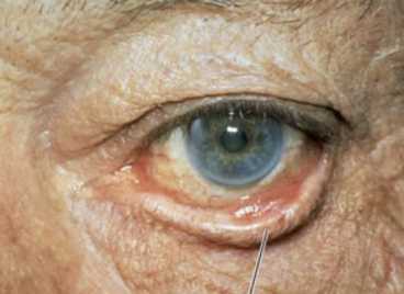
entropion
drooping of top eyelid
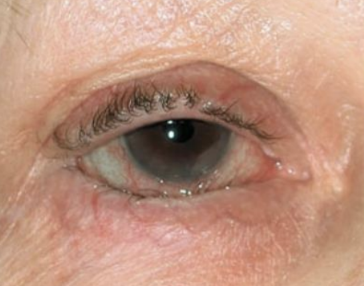
blepharitis
inflammation of inferior part of the eye where eyelashes grow

dacryocystitis
inflammation and infection of lacrimal sac caused by blockage in the nasolacrimal duct that leads to fluid stagnation

chalazion
chronic painless lump

hordeolum (styes)
acute painful bacterial infection → warm compress

conjunctivitis
inflammation of conjunctiva: beefy, red-looking vessels due to infection or chemical irritation
symptoms: itching, burning, foreign body sensation, eyelids stuck together

subconjunctival hemorrhage
due to increased pressure in eyeball → forceful vomiting → burst blood vessels

battle sign
bruising behind ear in the mastoid region → indicates basilar skull fracture
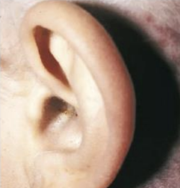
sebaceous cysts
smooth, round nodule on ear
commonly behind postauricular fold
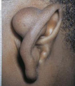
tophi
small, whitish yellow, hard nodules over helix or antihelix
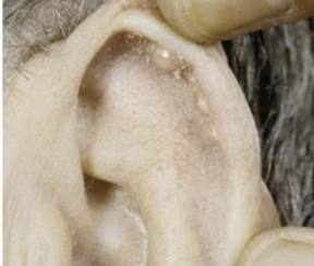
keloid
hypertrophic scar formed with excessive collagen which appear as a smooth, elevated nodule with a well-defined border on earlobe

chondrodermatitis nodularis helices
small, painful nodule on helix or antihelix

carcinoma
raised nodule with pearly translucent border on ear

cold sores
HSV-1
painful, fluid-filled blisters around lips

angular cheilitis
inflammatory condition that causes cracking, redness, and soreness at corners of mouth

candidiasis
(thrush) fungal infection of oral cavity
white patches on tongue, inner cheeks, or other oral surfaces
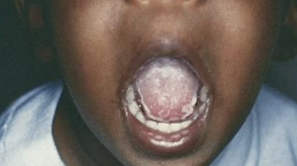
cleft lip and palate
craniofacial differences during embryologic growth

ankyloglossia
tongue-tied where tongue connects to floor of mouth

glossitis
inflammation of the tongue; swelling, redness, smooth appearance

macroglossia
abnormally enlarged tongue that interferes with breathing, eating, and speech

pharyngitis
inflammation of pharynx with bright red throat

umbilical hernia
soft skin covered mass protrusion → incomplete closure of ring
common in newborns but disappears by age 1

epigastric hernia
protrusion of abdominal structure with a hole in musculature = fatty nodule
self-reducing at the muscular wall of the abdomen
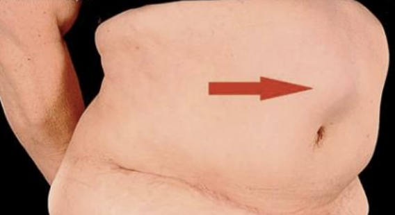
incisional hernia
bulge on or near an older operative scar
not usually noted until standing

abdominal aortic aneurysm (AAA)
pulsating mass with a bruit before the aorta splits
at risk of rupturing or disintegrating
located near the renal arteries but extends to umbilicus
femoral pulses decreases

annular
lesion pattern
circular, begins at center, and spreads outward

confluent
lesion pattern
run together, overlap, or border each other
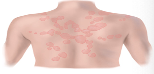
discrete
lesion pattern
distinct, individual lesions that remain separate
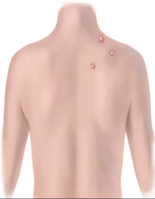
gyrate
lesion pattern
twisted, coiled, spiraled, snake-like

grouped
lesion pattern
cluster of lesions
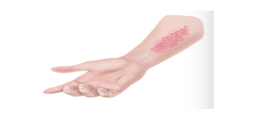
linear
lesion pattern
streak, line, stripe

target
lesion pattern
concentric rings

zosteriform
lesion pattern
linear arrangement along a unilateral nerve root

polycyclic
lesion pattern
annular lesions that grow together

macule/patch
primary lesion
flat, change in color
patch: when it is greater than 1cm

papule
primary lesion
elevated, solid, circumscribed, small, less than 1cm

nevus (mole)
primary lesion
clump of melanocytes

plaque
primary lesion
papules that overlap to become greater than 1cm

nodule
primary lesion
solid, elevated, hard or soft, larger than 1cm, deeper than a papule

wheal
primary lesion
superficial, raised, transient, or erythematous; irregular in shape
urticaria = overall condition (hives)

vesicles/bulla
primary lesion
elevated, free fluid
bulla: when greater than 1cm

cyst
primary lesion
encapsulated, fluid-filled in dermis or SUBQ tissue
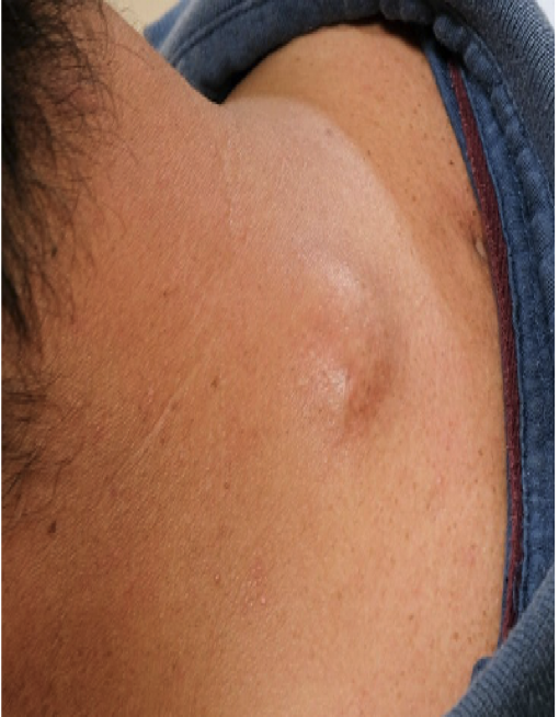
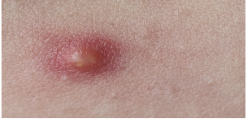
pustule
primary lesion
pus-filled, white top
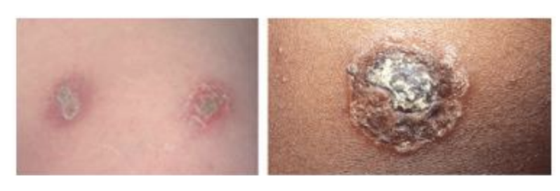
crust
secondary lesion
thickened, dried, and exudate when a vesicle or papule dries out
scale
secondary lesion
flake of skin due to dried skin

fissure
secondary lesion
linear crack with abrupt edges, extends into dermis

erosion
secondary lesion
shallow, scooped out, superficial, no bleeding, no scar after healing

ulcer
secondary lesion
deeper, into dermis, irregular shape, bleeding, can scar after healing

excoriation
secondary lesion
self-inflicted, superficial

scar
secondary lesion
replaced collagen with scar tissue
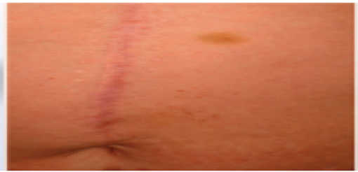
atrophic scar
secondary lesion
depressed skin without loss of tissue

keloid
secondary lesion
benign, excess of scar tissue

lichenification
secondary lesion
prolonged itching thickens the skin, tightly packed papules → raised area of skin

stage 1 pressure ulcer
non-blanchable erythema; redness does not turn white with pressure

stage 2 pressure ulcer
partial thickness skin loss; lost epidermis, exposed dermis
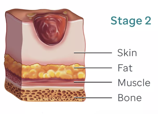
stage 3 pressure ulcer
full thickness skin loss; lost dermis, exposed SUBQ

stage 4 pressure ulcer
full thickness skin/tissue loss; exposure of muscle, tendon, or bone

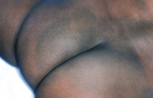
mongolian spots
hyperpigmentation common in black, asian, native, and latino patients
blue, black to purple macular area
often located on buttocks or sacrum
differentiate from bruising → inappropriate observation if no abuse associated
café au lait spots
large, round/oval patches
light brown pigmentation
neurofibromatosis: if more than 6 are greater than 1.5cm

harlequin color change
normal flushing
red or pale color down the midline or horizontal line

erythema toxicum
self-limiting
common rash in the first 3 to 4 days of life
tiny red macules and papules on cheeks, trunk, chest, back, or buttocks
idiopathic and there is no treatment

cutis marmorata
transient mottling; discoloration that comes and goes
response to being cold

acne
open and closed comedones
severe: papules, pustules, nodules → painful

solar lentigines
liver spots → small, flat, round macules
cluster of melanocytes due to sun exposure

keratosis
raised, thickened area of pigmentation
crusted, scaly, wart-like

actinic keratosis (AK)
red, tan, scaly plaques
sun-exposed areas

petechiae
little pin pricks, often due to popped capillaries

bruising
check for ecchymosis/contusion/hematoma

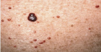
cherry (senile) hemangiomas
small, 1 to 5mm, smooth, and slightly raised bright red dots