RADT 113: Lower Limb and Pelvic Girdle TEST 5
 Call Kai
 Learn
Learn Practice Test
 Spaced Repetition
 Match
Match Flashcards
 Knowt Play
1/199

There's no tags or description
Looks like no tags are added yet.
Name | Mastery | Learn | Test | Matching | Spaced | Call with Kai | Chat |
|---|
No analytics yet
Send a link to your students to track their progress
200 Terms
Femur
Patella
Tibia
Fibula
Foot
What are the bones of the lower limb?
Femur
______:
thigh bone
2 femora - R & L
long bone
largest bone in the lower limb
longest, heaviest and strongest bone in the body
has a proximal end, body, and distal end
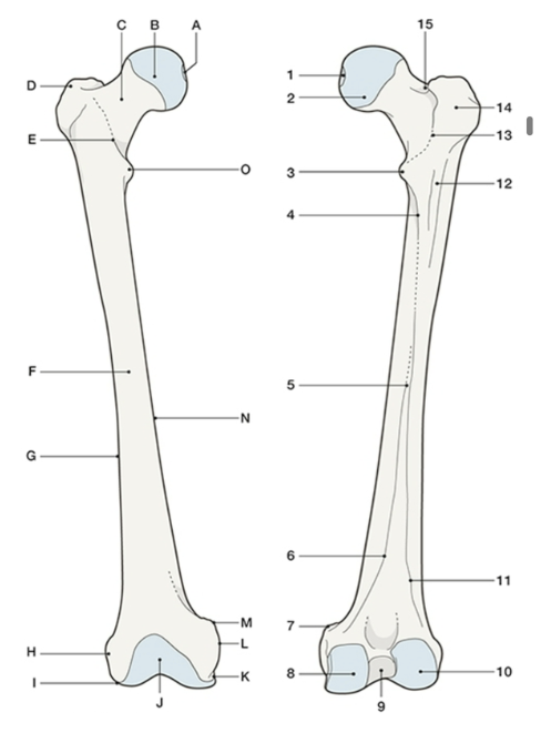
head
Femur - Proximal End:
________- the upper expanded rounded end that articulates with the acetabulum to form the hip joint
Not palpable
B / 2

Acetabulum
Femur - Proximal End:
What does the head of the femur articulate with to form the hip joint?
fovea
Femur - Proximal End:
The pit of the femoral head (_____) is a small but definite hole at the center of the head.
The ligament of the femoral head is attached to this.
A / 1

Neck
Femur - Proximal End:
________- the constricted part connecting the head to the body of the
femur
C

greater trochanter
Femur - Proximal End:
_________ __________- a large prominence on the lateral surface of the
upper femur
D / 14
lesser trochanter
Femur - Proximal End:
________ ____________- a much smaller rounded process that extends
medially at the junction of the neck and shaft
O / 3

intertrochanteric crest
Femur - Proximal End:
______________ _______- a ridge of bone that passes obliquely between
the trochanters on the posterior margin
13

intertrochanteric line
Femur - Proximal End:
_______________ _____ - a ridge that extends obliquely between the
trochanters on the anterior margin
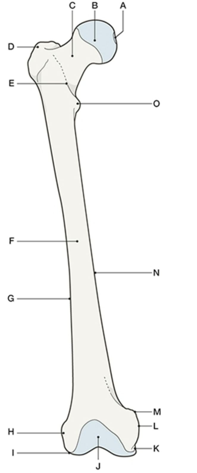
E
shaft
Femur - Body:
_______: the long cylindrical part that becomes broadened as it approached the knee
F

linea aspera
Femur - Body:
_____ _____ - a double bony ridge that passes longitudinally down
the posterior surface of the body of the femur
It divides below into 2 ridges that are directed to the margins of the femur
5

medial condyle
Femur - Distal:
___________ __________: rounded knoblike process that forms the medial part of the
distal end of the femur.
Its smooth surface articulates with the medial condyle of the tibia at the knee joint
K / 8

Lateral condyle
Femur - Distal:
________ __________: a similar rounded process forming the lateral part of the distal
end of the femur.
It articulates with the lateral condyle of the tibia forming part of the knee joint.
I / 10

medial epicondyle
Femur - Distal:
________ ________: a large prominence on the medial surface of the lower femur just above the medial condyle
L

Lateral Epicondyle
Femur - Distal:
_________ __________: a smaller rounded process on the lateral surface of the
lower femur above the lateral condyle.
H

intercondylar notch/ fossa
Femur - Distal:
____________ ___/___- a deep notch located between the inferior &
posterior parts of the femoral condyles
9

Adductor tubercle
Femur - Distal:
_______ ________- a small process on the medial surface above the medial epicondyle
M / 7

Patellar articular surface
Femur - Distal:
______ ________ _______ – the smooth anterior surface of the distal part
of the femur between the anterior parts of the femoral condyles. It forms
a joint with the patella
J
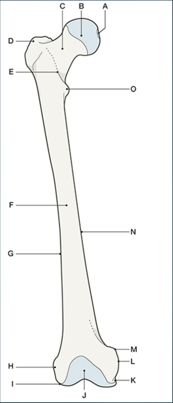
Popliteal Surface
Femur - Distal:
_______ _________-the flat area on the dorsal surface of the lower femur
between the divided ridges of the linea aspera
patella
__________: Aka knee cap
2 patellae, a R & L
Sesamoid bone
Lies anterior to the lower end of femur
It is palpable, and has a base and apex
sesamoid
Patella:
What type of bone is the patella?
base
Patella:
_______- the proximal slightly rounded upper border
A

apex
Patella:
_______- the lower pointed end
B

Facet for the lateral condyle of the femur
Patella - posterior:
_____ for the ____ _____ of the ____ - large, on posterior aspect
1

Facet for the medial condyle of the femur
Patella - posterior:
_____ for the ____ _____ of the ____ - smaller, on posterior aspect
3

Tibia
Shin bone
Long bone
Lies medial to the fibula
Expanded at the upper (knee) and lower (ankle) ends
Lies close to the skin surface
Tubercles of intercondylar eminence
Tibia - proximal end:
a small double pointed process on the proximal end of the tibia located between the articulation surfaces of the medial and lateral condyle of the tibia
Slightly posterior to the midpoint
2 small tubercles can be identified there
Extends into the intercondylar notch of the femur
C / 2

Medial condyle
Tibia - proximal end:
medial part of the upper expanded end of the tibia
Smooth, slightly concave proximal surface articulates wiht the medial condyle of the femur at the knee joint
D / 3

Lateral condyle
Tibia - proximal end:
the lateral part of the upper expanded end of the tibia
Smooth superior surface articulates with the lateral femoral condyle at the knee joint
B / 1

Tibial Tuberosity
Tibia - proximal end:
a round prominence on the anterior surface of the tibia below the condyle
Can be felt through the skin
E

Fibular Facet
Tibia - proximal end:
small smooth rounded area on the posterolateral border of the lateral tibial condyle
A

Fibula
Tibia - proximal end:
what does the fibulae facet articulate with to from the proximal tibiofibular joint?
Body
Tibia - _____:
long study part that becomes flattened as it approaches the ankle
Anterior border
Tibia:
G

Medial surface
Tibia:
H

Medial border
Tibia:
5

Inferior articular surface
Tibia - distal:
Smooth surface on the distal end of the tibia that articulates with the talus, forming the ankle joint
Medial Malleolus
Tibia - distal:
projects down from the margin of the distal end of the tibia
Reaches one inch below the level of the ankle joint
Forms a large prominence on the medial border of the ankle joint that is visible and palpable
I / 8

Fibular notch
Tibia - distal:
concave depression on the lateral surface of the tibia just above the ankle joint
Articulates the the medial border of the lower fibula to form the distal tibiofibular joint
J / 7

Fibular notch
Tibia - distal:
what the fibula interact with to form the distal tibiofibular joint?
Fibula
calf bone
Long bone
Long slender bone on the lateral side of the tibia
Ankle joint
Tibia:
What joint does the fibula form?
Ankle joint
Tibia:
does the tibia form the ankle joint or the knee joint?
Head
Fibula:
its upper expanded end of that articulates with the fibular articular surface of the tibia
K / 9

Apex/ Styloid process
Fibula:
the pointed upper end of the head
Body
Fibula:
long and slender
Lateral Malleolus
Fibula:
the distal expanded end of the fibula.
It extends down on the lateral margin of the ankle joint for about three quarters of an inch.
Its tip is at a lower level than that of the medial malleolus.
Along with the medial malleolus it forms the ankle mortice.
N / 12

Ankle mortice
Fibula;
What does the medial and lateral Malleolus form together?
Lateral surface
Fibula
m

Posterior border
Fibula
11

7
How many tarsal bones does the foot have?
Short bones
What types of bones are the tarsals?
Tarsals
What forms the posterior part of the foot?
Anterior
The (anterior/posterior) group of tarsals forms a joint with a metatarsal bone
Talus
Calcaneus
What are the proximal tarsal bones?
Navicular
Medial cuneiform
Intermediate cuneiform
Lateral cuneiform
Cuboid
What are the distal tarsal bones?
Talus
Tarsals:
It lies between the distal end of the tibia and the calcaneus, transmitting the body weight to the calcaneus
H

trochlea articular surface
Tarsal:
The _____ _____ _____ is the upper smooth convex surface of the talus that articulates with the lower end of the tibia.
The medial and lateral surfaces of the body below the trochlea are smooth and articulate with the medial and lateral malleoli forming the ankle mortice.
Body
Tarsals - talus:
What part of the talus articulates with the medial and lateral malleoli forming the ankle mortice?
Head
Tarsals - talus:
is the rounded anterior end of the talus.
Its convex anterior surface articulates with the posterior surface of the navicular bone
Inferior
Tarsals - talus:
The (inferior/superior) surface of the talus has 3 articular surfaces that form joints with smooth surfaces on the calcaneus.
The middle one lies above the sustentaculum tali.
Calcaneal Tuberosity
Tarsals - calcaneus:
its enlarged posterior end- visible and palpable
sustentaculum tali
Tarsals - calcaneus:
a small but definite shelf of bone that projects medially from the medial surface of the calcaneus immediately below the talus, which it helps to support
Navicular
Tarsals:
boat shaped
Lies anterior to the talus and behind the 3 cuneiform bones, on the medial side of the ankle
G

Cuboid
Tarsals:
lies on the lateral side of the foot and has the calcaneus behind it, with the fourth and fifth metatarsals in front
J

Medial cuneiform
Tarsals:
first cuneiform-
located on the medial border of the foot anterior to the navicular and posterior to the first metatarsal bone
D

Intermediate cuneiform
Tarsals:
second cuneiform-
located lateral to the medial cuneiform,
posterior to the second metatarsal bone and anterior to the navicular bone
E

Lateral cuneiform
third cuneiform-
lies lateral to the intermediate cuneiform,
posterior to the third metatarsal, and in front of the navicular bone
F

cuneiform
Tarsals:
All 3 _______ bones articulate with the navicular bone and with a metatarsal bone
5
How many metatarsals are in one foot?
First
Second
Third
Fourth
Fifth
How are the metatarsals named from medial/ great toe side of the foot to the little toe?
Fifth
Which toe has a tuberosity on the lateral margin of the bone?
joints
The bases of the metatarsals form _____ with the anterior tarsal bones
bases
The heads of the metatarsals articulate with the ______ of the proximal phalanges of the toes
14
How many phalanges are in one foot?
5
How many digits are in one foot?
Two
How many phalanges does the big toe (hallux) have?
3
How many phalanges do digits 2 through 5 have each?
Shaft of distal phalanx of great toe
A

Shaft of proximal phalanx of great toe
B

Shaft of 1st metatarsal
C

Base of 5th metatarsal
K

Shaft of 5th metatarsal
L

Head of 5th metatarsal
M

Shaft of proximal phalanx of 5th toe
N

Shaft of middle phalanx of 4th toe
O - part

Head of distal phalanx of 3rd toe
part
P

Interphalangeal joint of great toe (synovial hinge joint)
1
list joint type too

1st metatarsophalangeal joint (synovial ellipsoid joint)
2

5th distal interphalangeal joint (synovial hinge joint)
3

2nd proximal interphalangeal joint (synovial hinge joint)
4

Knee joint
Joints of the Lower Limb:
Formed by the medial and lateral condyles of the femur articulating with the medial and lateral condyles of the tibia, and the patellar surface of the femur articulating with the patella
Superior tibiofibular joint
Joints of the Lower Limb:
Located between the adjacent upper margins of the tibia and fibula
Formed by a fibular articular surface on the upper posterior margin of the lateral tibial condyle articulating with the articular surface on the medial margin of the head of the fibula
Inferior tibiofibular joint
Joints of the Lower Limb:
Located between the inferior margins of the tibia and fibular above the ankle joint
Formed by the medial surface of the lower part of the fibula articulating with the fibular notch of the tibia, on its lateral side
Ankle Joint
Joints of the Lower Limb:
Located at the distal ends of the tibia and fibula where they join the foot
Formed by the inferior articular surface of the tibia articulating with the trochlea of the talus and the medial and lateral malleoli with the medial and lateral surface of the talus
Intertarsal joint
Joints of the Lower Limb:
located between adjacent tarsal bones
5
Joints of the Lower Limb:
how many tarsometatarsal joints are in one foot?
Tarsometatarsal joint
Joints of the Lower Limb:
Located between the tarsal and metatarsal bones of the foot
Formed by the medial cuneiform articulating with the base of the 1st metatarsal, the intermediate cuneiform with the base of the 2nd metatarsal, the lateral cuneiform with the base of the 3rd metatarsal, and the cuboid with the bases of the fourth and fifth metatarsals
Intermetatarsal joint
Joints of the Lower Limb:
4 in each foot
Gliding joint
Located between the bases of the adjacent metatarsal bones