Anatomy
 Call Kai
 Learn
 Practice Test
 Spaced Repetition
 Match
 Flashcards
 Knowt Play
1/111

There's no tags or description
Looks like no tags are added yet.
112 Terms
What are the main paranasal sinuses and where do they drain within the nasal cavity?
frontal sinuses, ethmoid sinuses, sphenoid sinuses and maxillary sinuses
frontal, maxillary and anterior ethmoid drain into the middle meatus
posterior ethmoid drains into the superior meatus or sphenoethmoidal recess
sphenoid sinus drains into the sphenoethmoidal recess


If a nasal tube were inserted as part of a procedure, which meatus and anatomical pathway should the tube follow to avoid trauma?
Middle meatus used as busiest drainage area
Tube should be advanced along floor of nasal cavity away from middle turbinate and cribriform plate, to the posterior opening into pharynx.
Which arteries supply the nasal septum and lateral wall, and why is Kiesselbach’s area clinically significant?
Sphenopalatine artery - supplies blood to lateral wall of nose and nasal septum
Anterior ethmoidal artery - supplies upper portion of nasal cavity
Posterior ethmoidal artery - supplies ethmoidal air cells and superior portion of nasal septum
Greater palatine artery - supplies hard palate and blood flow in nasal cavity
Superior labial artery - supplies anterior portion of nasal septum
These arteries form a network at Kiesselbach’s plexus, which is crucial for blood supply to the nasal septum and a common site for nosebleeds.


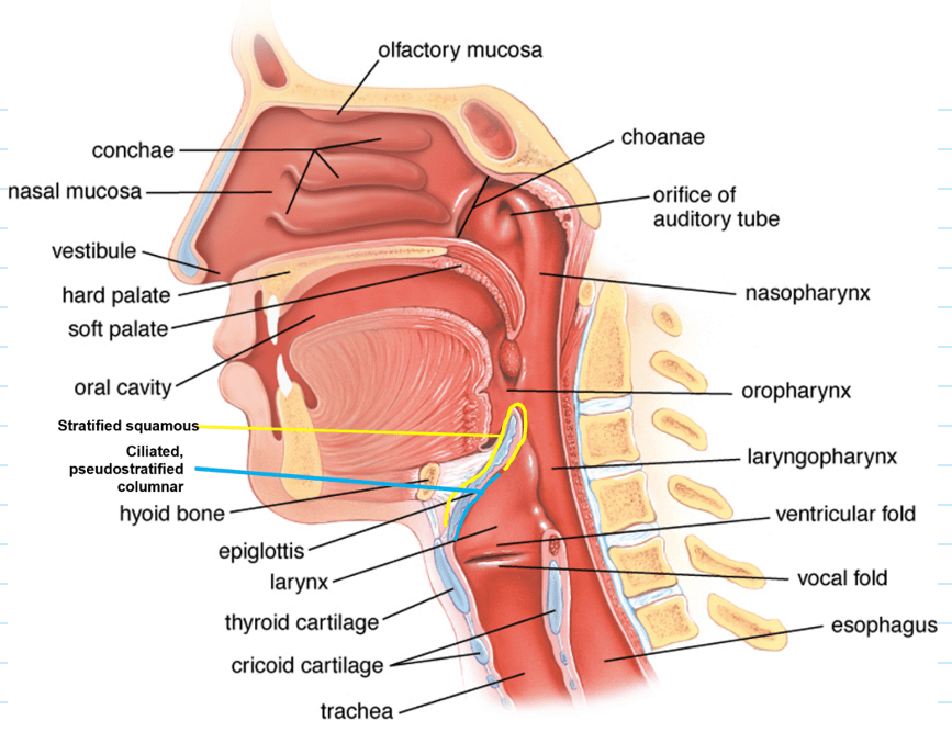


Name structures around pharynx (yellow), pharynx structures (blue) and types of epithelium (green)


Anatomical position of nasopharynx and what major features are found in the region? What type of epithelium is found here?
the nasopharynx is the uppermost part of the pharynx, located posterior to the nasal cavity and extending from the base of the skull to the soft palate
features in the region: pharyngeal tonsils, auditory tube openings, tubal tonsils, pharyngeal recess
nasopharynx is lined with pseudostratified ciliated columnar epithelium - facilitating mucus transport and airway defense


Anatomical position of oropharynx and what boundaries define the region? What type of epithelium is found here?
the oropharynx lies between the soft palate and the upper border of the epiglottis, serving as a passage for both air and food
features: base of the tongue (anterior), palatine tonsils (located in lateral walls), soft palate and uvula (superior boundary), posterior pharyngeal wall (posterior boundary)
oropharynx is lined with stratified squamous epithelium - providing protection against mechanical stress from food
Anatomical position of the laryngopharynx and key structures found in the region
the laryngopharynx extends from the upper border of the epiglottis to the cricoid cartilage, posterior to the larynx
structures: epiglottis (anterior boundary), piriform fossae (lateral recesses), posterior pharyngeal wall (posterior boundary)
laryngopharynx is lined with stratified squamous epithelium - protecting against abrasion from ingested material


At what vertebral level do the trachea bifurcate?
T4/T5, Manubriosternal angle, Angle of Louis
Foreign bodies that fall down the trachea are more likely to enter which bronchus and why?
right bronchus as it’s shorter, more vertical and fatter than the left
also there is a ridge in the middle at the base of the trachea, called the carina of the trachea - lies slightly to the left of the base
hence easier to fall into the right bronchus (in adults)
How do the bronchi divide?
17-23 divisions - alveolar ducts and alveoli
primary bronchi then secondary bronchi then tertiary bronchi then bronchioles

What has been removed? What can you identify?
ribcage/ thoracic cage have been removed, as well as intercostal muscles
as well as some overlying muscles such as pectoral muscles, then some fascia and skin
sternum has been removed as well as some large blood vessels
heart and lungs have been exposed
diaphragm inferior to heart and lungs
liver inferior to diaghragm
What are the three compartments of the thoracic cavity?
two pulmonary cavities, containing lungs and pleurae
central mediastinum, containing all other thoracic structures (including the heart)

Identify structures 1-9 on this transverse slice of CT scan.
sternum
vertebra
rib
rib
rib
rib
pulmonary cavity - contains lung
pulmonary cavity - contains lung
central mediastinum
Why is the pleura described as serous?
secreting fluid (pleural fluid)
Label each pleura based on colour used to highlight it:

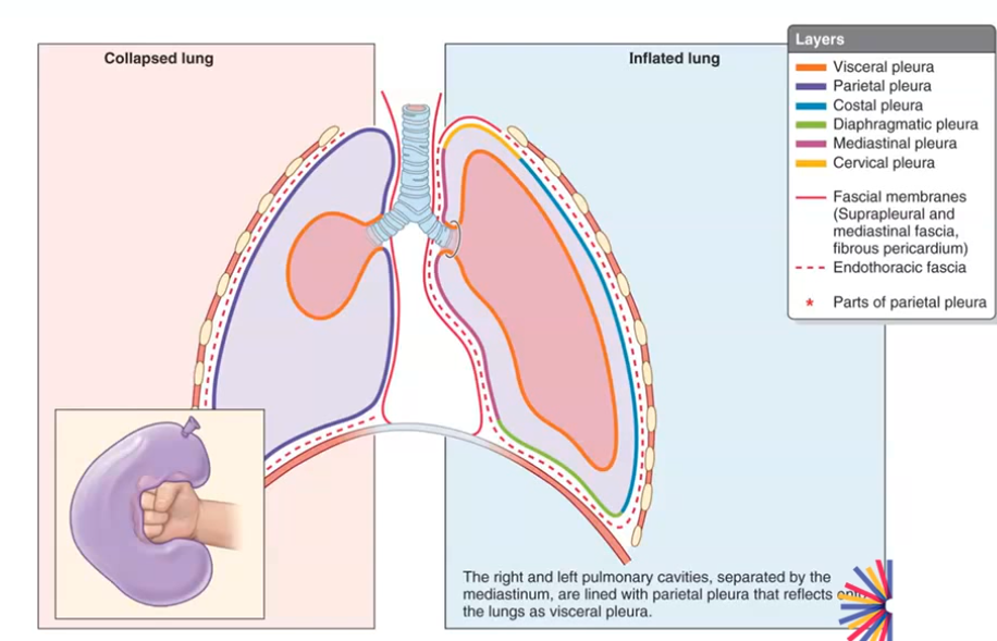
Difference between visceral pleura and parietal pleura. Where do they join?
visceral pleura covers the surface of the lungs
parietal pleura lines the pulmonary cavities - 4 different regions: cervical pleura, mediastinal pleura, diaphragmatic pleura, costal pleura
pleural membranes are continuous - join at the hilum
pleura permits smooth gliding - serous fluid
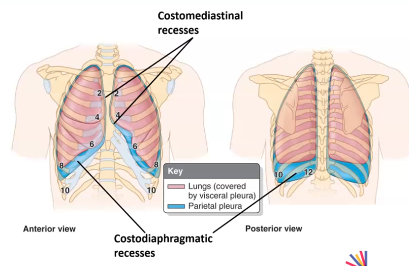
What are the costomediastinal and costodiaphragmatic recesses and role in inspiration and expiration?
spaces in the pleural cavity - between visceral and costal pleura
costodiaphragmatic recess - found inferiorly at the junction where the costal pleura meets diaphragmatic pleura
costomediastinal recess - found anteriorly behind the sternum, where the costal pleura reflects to become mediastinal pleura. it is larger on the left side to accommodate the cardiac notch of the left lung and heart
lungs expand into recesses during inspiration and retreat from them during expiration
Describe the surfaces of the lungs.

apex - extends into root of neck above clavicles
diaphragmatic surface - broad inferior surface in contact with diaphragm
costal surface - area in contact with ribs
mediastinal surface - medial surface of lungs surrounding mediastinum
hilum - point on mediastinal surface at which structures leave and enter lung (not shown)
Describe the borders of the lungs.
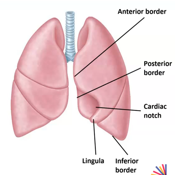
anterior border - where costal and mediastinal surfaces meet anteriorly
posterior border - where costal and mediastinal surfaces meet posteriorly
inferior border - circumscribes diaphragmatic surface
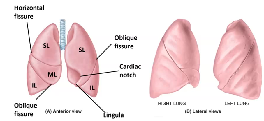
cardiac notch - indents anterior border of left lung (makes room for heart)
lingula - process of left superior lobe that extends below cardiac notch
Surface anatomy of the lungs (fissures and lobes).
right lung has two fissures (horizontal and oblique) and three lobes (superior, middle and inferior)
left lung has one fissure (oblique) and two lobes (superior and inferior)

What lobe of the lung is affected in this X-ray?
Middle lobe of the right lung - white mass that shouldn’t be there - it is full of fluid
Where would you listen to auscultate anteriorly - apex of right lung, superior lobe of right lung, middle lobe of right lung and inferior lobe of right lung?
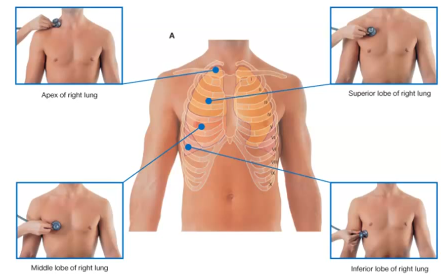
apex of lung - just above clavicle
superior lobe - between ribs 2 and 3 on both sides
middle lobe of right lung - at ribs 4 to 5, medial to nipple
inferior lobe - inferiolateral to nipple at ribs 6-7
Where would you auscultate posteriorly to listen to: apex of left lung, superior lobe of left lung and inferior lobe of left lung?

Relationship of ribs and lungs - lung borders coincide with which ribs?

mediastinal border coincides with 2nd and 4th ribs in sternal line
Hila of the lungs - what is it and importance of it?
wedge-shaped area on mediastinal surface
where structures forming the root of the lung enter and exit
enclosed by area of continuity of visceral and parietal pleura - more pleura here than needed to cover up the hilum, hangs down in a pulmonary ligament
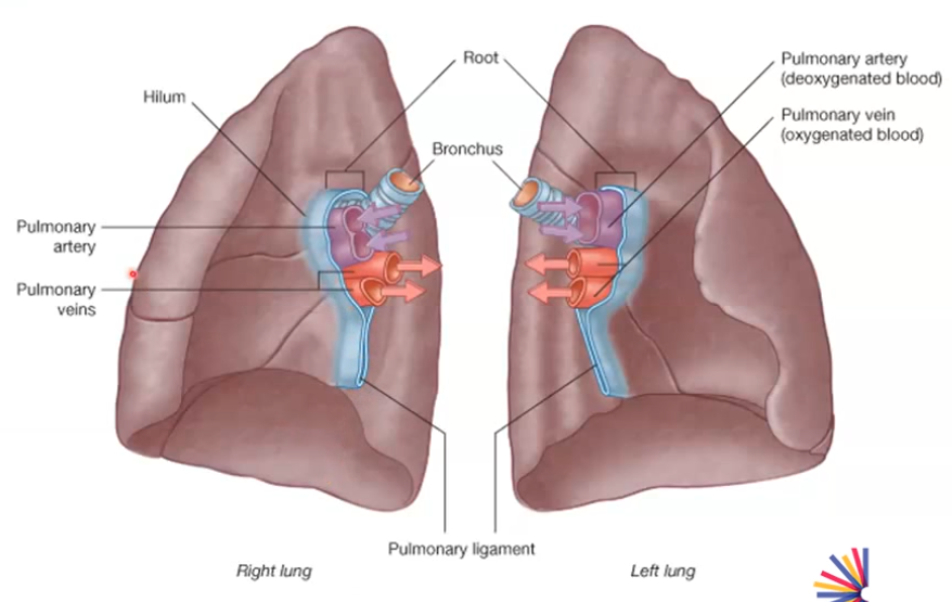
Contents of the hilum?
principal bronchus
pulmonary artery
pulmonary veins
bronchial artery
bronchial vein
nerves - via pulmonary plexus
lymphatics

Describe the arrangement of the structures in the hila
Relationship between pulmonary artery and bronchus = RIGHT ANTERIOR LEFT SUPERIOR
Pulmonary artery is anterior to bronchus in right hilum but superior to it in left hilum
bronchi have rings of cartilage and are easier to identify
in the right side of hila, the bronchi have already divided. anterior to the bronchus in the hilum is the pulmonary artery, then pulmonary veins are inferior to the pulmonary artery
in the left side of hila, pulmonary artery is superior to the bronchus. pulmonary veins can then be located.


Identify as many structures as possible on chest radiograph.

hemidiaphragm = half diaphragm curve
What is the mediastinum and its boundaries?
middle part of the thoracic cavity surrounded by pulmonary cavities
boundaries:
thoracic inlet (root of neck)
diaphragm
mediastinal pleura (bounded on either side of mediastinum)
What are the main contents of the mediastinum?
heart and its vessels
oesophagus
trachea
phrenic and cardiac nerves
thoracic duct (largest lymphatic vessel of lymph system)
thymus
lymph nodes of central chest
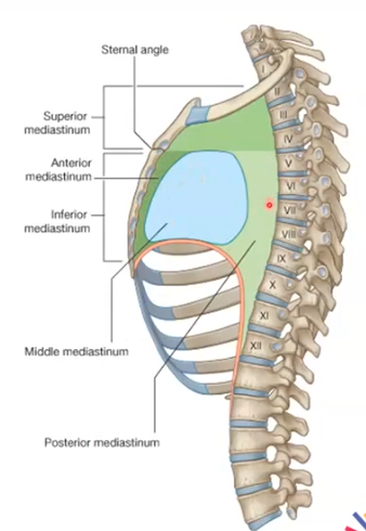
How is the mediastinum divided?
divided into superior and inferior parts divided by the thoracic plane, inferior mediastinum is larger than superior.
the inferior mediastinum is further divided into anterior, middle and posterior mediastinum
thoracic plane at the level of:
manubrio-sternal joint
2nd costal cartilage
T4/T5 invertebral disc
What are the core contents of the superior mediastinum?
Aortic arch and its branches:
brachiocephalic artery
left common carotid artery
left subclavian artery
brachiocephalic veins
trachea
oesophagus*
thoracic duct*
vagus and phrenic nerves*
*also in other mediastinal divisions

What are the boundaries of the divisions of the inferior mediastinum?
Anterior mediastinum - between sternum and pericardium
Middle mediastinum - bounded by pericardium (contains heart)
Posterior mediastinum - between pericardium and thoracic vertebrae
What are the contents of the anterior mediastinum?
thymus gland in children
fibrofatty connective tissue in adults
lymph nodes
sternopericardial ligaments
internal thoracic vessels, branches and tributaries
anterior mediastinum is very narrow

What is shown in this prosection?
Transverse section of chest
sternum seen anterior
posterior - vertebra
posterior to the sternum the anterior mediastinum can be seen - very narrow
heart in the middle mediastinum, and around the heart, fibrous sac of pericardium can be seen
lungs can be seen as well as trachea and windpipe
What are the contents of the middle mediastinum?
pericardium
heart
phrenic nerves
deep cardiac plexus
pulmonary vasculature
adjoining great vessels
What is the pericardium?
Double-walled, fluid-filled sac that surrounds and protects the heart and the roots of the major blood vessels
What are the contents of the posterior mediastinum?
oesophagus
vagi on oesophagus
thoracic duct
descending thoracic aorta
azygos vein
hemiazygos and accessory hemiazygos veins
lymph nodes

Label features of aorta.


Label structures A-E in this CT scan
A+B - brachiocephalic veins
C - brachiocephalic artery
D - left common carotid artery
E - left subclavian artery

Label structures 1-5 in this CT scan.

AAo - ascending aorta
DAo - descending aorta
LA - left atrium
RA - right atrium
RV - right ventricle

Label structures 1 and 2 in this CT scan.

SVC - superior vena cava
What is the lung parenchyma?
The lung parenchyma is the functional tissue of the lungs responsible for gas exchange. It includes the alveoli, respiratory bronchioles, alveolar ducts, alveolar sacs, pulmonary capillaries, and the interstitial framework that supports them.
What are the main functional roles of the pericardium?
pericardium is a double-walled sace that encloses the heart and roots of the great vessels
it anchors the heart in the mediastinum and attaches to the diaphragm, sternum and great vessels to prevent excessive twisting or displacement during body movement
inner serous layers secretes small amount of pericardial fluid into the pericardial cavity, which acts as a lubricant to minimise the friction generated as the heart continuously beats and slides against neighbouring tissues
shields heart muscle from mechanical trauma and serves as anatomical barrier to block spread of infections from nearby structures like the lungs
tough, inelastic outer fibrous layer places a physical constraint on how rapidly or large the heart chambers can dilate, protecting heart from acute volume overload
Effects of inflammation or fluid accumulation in pericardial cavity
inflammation causes inner and outer layers of pericardium to become rough and swollen so when heart beats the layers rub against each other, generating sharp chest pain that lessens when sitting up and leaning forward
excess fluid in pericardial space squeezes the heart chambers, restricting heart from expanding or filling with blood properly during diastole, leads to a lower stroke volume and less blood pumped to body, triggering a fast heart rate (tachycardia) as the body tries to compensate
What is meant by a sinus in anatomical terms?
hollow cavity, space, channel or pocket within a bone, organ or tissue
Where is the transverse pericardial sinus and what is the clinical significance of this passage?
short, tunnel-like passage inside pericardial cavity of the heart
anterior and superiorly: ascending aorta and pulmonary trunk
posteriorly: superior vena cava and left atrium
clinical significance - used in surgery during cardiac procedures to slide a surgical clamp around the aorta and pulmonary trunk to isolate the major blood vessels and temporarily control blood flow

What is the relationship between the fibrous pericardium to the diaphragm and great vessels and why is this important for the movement of the heart during respiration?
fibrous pericardium is a tough, cone-shaped sac that anchors the heart
broad, lower base of pericardium fuses directly with the central tendon of the diaphragm - because of this, the position and movement of heart are influenced by diaphragmatic motion (breathing)
superiorly, the apex of the fibrous pericardium turns upwards and is continuous with the outer connective tissue layer of the roots of the great vessels, hence the vessels are enclosed (aorta, pulmonary trunk/arteries, superior and inferior venae cavae and pulmonary veins)
by blending with these blood vessels, the fibrous pericardium suspends and holds the heart cranially, preventing heart from twisting or moving excessively during deep breaths, sudden body movements or changes in posture
Why are pulmonary veins often coloured purple rather than blue or red in anatomy textbooks?
as they carry deoxygenated blood, which in reality appears deep red-purple or maroon
Why do the atria have both rough and smooth internal surfaces? Describe how this reflects the embryological development of the heart.
smooth area is where major systemic veins (superior and inferior venae cavae, and coronary sinus) empty blood into heart with minimal resistance
in right atrium, smooth posterior wall develops from sinus venosus of primitive embryonic heart
in left atrium, smooth walls develops by incorporation and absorption of primitive pulmonary veins
rough surface helps auricles provide extra contractile strength and volume capacity to help squeeze blood into the ventricles during atrial systole, while preventing excessive blood stasis
rough sections, marked by pectinate muscles, are derived from primitive atrium
What is the functional significance of the fossa ovalis? Describe how this structure changes from fetal life to after birth in a healthy individual.
fossa ovalis - small, oval-shaped depression in the right atrium of the heart on the interatrial septum (wall separating right and left atria)
it is the closed-off remnant of the foramen ovale, an opening that allowed oxygenated blood to bypass the non-functional fetal lungs during development
when a baby takes its first breath, lung pressure drops and pushes a flap of tissue (septum primum) shut against the wall. over time, this flap fuses completely to form the depression of the fossa ovalis

What is the clinical significance of the fossa ovalis?
in about 25% of people, the fossa ovalis fails to fuse completely, leaving a small, tunnel-like gap called a patent foramen ovale. often asymptomatic, however PFO can sometimes allow blood clots or bubbles to pass from the right side of the heart to the left.
as it is thin and centrally located, it is the primary site used for transseptal punctures (accessing left atrium from the right atrium during cardiac procedures)
What are the papillary muscles, and how do they contribute to the function of the atrioventricular (AV) valves?
small cone-shaped muscular projections located inside ventricles of the heart
they connect to the cusps of the atrioventricular valves - mitral vale on left side and tricuspid valve on right side - via thin fibrous cords called chordae tendineae
they prevent backflow - when ventricles contract, the papillary muscles contract at the same time to pull tight on the chordae tendineae - which stops the valve leaflets from folding backward into the atria, preventing blood from leaking backward
What might happen if the papillary muscles or chordae tendineae are damaged? Name a common cause of damage.
weakness, dysfunction or rupture of papillary muscles can lead to severe mitral or tricuspid regurgitation, where blood flows backward into the heart instead of moving forward to the body - severe valve regurgitation
Heart attack (myocardial infarction) is a cause of damage as blocked coronary artery starves the papillary muscle of oxygen, causing the tissue to weaken and tear.
What roles does the ‘cardiac skeleton’ play in cusp integrity and in electric conduction?
cardiac skeleton - fibrous skeleton of the heart
it consists of dense connective tissue rings that encircle the atrioventricular and semilunar valves, providing strong, rigid points of attachment for the valve cusps
fibrous rings prevent the valves from stretching or deforming under high hemodynamic pressure during ventricular contraction
Role in electric conduction - dense collagen of the cardiac skeleton is non-conductive, and forms impermeable partitions that separate atrial myocardium from ventricular myocardium, blocking random electrical spread from atria to ventricles. Acts as an electrical insulator.
Which are the atrioventricular valves and which are the semilunar valves?
Atrioventricular - mitral and tricuspid
Semilunar - aortic and pulmonic
Where is the sinoatrial node located? What is the significance of the sinoatrial node in cardiac function?
located in the upper wall of right atrium, near entrance of superior vena cava
it is the natural pacemaker of the heart - generates rhythmic electrical impulses without needing external nerve stimulation, sets the heartbeat
How does the autonomic nervous system modulate sinoatrial (SA) node activity?
autonomic nervous system modulates SA node activity - sympathetic nervous system speeds it up during stress/exercise, parasympathetic nervous system slows it down during rest
What blood vessel gives off a branch to supply arterial blood to sinoatrial (SA) node?
most commonly the artery branches off the right coronary artery
less commonly branches off the left circumflex artery
the sinoatrial nodal artery supplies arterial blood to the sinoatrial node
How variable are the coronary vessels in the population?
typical anatomy seen in 97.9% of population - standard of right coronary artery and left main coronary artery, which splits into left anterior descending artery and left circumflex artery
anatomical variations such as branch trifurcations, differences in vessel length are very common
congenital anomalies such as structural deviations in origin, course or termination (e.g. coronary artery rising from wrong aortic sinus) occur in 1-2% of individuals
coronary dominance variabilities (determines supply to posterior heart):
right dominant (70-86% of people) - the posterior descending artery (PDA) arises from right coronary artery
left dominant (6-15% of people) - PDA arises from the left circumflex artery
co-dominant/balanced (3.8-9.4% of people) - both the right coronary artery and left circumflex artery contribute branches to the posterior interventricular groove
What is the clinical significance of variability in coronary vessels in the population?
while most common variations are harmless on their own, they dictate how much heart muscle is at risk during a blockage, can mimic heart disease, or cause life-threatening blood flow compression during exercise
left dominance vulnerability - people with left-dominant circulation face higher risks during coronary events, because larger portion of left ventricle depends on single main left system so a blockage in left coronary artery puts a much larger area of heart muscle at risk and leads to worse outcomes than in right-dominant hearts
when a coronary artery originates from opposite side of aorta and travels between aorta and pulmonary artery, vigorous exercise causes both great vessels to expand, squeezing the artery shut, leading to exercise-induced chest pain, fainting or sudden cardiac death
Why is cardiac pain frequently experienced in locations such as along the left upper limb?
due to a phenomenon called referred pain, where the brain misinterprets origin of distress signals because the heart and left arm share the same nerve pathways into the spinal cord
sensory nerves carrying pain signals from the heart enter the spinal cord at the exact same upper thoracic and lower cervical segments (levels C8 to T4) as nerves carrying impulses from inner arm, shoulder and chest wall
shared spinal pathways and due to visceral nerves from heart and somatic nerves from skin and muscles converging onto same secondary neurons that travel to the brain
What are the four cardiac valves?
Aortic
Pulmonary
Tricuspid
Mitral
Describe the auscultation sites for the four cardiac valves.
Aortic - right second intercostal space, adjacent to the sternal border
Pulmonary - left second intercostal space, adjacent to the sternal border
Tricuspid - lower left sternal border, typically around fourth or fifth intercostal space
Mitral - left fifth intercostal space, at the midclavicular line (cardiac apex)

An inhaled foreign body is more likely to enter the right main bronchus than the left. Why is this the case, and where might it typically become lodged?
due to the location of the carina. the tracheal bifurcation (carina) is often situated slightly to the left of the midline, making it more likely for foreign bodies to fall into the right main bronchus
most likely to lodge in the right bronchial tree, particularly the right main bronchus and right lower lobe bronchus
Why are respiratory bronchioles considered part of the respiratory zone, whereas terminal bronchioles are not?
respiratory bronchiole walls contain alveoli, allowing them to actively participate in gas exchange
terminal bronchioles don’t have alveoli in walls and their function is purely to conduct air, marking the end of the conducting zone
Histology of terminal bronchioles
lined with ciliated cuboidal epithelium and club cells
zero gas-exchange capability
Explain how the structure of alveoli makes them efficient for gas exchange.
walls of alveoli are made of single layer of thin squamous epithelium (type I pneumocytes) and they are tightly pressed against capillaries that also have walls only one cell thick - creates short diffusion distance for gases
presence of 300 to 480 million alveoli creates a huge combined surface area - allows more gas molecules to move across at the same time
every alveolus is wrapped in a dense web of tiny blood capillaries - continuous blood flow brings deoxygenated blood right to the air sac, maintaining a steep concentration gradient, forcing oxygen to diffuse into blood and CO2 to diffuse out into air
inner surface of alveoli is coated with surfactant produced by Type II pneumocytes - lowers surface tension to keep alveoli from collapsing when you breathe out
Explain how air entering the pleural cavity (pneumothorax) leads to lung collapse. What is the normal anatomical relationship between the lung and pleural layers?
when air enters the pleural cavity, it disrupts the normal pressure balance. usually the pleural space has a negative pressure which keeps the lung expanded against the inside of the ribcage.
when the negative pressure is lost, the lung’s natural elastic recoil causes it to shrink and collapse inward like a deflating balloon - pneumothorax, collapsed lung
the chest wall naturally wants to spring outward, while the lung naturally wants to pull inward due to its own elastic recoil - which is what creates this negative pressure
How would the presence of fluid in the pleural cavity (pleural effusion) affect lung expansion? Are there any areas where fluid may pool when the patient is upright?
normally, the fluid in pleural cavity is a very small amount to keep the lungs expanded - negative pressure in pleural cavity does this, as this pressure is more negative than the collapse pressure of the lungs
when a large volume of liquid accumulates, it disrupts the normal pressure balance, causing lung volume to shrink and chest wall to expand less efficiently
heavy fluid collection can press down on the diaphragm, making it harder for the diaphragm to move downward during inspiration
What are the five great vessels of the heart?
aorta
superior vena cava
inferior vena cava
pulmonary arteries
pulmonary veins
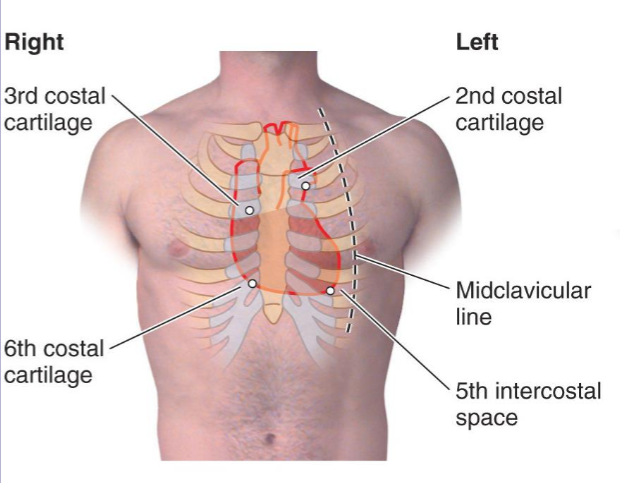
Describe the position of the heart in regards to where it lies in the thorax, including where the right and left side of the heart lie.
1/3 of heart right of midline, 2/3 of the heart to the left of midline
right side runs between 3rd and 6th costal cartilages in parasternal line
left side runs from 2nd CC to the 5th intercostal space near to midclavicular line
posterior surface is between T5 and T8 (thoracic vertebrae levels)
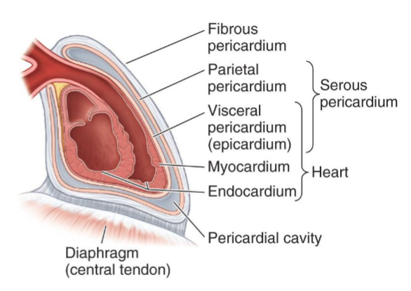
Describe the pericardium of the heart including its layers.
fibrous pericardium is outermost layer, parietal pericardium lines the inside of fibrous pericardium. parietal pericardium is continuous with the visceral pericardium.
parietal pericardium is also known as epicardium
then myocardium, then endocardium lines the myocardium
pericardial cavity is a potential space normally containing a small amount of serous fluid
pericardium fuses with central tendon of diaphragm
pericardial cavity is a potential space normally containing a small amount of serous fluid
What are the two pericardial sinuses and where are they?
two sinuses formed as pericardium wraps around heart - transverse and oblique
transverse pericardial sinus between ascending aorta and superior vena cava
oblique pericardial sinus - depression in posterior wall between the pulmonary veins and arteries

Label the cardiac borders:

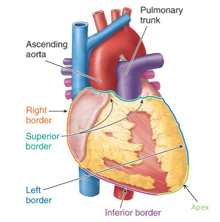
Label the cardiac surfaces:


Label the cardiac borders in a chest radiograph


aortic knuckle - contour of aortic arch seen in an antero-posterior radiograph
auricle - protrusion of the surface wall of each atrium
pulmonary trunk branches into left and right pulmonary arteries
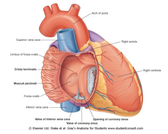
Describe anatomy of the right atrium of heart.
forms the right border
superior vena cava, inferior vena cava and coronary sinus drain here
it has a smooth posterior wall, known as sinus venarum
fossa ovalis on sinus venarum
rough anterior wall with pectinate muscles
the crista terminalis separates rough and smooth walls
the sulcus terminalis does this on the exterior
RA empties into RV via triscuspid valve
What part of the embryological heart tube gives rise to the ascending aorta and pulmonary trunk?
Truncus arteriosus
What part of the embryological heart tube gives rise to the smooth (superior) portions of the right and left ventricles?
Bulbus cordis
What part of the embryological heart tube gives rise to the trabeculated portions of the right and left ventricles?
Primitive ventricle
What part of the embryological heart tube gives rise to the rough portions of the right and left atria (pectinate muscle)?
Primitive atrium
What does the sinus venosus of the embryological heart tube develop into?
Smooth portion of right atrium, coronary sinus, oblique vein of left atrium
Label right atrium:
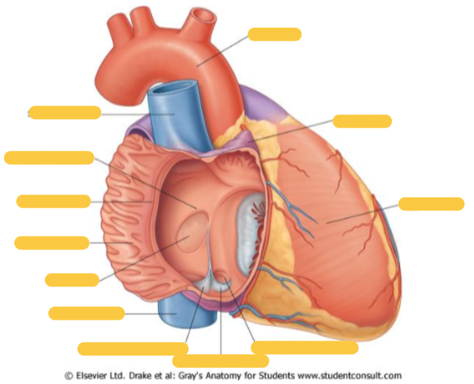
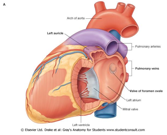
Label left atrium:


Label left ventricle:
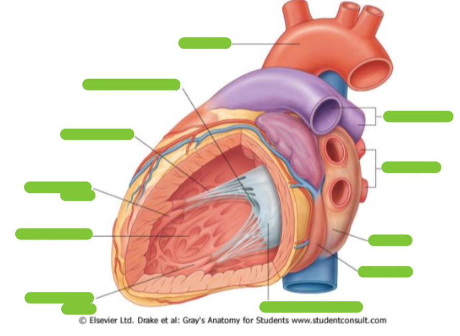
Label right ventricle:

Include names of cusps of the tricuspid and pulmonary valves

Describe the anatomy of the right ventricle.
receives blood from right atrium through the tricuspid valve
blood then follows a horseshoe shape via the infundibulum (conus arteriosus - smooth region of right ventricle) and through the pulmonary valve to the pulmonary artery
it has prominent trabeculae carnae
contains chordae tendineae attached to cusps of tricuspid valve and posteriorly to papillary muscles
contains septomarginal trabecula (moderator band) - prominent condensation of trabeculae carneae

What is the interventricular septum?
thick, triangular wall of cardiac tissue that separates right and left ventricles of the heart
muscular portion - makes up majority of the septum, it is thick, muscular and divided into inlet, trabecular and infundibular parts
membranous portion is a small, thin, fibrous region located near heart’s upper base near aortic and tricuspid valves
blood supply - left anterior descending artery and posterior interventricular artery
What is the septomarginal trabecula (moderator band)?
prominent muscular ridge in right ventricle that extends from lower portion of the interventricular septum to base of the anterior papillary muscle.
it is a condensation of the trabeculae carneae
carries right branch of atrioventricular bundle directly to anterior papillary muscle, transmitting electrical impulses quickly and ensuring synchronised contraction of the ventricle during systole
Describe the anatomy of the left atrium.
It forms the posterior surface/ base of the heart. The four pulmonary veins drain here, bringing oxygenated blood from the lungs.
It contains both smooth and rough areas (pectinante muscle in rough area)
Contains foramen ovale (remnant from embryological development)
Empties into LV via mitral valve. Mitral valve is bicuspid.

Describe the anatomy of the left ventricle.
Blood from LA through mitral valve.
Blood to aorta via aortic valve
Blood moves in a U shape to aortic vestibule then out via aortic valve, to be pumped into the aorta.
Contains prominent trabeculae carnae
Papillary muscles and chordae tendineae
it is much larger than the right ventricle as well as the wall being much thicker due to distance that it has to pump blood

What are the four cardiac valves?
Atrioventricular - tricuspid and mitral valves, which open in diastole when atria contract
Semilunar valves - aortic and pulmonary valves, which open in systole when ventricles contract
How many cusps does each cardiac valve have?
tricuspid - 3
mitral - 2
aortic - 3
pulmonary - 3
What is the function of the fibrous skeleton of the heart?
anchors cardiac muscle
keeps atrioventricular and semilunar valves patent and in right shape
provides attachment for valve cusps and leaflets
electrical insulation between the atria and ventricles (independent contraction)
How do the atrioventricular valves open and close?
papillary muscles are attached to tendinous cords which are attached to each cusp
when ventricles relax, blood from body and lungs fills atria, causing atrial pressure to rise above ventricular pressure - this pressure pushes the AV valve cusps down into the ventricles.
as the papillary muscles are also relaxed and chordae tendineae remain slack, the valve leaflets swing open freely so blood flows into ventricles.
papillary muscles contract at the same time the ventricles do
Contraction of the papillary muscles pulls on the tendinous cords, anchoring the cusps when ventricular pressure during ventricular systole causes the cusps to slam shut.
