RBC evaluation
 Call Kai
 Learn
 Practice Test
 Spaced Repetition
 Match
 Flashcards
 Knowt Play
1/56

There's no tags or description
Looks like no tags are added yet.
57 Terms
erythrocyte function
• Transport Oxygen to tissue
• Transport Carbon Dioxide to lungs
• Buffer H+
PCV
High-speed centrifugation to separate blood into cell layers based on
density
impedance
Measures the electrical resistance of cells in a fluid as they pass through an aperture
optical flow
Cells categorized based on light scatter and internal complexity
outer lipid bilayer function
• Maintain deformability and integrity of RBC
• Semi-permeable for gas exchange
• Maintains environment for Hgb to function
erythrocyte antioxidant pathways
• RBC membrane and hemoglobin are particularly susceptible to oxidative injury
• RBCs generate protective reducing agents to neutralize potentially harmful effects. (antioxidants)
• Enzymes:Glutathioneperoxidase,superoxidedismutase,catalase,etc • Antioxidants:Glutathione,VitaminC,VitaminE,etc
hemoglobin function
transport of oxygen from the lungs to tissues
transport CO2 from tissue to lungs
carbonic anhydrase
enzyme that converts CO2 to H+ and HCO3- to be transported to the lungs
carbaminohemoglobin
CO2 binds directly to hemoglobin, 20% of CO2 is transported this way
oxygen carrying capacity
RBC
Hgb
HCT
PCV
wintrobes RBC indices
MCV
MCHC
MCH
used to characterize RBC
erythropoietic activity
reticulocytes
RDW
nRBC
PCV
percentage of packed RBCs within a given volume of blood (centrifugation)
HCT
Calculated value generated by hematology analyzer
only reliable if MCV and RBC are accurate
should be within 3% of PCV
hemoglobin
Measured by lysing the RBCs -> oxidizing Hgb -> Formation of Heme products that are detected spectrophotometrically
Any substance that interferes with sample turbidity can cause false elevation in Hbg
• Lipemia(post-prandialsampling)
• Intravascular hemolysis (hemoglobinemia)
• Heinz bodieshgb is 1/3 of the HCT
RBC
measured by
hemocytometer
automated impedance counter
automated flow cytometer
RBC agglutination or clotting can falsely decreased rbc conc
hemocytometer
manual method
automated impedance counter
Cells are drawn through an aperture and cause electrical resistance
Degree of resistance differentiates Plt, RBC, and WBC
automated flow cytometer
Cells individually pass through a laser beam
Size, number, and internal complexity are determined by scatter of the laser beam
MCV
mean corpuscular volume - mean vol per RBC
MCH
mean corpuscular hemoglobin
mean hgb per RBC
MCHC
mean corpuscular hemoglobin concentration
mean Hgb concentration per RBC
macrocytosis
increased MCV (larger erythorcytes)
microcytosis
decreased MCV (smaller erythrocytes)
macrocytosis is caused by
artifacts, prolonged storage, hyperosmolality, breed associated, regenerative anemia, etc
microcytosis is caused by
artifact (excess EDTA), hyponatremia, young animals, breed associated, iron deficiency and portosystemic shunt.
hyperchromasia
increased MCHC, always an artifact
hypochromasia
decreased MCHC, due to regenerative anemia, iron deficiency (most common)
erythropoietic activity
reticulocytes, RDW, nRBC
reticulocytes
non-nucleated, immature RBCs with stainable cytoplasmic RNA
used to categorize anemias as regenerative or non regenerative
assessed by
1. manual counts, incubated with methylene blue to precipitate DNA, per 1000 rbc
2. routine blood smear (wright stain)- polychromasia due to increased RNA content
3. automated counts and absolute reticulocyte concentration to evaluate marrow response to anemia
no
do horses release reticulocytes?
2
how many stages of reticulocytes are observed in cats?
aggregate and punctate
stages of reticulocytes in cats
cattle
Reticulocytes do not occur in high numbers until the anemia is severe
dogs
Normal to have low numbers in health (~1.5% of erythrocytes)
Reticulocytosis without anemia: corticosteroids, NSAIDs, catecholamine-induced splenic contraction, etc
RDW
Calculated value that reflects the variation in RBC volume (anisocytosis)
• Younger RBCs will have high MCV -> if they are in high enough concentration, they will increase the RDW
nRBC
nucleated red blood cell in circulation
• Assessed on a blood smear
reveals regenerative anemia, bone marrow injury, splenic function, heat stroke, lead poisoning, etc
Erythrocyte Blood Smear Assessment
Density
Arrangement
Size and variation in size
Shape changes
Inclusions
density
• Crude visual assessment of hematocrit
arrangement
rouleau= linear aggregates/ coin stacks cause by charge interactions (normal in cats and horses)
agglutination- grape clusters caused by IGs bindng to RBC antigens
normal shape
• Discocytes:Biconcave disc with central pallor
• Elliptocyte/Ovalocyte: Elongated RBC with no central pallor (camels)
• Nucleated elliptocyte RBC
poikilocytes
abnormal shape associated with underlying pathology

acanthocyte
Irregularly spaced projections of various lengths

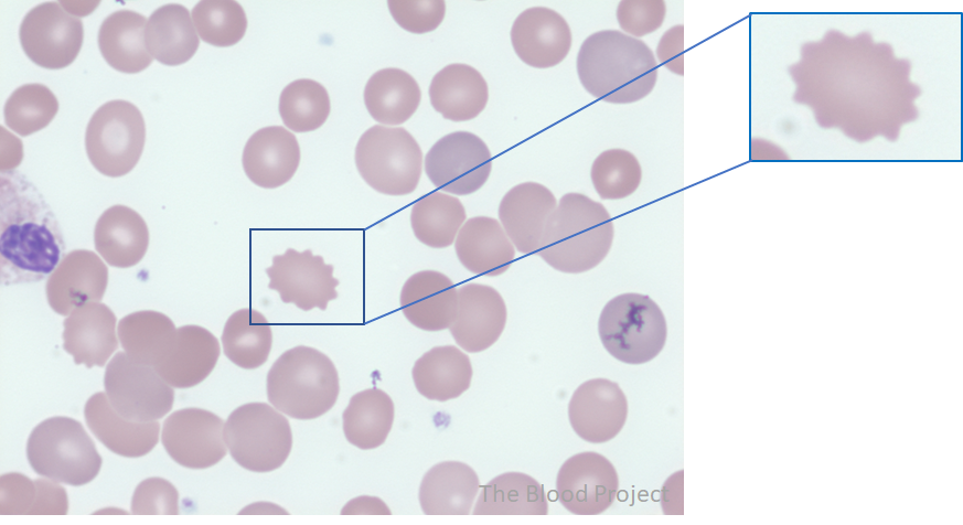
echinocyte
burr cell
regularly spaced pointy projections
codocyte
target cell
• Central focus of Hgb that is surrounded by a ring of pallor that separates it from peripheral Hgb

dacrocyte
teardrop to pear shaped

eccentrocyte
bite cell
eccentric dense-staining Hgb and adjacent clear space

leptocyte
thin cell that contains less Hgb

elliptocyte
elongated RBC

schistocyte
RBC fragment

spherocyte
Round RBC with no central pallor and decreased cell diameter

stomatocyte
Elongated area of cytoplasmic pallor (slit- like or mouth-like)

color
Changes in color are affected by the RNA and Hgb content
ghost cells
RBC cell membrane with little to no residual cytoplasmic Hgb
basophilic stippling
• Seen with regenerative anemia in cattle and plumbism

heinz bodies
• Oxidized Hgb that precipitates and binds to the RBC membrane
• Highlighted with new methylene blue stain

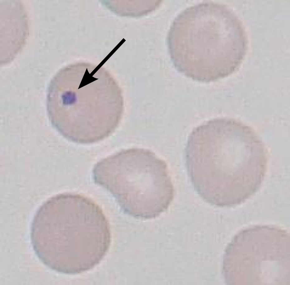
howell jolly bodies
• Small nuclear remnant, typically a homogeneous dark purple circle
• Low numbers can be seen in healthy animals and in splenectomized animals