Speech Science Midterm
 Call Kai
 Learn
Learn Practice Test
 Spaced Repetition
 Match
Match Flashcards
 Knowt Play
1/143

There's no tags or description
Looks like no tags are added yet.
Name | Mastery | Learn | Test | Matching | Spaced | Call with Kai | Chat |
|---|
No analytics yet
Send a link to your students to track their progress
144 Terms
Speech Breathing
Regulation of breathing for voice and speech production; SLPs work w/ clients to work with clients with speech and voice problems
Breathing
Inhalation (inspiration) and Exhalation (expiration)
Respiration
Gas exchange on the cellular level; bringing O2 in and CO2 out via simple diffusion
Ventilation
Moving air in and out of the lowering airway
Boyle’s Law
as volume of a space increases, the pressure decreases (and vice versa)
Lung Expansion
Pressure drop within the lungs (negative pressure) - air flows in
Lung Contraction
Pressure increase within the lungs (positive pressure) - air flows out
Pulmonary System
Lungs and airways (URS and LRS)
Chest Wall System
Ribs, abdomen, and diaphragm
LRS
Larynx, alveoli, bronchial tree; begins with trachea, 10-16 cm long w/ 20 rings to provide flexibility and support
Anatomy of lower airway
rib cage - 12 sets of ribs
trachea - lined with cilia to filter pollutants
bronchial tree
lings - 5 lobes (90% air)
Lungs
Bronchial Tree: each subdivision yields smaller bronchioles
level of gas exchange = alveoli
diffusion must occur quickly
large surface area with permeable membrane for diffusion w/ the concentration gradient
Alveolar Sacs
Respiratory bronchioles open into alveolar ducts leading into alveolar sacs
kept inflated by surfactant
Muscles for breathing
Neck, thorax, back, abdomen (function - inhalation and exhalation)
inhalation: diaphragm and external intercostals
exhalation: internal intercostals
** Rib cage expands in 3 directions (F-B, S-S, T-B)
Major muscle of inhalation
Diaphragm (regulates volume of thoracic cavity)
External intercostals
Pull the rib cage up and out during inspiration; increasing volume of thoracic cavity
Internal intercostals
Pull rib cage down; decreasing volume of thoracic cavity
Three functions of all muscles of breathing
1) Breathing (changing volume of lungs)
2) Posture (core stabilization); may increase efficiency
3) Movement (bending, turning, etc)
Breathing at rest
Quiet tidal breathing
Two forces enable lungs to change volume
1) Restorative forces of tissues
2) Plural linkage
Pleural linkage
Parietal and visceral pleura
Negative intrapleural space (w/ fluid)
Maintains the expansion of the lungs and the compression of ribcage
Transpulmonary pressure
Difference between pressure in lungs (alveoloar) and intrapleural pressure
Breath cycle
1) Diaphragm contacts (flattens downward)
2) PM stretched causing drop in pressure
3) Abdominal structures are compressed and abdomen protrudes outward
4) External intercostals contract a bit to stiffen walls of thorax which increases efficiency of diaphragm. (Large movement of external intercostals not necessary in tidal
breathing.)
5) Air rushes inward to equalize pressure
6) The expansion of the lungs and the lowered intrapleural pressure increase elastic recoil force
7) Diaphragm & external intercostals relax and together with recoil forces, lungs & thoracic cavity decrease in volume and air rushes out
EXHALATION IS PASSIVE IN TIDAL BREATHING
Average number of breaths per minute in tidal breathing
12-15 breaths
When air pressure (𝑃𝑎𝑙𝑣) inside the lungs is negative
air is forced to enter the respiratory system (because air moves from areas of high pressure to areas of low pressure)
When air pressure (𝑃𝑎𝑙𝑣) inside the lungs is positive
air is forced out of the respiratory system into the atmosphere
To decrease (Palv)
Must increase the volume of the thoracic cavity and lungs; contract the diaphragm, which flattens out and increases the vertical dimension of the thorax
▪ External intercostals pull the ribcage up and out
▪ The lungs, attached by pleural linkage, are pulled in the same direction as the
thoracic cavity and therefore increase in volume
To increase (Palv)
Must decrease the volume of the thoracic cavity and lungs; relax the diaphragm back to its dome-shaped position
▪ We relax the external intercostals, allowing the rib cage to return to its original
position
At the end of each inhalation and exhalation cycle
There is a brief moment in which air does not move into or out of the
respiratory system
- 𝑃𝑎𝑙𝑣 equals 𝑃𝑎𝑡𝑚𝑜𝑠 - (REL)
Lung volume
Single, non overlapping value
Lung capacity
2+ volumes
Tidal volume (TV)
the volume of air that we breathe in and out during a cycle of respiration
Inspiratory reserve volume (IRV)
The amount of air that can be inhaled above TV
Expiratory reserve volume (ERV)
The amount of air that can be exhaled below the TV
Residual volume
the amount of air always in the lungs that cannot be exhaled voluntarily
-lung tissue is always slightly stretched due to pleural linkage; therefore, there must be
some air pressure in the lungs
Vital capacity
The maximum amount of air a person can exhale after a maximum inhalation (the total amt.of air available for ALL purposes)
VC = IRV + TV + ERV
Functional residual capacity
The amount of air remaining at the resting expiratory level or end expiratory level
FRC = ERV + RV
Inspiratory capacity
The amount of air that can be inhaled from the end-expiratory level
IC=TV+IRV
Total lung capacity
The total amount of air the lungs can hold
TLC = TV + IRV + ERV + RV
Relaxation Pressure
Act to return the breathing system to REL
positive when it acts to decrease lung pressure
negative when it acts to expand lung volume
Active forces
Muscle contraction
Passive forces
Gravity, elastic recoil, surface tension of alveoli
Relaxation curve
Relaxation pressure as a function of lung volume displayed graphically
Thorax
Compressed spring & its elastic recoil exerts an expansion force
Lungs
Stretched spring with elastic recoil exerting a compression force
Relaxation Curve
Pressures generated by the elastic recoil forces at different
percentages of vital capacity

A on the RC
At the bottom of vital capacity, the elastic recoil forces acting on the lungs to decrease their overall volume are minimal
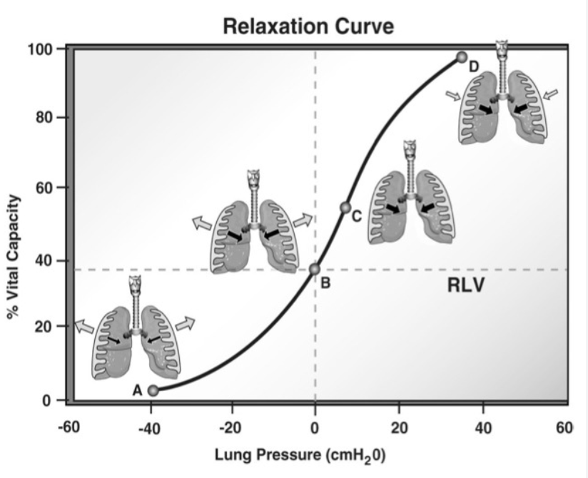
B on RC
At resting lung volume, the relaxation pressure is balanced such that the elastic recoil forces acting to expand the chest wall are equal to the forces acting to decrease lung volume.
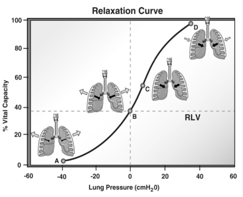
C on RC
As vital capacity increases, a point is reached in which the chest wall is at its resting size.

D on RC
At max vital capacity, elastic recoil forces of the chest wall and lungs are acting to decrease overall lung volume

Resting expiratory level (38% vital capacity)
Forces are balanced
Curve below RLV
Relaxation pressures working to increase lung volume (negative)
Curve above RLV
Relaxation pressures working to decrease lung volume (positive)
Average rate of breathing for speech breathing
12 to 20 breaths/minutes, but the balance of inspiration to expiration differs
Tidal breathing I/E phases
40% (Inspiration) 60% (Expiration)
Speech breathing I/E phases
10% (Inspiration) 90% (Expiration with speech)
Hixon, Mead, & Goldman (1976)
Passive relaxation pressure alone is never the solitary force managing speech breathing. Active muscle force always contributes.
▪ Diaphragm, rib cage inspiratory muscles and abdominals
are all active to varying degrees at the beginning of an
utterance. Abdominals are more continuously active in
speech breathing than in tidal breathing.
direct relationship between lung volume and and inspiratory muscle force
indirect relationship between lung volume and expiratory muscle force
Adaptive Control
Changeable internal & external demands require speech breathing system to be highly
adaptable
Body Type
Endomorphs use greater abdominal wall activity over that of the rib cage compared to ectomorphs
Additional mass on abdomen exerts a pulling force that tugs on diaphragm (flattening effect), inhibiting rapid contraction of diaphragm needed for speech
Cognitive-Linguistic Variables
Basic mechanisms of speech breathing are preserved, but timing of inspiratory & expiratory events are altered.
▪ Inspiration occurs mainly at grammatical and semantic boundaries.
▪ Length of phrase & depth of inspiration are directly related.
▪ Cognitive load influences timing
Speech breathing personality
Different styles of breathing relative to activity of the thorax and abdomen
Clavicular breathing
Over-activation of sternocleidomastoid and other shoulder muscles with lesser activation of diaphragm. Lateral expansion of thorax is limited, with emphasis on vertical expansion.
▪ Marked by distinct upward movement of the shoulders or clavicle.
Individuals with neurologic or lung disease may use to compensate for
impaired function (highly inefficient for lung expansion in health
individuals)
Chest (pear shape up, up & in, high breathing)
Activation of abdominal muscles limits downward excursion of diaphragm, but
▪ Antero-posterior expansion of chest wall exerts lateral pull on
diaphragm, allowing vertical expansion of lungs.
▪ May be advantageous in vocal tasks demanding increased breath support, such as singing
Diaphragmatic (pear shape down, down & out, low breathing)
Antagonist muscle contraction of abdomen and thorax are minimized (except for postural stabilization)
▪ As a result the abdominal contents are distended outward, offering less resistance to the diaphragm, which is allowed to contract and flatten.
▪ Optimal style, the target pattern in speech and voice therapy
Respiratory Demands
Our bodies must integrate the demands of speech production with the metabolic demands of gas exchange, of getting oxygen into our blood so that we stay alive
Hyperventilation
over-breathing; excess of CO2 (dizziness, blurred vision, numbness & tingling)
Hypoventilation
(under breathing; too little O2 (impaired cellular respiration, tissue damage, death)
In speech breathing (compared to tidal)
Ventilation rates higher
▪ Slight hypoventilation
▪ Irregular breathing, including breath-holding after speaking for short periods to normalize blood gas
During exercise
Larger amounts of air must be moved faster
Length of phrase breath group decreases.
▪ Expiratory time decreases.
▪ Larger lung volumes used.
▪ Rate of speech unaltered
Neural Control of Breathing
Sensorimotor pathways that mediate respiration for metabolic demands are modulated during speech production
Breathing is a reflex arising from the respiratory center in the medulla oblongata.
▪ Motor control: Phrenic and intercostal nerves and other spinal nerves
Sensory receptors
▪ Chemoreceptors (sensitive to CO2, pH, O2)
▪ Mechanoreceptors (tissue distortion)
▪ Muscle spindles (in muscles: respond to stretch)
▪ Stretch receptors in bronchioles & alveoli
Airflow
volume of air moving through a given cross section of tube per unit of time; driving pressure/resistance
Relationship between airflow and driving pressure
Airflow directly proportional driving pressure
▪ double pressure, double flow
▪ halve pressure, halve flow
Resistance
Opposition to movement (airway, elastic, viscosity)
airflow is inversely proportional to resistance
Airway resistance
Narrowness of tubes that make up the airway;
Doubling the airway diameter decreases resistance by a factor of 16 (and vice versa)
Larger lung volume (inhalation) distends airway, lowering resistance
Turbulent flow is most commonly found in
At high velocity and/or through tubes with irregular walls
▪ Found in the largest airways, such as the trachea
▪ Requires greater driving pressure
▪ Flow is not directly proportional to driving pressure
Elastic Resistance
Work required to prevent tissue from springing back once deformed (in order to change
the volume of the thoracic cavity and lungs)
Quiet breathing: work of breathing is ~5% of our total metabolic rate
Viscosity
Measure of internal friction of fluid
▪ In a sauna, you may have noticed more difficult to breathe. That is because the extra moisture adds to the overall viscosity of the air.
Phonation
Process of sound generation by vocal fold vibration
Vocal folds vibrate rapidly open and closed to produce puffs of air consisting of a oscillating air molecules that together produce a sound pressure wave that travels up the vocal tract
Larynx
From the root of the tongue to the trachea in the anterior neck or throat
▪ Level of C4–C6
Airway protection
▪ Throat-clearing, coughing, Valsalva maneuver
▪ Participate in regulation of airflow during breathing
▪ Stabilize upper body during movement (such as lifting a
heavy object, running upstairs)Phonation!
3 unpaired cartilages
Cricoid, thyroid & epiglottis

Extrinsic
have at least one attachment point outside of the larynx (e.g., hyoid) and anchor the larynx
Intrinsic
confined entirely within the larynx
Conus elasticus
▪ Medial cricothyroid ligament
▪ Lateral cricothyroid membrane (free border superiorly = vocal ligament)
Quadrangular Membrane
Superior Free margins = aryepiglottic folds
▪ Inferior free margins = ventricular ligaments (false vocal folds)
The larynx contains 3 sets of valves or folds
The aryepiglottic, the false (ventricular), and the true vocal folds
Aryepiglottic folds
Run from the sides of the epiglottis to the apex of each arytenoid cartilage.
They contract to pull the epiglottis backward and to close the entrance of the larynx during swallowing
False vocal folds
Capable of limited movement-they close during swallowing and effortful activity
True vocal folds
most complex valves and are the vibrating element of the larynx
Vestibule
Supraglottic space
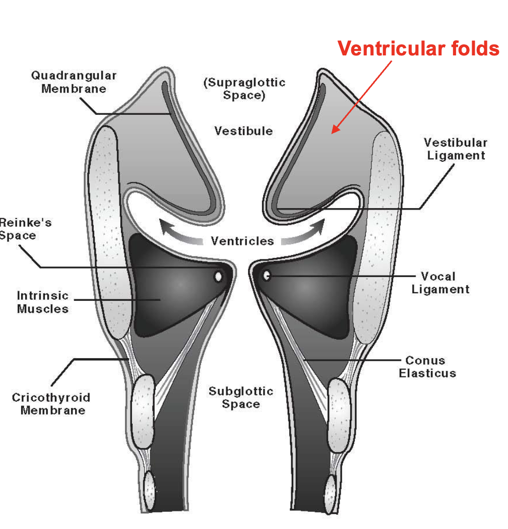
Ventricles
Contain mucous glands to lubricate vocal folds

Glottis
Space between the true vocal folds
lateral cricoarytenoid muscle (LCA)
One of the two vocal fold adductors; It closes the glottis

Interarytenoid muscle (IA) (transverse and oblique)
vocal fold adductor/closes the glottis
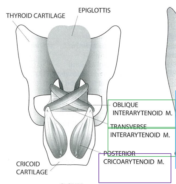
Posterior cricoarytenoid muscle (PCA)
Only muscle that abducts (opens) the vocal folds

Cricothyroid muscle (CT)
Consists of pars recta & pars oblique; regulates vocal pitch by elongating and tensing the vocal cords

Extrinsic Muscles
Connect cartilages to external point of attachment
▪ Include many infrahyoids; muscles of jaw & strap (large shoulder & neck)
▪ Larynx is suspended from above by extrinsic muscles (as well as attached inferiorly
by ligaments)
Subglottal space
Space under the vocal folds
Supraglottal space
Space above vocal folds

Vocal fold histology (superficial to deep)
Epithelium: stratified squamous cell; plicae
▪ Lamina Propria - 3 layers critical to vocal fold vibration
▪ Superficial (Reinke’s space): consistency like gelatin
▪ Intermediate: composed of elastin
▪ Deep: composed of collagen
▪ Muscle (TA)
