Growth Plate and Pelvic Fractures
 Call Kai
 Learn
Learn Practice Test
 Spaced Repetition
 Match
Match Flashcards
 Knowt Play
1/103

There's no tags or description
Looks like no tags are added yet.
Name | Mastery | Learn | Test | Matching | Spaced | Call with Kai | Chat |
|---|
No analytics yet
Send a link to your students to track their progress
104 Terms
Growth plate fractures
fracture to the growth plate of developing animals; can interfere with normal bone development
Growth plate
aka physis; where cartilage is transformed into bone during development
A Growth plate is surrounded by what two bony landmarks?
epiphysis and metaphysis
What makes growth plates susceptible to injury?
because its cartilage is considerably weaker than the surrounding epiphyseal and metaphyseal bone
What is the process of a growth plate becoming a bone called?
endochondral ossification
Four components of a growth plate:
1.) outer fibrous layer
2.) cartilaginous component
3.) bony component
4.) vascular supply
outer fibrous layer of a growth plate
fibrous layer that surrounds the growth plate and provides support and protection
Two structures associated with the outer fibrous layer of a growth plate:
1.) Groove of Ranvier
2.) Ring of LaCroix
Groove of Ranvier
region of chondrocytes located around the periphery of the growth plate; contributes to cartilage growth at the physis
Ring of LaCroix
structure located between the groove of Ranvier and metaphysis; provides mechanical support to help stabilize and protect the growth plate
Cartilaginous component of the growth plate
where chondrocytes proliferate and eventually mineralize to be replaced by bone
The cartilaginous component has four zones:
1.) resting (reserve) zone
2.) proliferative zone
3.) hypertrophic zone
4.) mineralization zone
resting (reserve) zone
closest to epiphysis; contains small, immature chondrocytes that serve as a reservoir to contribute to continued bone growth
What is the clinical significance of the resting (reserve) zone?
damage to these cells can interfere with continued growth; fractures that spare this zone have a more favorable potential for continued bone growth than injuries that destroy it
proliferative zone
where chondrocytes begin actively dividing and organizing themselves into longitudinal columns
hypertrophic zone
chondrocytes become substantially larger as they mature
mineralization zone
hypertrophic chondrocytes eventually die, and the mineralized cartilage provides a scaffold for the deposition of new bone by osteoblasts
Which cartilage zone is the weakest? Why?
hypertrophic zone; as chondrocytes become larger, the supporting extracellular space decreases, making the area weaker and more susceptible to fracture
The growth plate has a relatively _________ vascular supply
limited
Which two cartilage zones do not have any blood vessels?
proliferative and hypertrophic zones
Two types of growth plates based on location:
1.) pressure growth plates
2.) traction growth plates
pressure growth plates
located at either end of long bones and are responsible for most of the bone's longitudinal growth
True or false: the contribution towards the growth of a long bone is even between a distal and proximal pressure growth plate
false!! (except rare occasions)
ex: humerus in dogs, the proximal physis contributes the majority of the growth, while the distal physis contributes only a small portion
Why is the the uneven contribution towards the growth of a long bone between pressure physes clinically important?
damage to a physis responsible for a large proportion of bone growth can produce more severe growth disturbances than damage to a physis that contributes relatively little
traction growth plates
occur at sites where muscles, tendons, or ligaments attach to bone; contribute to development of bony prominences
traction growth plates develop from _______ centers of ossification
secondary
Examples of traction growth plates
-tibial tuberosity
-olecranon
-greater trochanter
The age at which growth plates close varies according to...
species, breed, bone, and the specific physis involved.
Growth plates are relatively weak compared to the surrounding bone because...
because they consist primarily of cartilage and extracellular matrix (esp hypertrophic zone)
Therefore, a force that might not fracture mature bone can cause a fracture through an open growth plate
Growth plate injuries can occur due to _____ energy trauma or ______ energy trauma
high; low
high energy trauma
things that do substantial damage such as motor vehicle accidents, impalement injuries, and kicks from other animals
low energy trauma
injuries that occur after minor incidents, like falling from a low height, stepping off a step, or being stepped on
Salter-Harris Fracture Classification system
categorizes fractures according to which anatomical regions of the developing bone are involved
Six types of growth plate fractures according to the Salter-Harris Fracture Classification system:
1.) Salter-Harris Type I
2.) Salter-Harris Type II
3.) Salter-Harris Type III
4.) Salter-Harris Type IV
5.) Salter-Harris Type V
6.) Salter-Harris Type VI
Salter-Harris Type I fracture
fracture that passes directly through the growth plate without going into either the epiphysis or metaphysis

Salter-Harris Type I fractures commonly occur in what cartilaginous zone?
the hypertrophic zone
*but can occur at other levels within the physis
What is one benefit of a Salter-Harris Type I fracture?
Because the fracture often passes only through the hypertrophic zone, the resting or germinal zone remain intact, will will allow for continued longitudinal growth
Radiographic appearance of a Salter-Harris Type I fracture
May not be apparent on initial radiographs
-look for subtle changes like widening of physis
Common location of a Salter-Harris Type I fracture
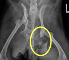
Capital Femoral Physeal Fracture

Capital femoral physis
located at the femoral head
Salter-Harris Type II fracture
fracture across the physis and extending into the metaphysis

Salter-Harris Type II fractures are the most ___________ growth plate fractures in veterinary medicine
common!
Common location of a Salter-Harris Type II fracture
distal femoral physis

Salter-Harris Type III fracture
fracture across the physis which extends into the epiphysis (and therefore articular surface)
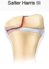
Why might a Salter-Harris Type III fracture be particularly concerning?
-they involve the resting or germinal zone of the physis, so damage to this zone may interfere with continued longitudinal growth
-additionally, can extend into the articular surface and could cause joint incongruity and osteoarthritis
Common locations of a Salter-Harris Type III fracture
condyles of long bones, particularly the femoral condyles and humeral condyles

Salter-Harris Type IV fracture
fracture through the metaphysis, physis and epiphysis (and therefore articular surface)

Why might a Salter-Harris Type IV fracture be particularly concerning?
same reasons as a type III
-they involve the resting or germinal zone of the physis, so damage to this zone may interfere with continued longitudinal growth
-additionally, can extend into the articular surface and could cause joint incongruity and osteoarthritis
Common location of a Salter-Harris Type IV fracture
distal humerus

What dog breed commonly experiences Salter-Harris Type IV fractures?
brachycephalic or toy breeds (Bulldogs, French Bulldogs, etc)
Salter-Harris Type V fracture
crush injury to physis that "erases" the growth plate

Why can a Salter-Harris Type V fracture be concerning?
Compression can destroy the resting or germinal zone; because these cells are responsible for supporting continued growth, damage may result in premature physeal closure and permanent growth disturbances
If the entire physis closes prematurely, longitudinal growth may stop.
If only one side closes, the unaffected side may continue growing, producing an angular deformity.
Common location of a Salter-Harris Type V fracture
distal ulna
What complication can occur from a Salter-Harris Type V fracture regarding the distal ulna?
Premature Closure of the Distal Ulnar Physis
Premature Closure of the Distal Ulnar Physis
most of the growth of the ulna comes from the distal ulnar physis, so damage and premature closure to this plate can limit it's ability to continue growing
While the ulna may stop growing normally from premature closure of the distal ulnar physis, the radius will...
continue growing normally
What might result when the ulna stops growing but the radius continues growing?
Instead of growing normally along its long axis, the radius becomes progressively curved
The ulna acts as a tether while the radius continues attempting to grow, producing a bowstring effect

Which two Salter-Harris Fractures are intra-articular?
Salter-Harris Type III and Salter-Harris Type IV (since they can both extend past the epiphysis and affect the joint)
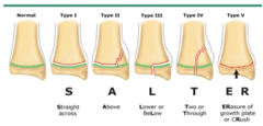
Mnemonic to remember growth plate fracture classifications:
SALTER
S traight across (type I)
A bove (type II)
L ower (type III)
T two/through (type IV)
ER asure of growth plate (type V)
***A and L only work if looking at a distal physis; will need switched if looking at a proximal physis
Salter-Harris Type VI fracture
involve damage to the peripheral portion of the growth plate, including structures associated with the perichondrial ring (Groove of Ranvier)
Four complications of a growth plate injury:
1.) Growth arrest
2.) Valgus deformity
3.) Varus deformity
4.) Osteoarthritis
Growth arrest
Damage to the germinal zone may cause the physis to close before skeletal maturity, can resulting in a shortened bone
Valgus deformity
lateral deviation of the limb distal to deformity; occurs when only part of the physis is damaged

Varus deformity
medial deviation of the limb distal to deformity; occurs when only part of the physis is damaged

Osteoarthritis
occurs with fractures involving the joint surface (type III and IV)
Pelvic fractures
relatively common fractures that are typically associated with high energy trauma
An important concept in pelvic fracture evaluation is that the pelvis functions as a ______-like structure
box
How is the pelvis a box-like structure?
the components of the pelvis are interconnected, so displacement at one portion of the pelvis means there will likely be displacement at another location
Therefore, if a pelvic fracture is visibly displaced, there must usually be at least _______ sites of disruption
two
Exceptions to the Pelvic Box Principle
-in young animals, the pelvis is flexible, so injury can occur in one place without a second injury
-isolated acetabular stress fractures
There are often concurrent injuries associated with pelvic fractures. Why?
they generally result from high energy trauma, so the force responsible can also injure other body systems
In what region of the body will there be an especially high chance of concurrent injury associated with a pelvic fracture?
the thorax
*should always radiograph thorax in addition to pelvis
Examples of thoracic concurrent injuries with pelvic fractures:
-Pneumothorax
-Rib fractures
-Hemothorax
-Diaphragmatic hernia
Other common concurrent injuries associated with pelvic fractures:
-Urinary tract trauma --> ruptured bladder, ruptured urethra, bladder herniation
-GI tract trauma --> ruptured GI tract, spleen, liver
-Orthopedic trauma --> spinal fractures, long bond fractures
-Integument --> full thickness wounds
For patient evaluation of a pelvic fracture, what should be prioritized?
pelvic fractures are generally not life threatening, so instead should focus on treating shock or other life threatening injuries (such as pneumothorax, bladder rupture, etc)
Important landmarks to palpate during pelvic evaluation:
1.) palpate ilial wings
2.) assess stability of sacroiliac joint
3.) palpate greater trochanter and location within pelvis (acetabulum)
4.) palpate ischial tuberosities for asymmetry
When performing radiographs for a pelvic fracture, it is important to ALWAYS take _____ views
two
Lateral radiographs of a pelvic fracture are performed how?
with the affected side down
-down hip is flexed, while upper hip extended ("running man" position)

V/D radiographs of a pelvic fracture are performed how?
either "frog leg" where legs are spread apart, or have legs fully extended

An important consideration when determining whether a pelvic fracture requires surgery is...
whether the fracture involves the weight-bearing axis of the pelvis
weight-bearing axis of the pelvis involves three structures:
1.) acetabulum
2.) iliac body
3.) sacroiliac joint
Fractures of the pelvis usually will or will not heal well? Why?
will!
the pelvis is surrounded by a large muscle mass that:
-provide stability and support
-provide substantial blood flow to the pelvis
additionally, the ilium contains a large amount of cancellous bone, which is rich in blood supply
Seven injuries of the pelvic bones:
1.) Pelvic canal narrowing
2.) Sacroiliac Luxation
3.) Iliac Wing Fractures
4.) Iliac Body Fractures
5.) Acetabular Fractures
6.) Ischial Fractures
7.) Pubic Fractures
Pelvic canal narrowing
occurs when fracture fragments heal in an abnormal position, reducing the diameter of the pelvic canal
Conditions that can result due to pelvic canal narrowing
-constipation
-dystocia
-urethral obstruction
Sacroiliac Luxation
traumatic separation of the ilium from the sacrum at the sacroiliac joint
For a sacroiliac luxation, the ilium typically becomes displaced _______ to the sacrum
craniodorsally
Sacroiliac Luxations can be associated with concurrent _______ damage
nerve
Two nerves located near the sacroiliac joint
Sciatic nerve and lumbosacral nerve roots
Neurologic deficits associated with sacroiliac luxations:
-pelvic limb weakness
-sciatic nerve deficits
-reduced anal tone
-urinary dysfunction
What is found on physical examination of a sacroiliac luxation?
asymmetry/abnormal movement of the iliac wings
What is found on radiographs of a sacroiliac luxation?
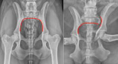
V/D radiographs are useful!!
Normally, a smooth line can be followed along the medial aspect of one ilium, across the ventral aspect of the sacrum, and onto the opposite ilium.
With SI luxation, this smooth line is disrupted.
Iliac Wing Fractures
non weight bearing, non articular, and not associated with any nerves; therefore, typically usually managed conservatively rather than surgically

Iliac Body Fractures
major component of the weight bearing axis, and commonly long oblique fractures

What type of fracture is common with an Iliac body fracture?
oblique fracture with craniomedial displacement
Treatment of Iliac body fractures
Require surgical stabilization involving plate fixation
Iliac body fractures are associated with three other fractures:
1.) ischium
2.) publis
3.) acetabulum
Acetabular Fractures
major component of the pelvic weight-bearing axis AND involve an articular surface; will feel crepitation on palpation of the hip

Acetabular Fractures typically have a more _______ prognosis. Why?
gaurded; a fracture involving the acetabulum can disrupt the articular cartilage, so the femur no longer articulates smoothly with the acetabulum
Can produce degenerative joint disease and lameness!
Ischial Fractures
not part of the primary weight-bearing axis, so of less concern; may return to normal alignment without on their own when other pelvic fractures are surgically repaired