Zoology Chp 38- Muscular System
 Call Kai
 Learn
 Practice Test
 Spaced Repetition
 Match
 Flashcards
 Knowt Play
1/59

There's no tags or description
Looks like no tags are added yet.
60 Terms
skeletal, smooth, cardiac
types of muscle tissue
muscle tissue
tissue that contracts when stimulated by signal then relax and lengthens
composed of cylindrical cells often called “muscle fibers” (muscle cells)
skeletal muscle
type of muscle
helps move the body and its parts
made of cells that can contract when stimulated
attached to bones
cardiac muscle
type of muscle
found only in the wall of the heart
plasma membranes of cells fused together so all cells contract as a unit
skeletal muscle
what kinds of muscle are voluntary muscle control
smooth and cardiac
what kinds of muscle are involuntary muscle control
skeletal muscle
muscle type that helps move body and parts
built of cells that can contract when stimulated attaches to bones
found in the arm
smooth muscle
muscle type found in internal organs
cardiac muscle
muscle type found only in the heart wall
skeletal muscle
striated
voluntary control
long, cylindrical cells
multinucleated
multiple cells (muscle fibers) fused together to meet cellular demands
attached to bones via tendons
location of skeletal muscles
myoblasts (stem cell)
muscle fiber is formed from…
myoblasts
what fuses together to form one long cell for the skeletal muscle
tendons
ties muscles to bones
ligaments
links bone to bone
cardiac muscle
striated
involuntary control
branched cell with intercalated discs
pumps blood through the heart
smooth muscle
non-striated (smooth appearance)
involuntary control
spindle-shaped cells
single nucleus per cells
moves substances through organs (peristalsis)
peristalsis
in smooth muscle
movement that squeezes and pushes things down a tube
organs, blood vessels, and airways
locations of smooth muscles
sarcolemma, sarcoplasm, myofibrils, sacroplasmic recticulum
structures within a skeletal muscle fiber
sarcolemma
plasma membrane of a muscle fiber
forms inward folds called T tubules
T tubules
carry electrical signal into the interior of muscle fiber
extensions of the sarcolemma
sarcoplasm
cytoplasm of muscle fiber
myofibrils
long, cylindrical structures inside muscle fiber
responsible for muscle contraction
made of myofilaments (thick and thin filaments)
sarcoplasmic recticulum
specialized form of smooth ER
surrounds each myofibril
myosin
what kind of protein does thick myofilaments contain
actin
what kind of protein do thin myofilaments contain
endomysium
thin layer of connective tissue that surrounds each individual muscle fiber (muscle cell)
helps mind muscles fibers together and transmit force
perimysium
connective tissue that surrounds each fascicle (bundle of muscle fibers)
helps organize fibers into functional bundles
contains blood vessels and nerves that supply muscle fibers
epimysium
entire muscle surrounded by a dense connective tissue
muscle → fascicle → muscle fiber → myofibril → sarcomere → myofilaments
levels of muscle organization in order
origin
one end of the muscle that attaches to bone and stays relatively motionless
insertion
one end of the bone that attaches to bone and moves the most
when the forelimb is pulled up the bicep contracts and the tricep relaxes
when the forelimb is pulled down the tricep contracts and the bicep relaxes
explain how muscles move bones using a lever system
bursa
small, fluid-filled sacs often found between tendons and bones
act as cushions and allow ease of movements
fascia
a type of connective tissue that extends beyond the muscle and becomes tendon
sarcolemma
plasma membrane surrounding muscle fiber
muscle fiber
cluster of myofibrils
surrounded by sarcolemma (plasma membrane)
fascicle
bundle of muscle fibers surrounded by perimysium
contains blood vessels and nerves
organizaes muscle fibers for coordinated contraction
myofilaments
actin and myosin
entire muscle
bundles of fascicles surrounded by epimysium then fascia
muscle fiber → endomysium → perimysium → epimysium → fascia
order from inside to outside of connective tissue layers
shorten
muscle contracts when cells…
sarcomere
basic unit of contraction
made up of thick and thin filaments arranged in overlapping array
glucose, glycogen, triglycerides, fatty acids
identify the four major fuel sources used by muscle cells
decreases
as length of exercise period increases, use of muscle energy stored…
increases
as length of exercise period increases use of energy sources from blood…
cellular respiration, fermentation, creatine phosphate pathway
the three pathways used to regenerate ATP
creatine phosphate pathway
ATP production pathway
simplest and most rapid way to produce ATP
immediate burst of energy that only lasts about 10 seconds
transfers phosphate to ADP→ produces ATP quickly
used for short explosive movements
fermentation
ATP production pathway- anaerobic metaboslism
produces ATP without oxygen
breaks down glucose to form lactic acid
short-term energy, leads to fatigue
process:
hormones signal muscle cells to break down glycogen making glucose available
glucose broken down through glycolysis into pyruvate (produces small amount of ATP)
pyruvate converted to lactic acid
cellular respiration
ATP production pathway- aerobic metabolism
requires oxygen
slower but efficient
produces large amounts of ATP
supports long-term, sustained activity
Slow-twitch fibers (Type I)
contract slowly, but keep going for a long time
use aerobic respiration for energy
rich in mitochondria, myoglobin (redder color), and blood supply
best for endurance activities
Fast-twitch fibers (Type II)
contract quickly and powerfully
use mostly anaerobic respiration
fatigue quickly, accumulate lactate
fewer mitochondria and less myoglobin (whiter in color)
best for short bursts of power
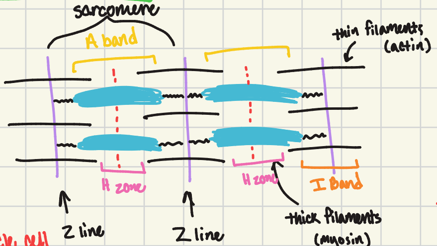
actin (thin) and myosin (thick) filaments, I band, A band, H zone, Z disc, m line, myosin heads
structures of a sarcomere
thin filaments
structure of the sarcomere
two strands of beads
beads are actin molecules which can contract
thick filaments
structure of the sarcomere
made of myosin molecules
sliding filament mechanism
When a sarcomere contracts, the Z lines move closer together and the I band and H zone get smaller. The A band stays the same width and, at full contraction, the thin filaments overlap.
fascia
sheet-like connective tissues that surround and separate muscles, muscle groups, bones, organs, and blood vessels throughout the body
one muscle (the agonist) contracts to create movement while the other muscle (the antagonist) relaxes and is being pulled
(biceps contracting while triceps relax)
Describe how antagonistic muscle pairs work together to produce movement.