D103 Final Review
 Note
 Call Kai
 Learn
 Practice Test
 Spaced Repetition
 Match
 Flashcards
 Knowt Play
1/119

There's no tags or description
Looks like no tags are added yet.
120 Terms
signal transduction importance
involved in development → issues can affect development and tissue homeostasis
signaling pathway composition
stimulus (intra or extra-cellular)
receptor
transduction cascade
effector
responses
stimulus aka ligand
hydrophilic molecules require transmembrane receptor
hydrophobic molecules require intracellular receptor
ligand-receptor interaction should have high specificity but low affinity → REVERSIBLE!
overall, should be made, mobilized, altered/degraded rapidly
receptor
ligand gated ion channel
gpcrs → use a GTPase to transduce signals
enzyme coupled receptors (rtks) → use a kinase to transduce signals
transduction cascade
mediated by proteins → pass a signal downstream
control them by altering activity → ie modifications like phosphorylating to inhibit/activate
how do intracellular components of a transduction cascade pass a signal?
want to induce a reversible protein conformational change
can have p-p interactions, ion-p interactions, GTP-effector interactions, or reversible modifications such as ubiquitination
protein domain
region of aas that can fold independently of the rest of the protein
serves as a point of recognition → mediates interactions between proteins
ex) sh2 domain binds to phosphorylated tyrosine residues in proteins
protein motif
conserved subset of aas within a domain, essential for domain function
domain role in signaling pathways
mediate interactions between intracellular signaling proteins
recruit pathway components and send signal downstream
ex) rtk has p-tyrosine sites on tail, can be recognized by sh2 domain on protein a → protein a can be recognized by the domain of protein 2 → etc etc
signal transduction complexity
one signal can activate multiple pathways
these pathways can share components
can be linear, convergent, or have multiple branches
contact-dependent signaling
signal cell has a membrane bound signal molecule, gets recognized by receptor on target cell
paracrine signaling
ligand released from signaling cell to neighboring target cell
synaptic signaling
neurotransmitter released from neuron, travels axon to synapse
endocrine signaling
hormones sent through blood to target tissue
NO → intracellular signaling
from l-arginine by NOS (endothelial NOS is in blood vessels)
regulated by Ca2+-calmodulin complex
ACh binds to GPCR → IP3 produced → IP3 heads to ER → Ca2+ release → calmodulin activation → NO production
NO heads to epithelial cell → guanylyl cyclase activated → GTP converted to cGMP → protein kinase G activated → myosin phosphorylation inhibited → vasodilation
angina treatment
angina → insufficient blood supply to heart
nitroglycerin can be broken down to NO
viagra inhibits cGMP phosphodiesterase → increases cGMP levels
steroid → intracellular signaling
steroids are v hydrophobic molecules
generally bound to plasma protein carriers, only free steroid hormones can participate in signaling transduction
receptors can be in cytoplasm or nucleus
also have some membrane bound receptors that are enyzme coupled and typically have rapid response
steroids bind to cytoplasmic receptors then get translocated into nucleus, or bind to nuclear receptors from the get go
translocation of ligand-receptor complex occurs bc the interaction causes a conformation change, revealing the NLS
steroid receptor complex then has transcription factor activity and can produce new proteins
slower than membrane bound
nuclear hormone receptors
have a variable region, a DNA binding domain, and a ligand binding domain
can bind to DNA and act as transcription factors
also located on inner leaflet of pm
activation of hormone receptors
can have basal condition bound to repressor complex
binding of ligand induces conformational change, loss of repressor and association of activator
androgen insensitivity syndrome
dysfunctional androgen receptor AR cannot recognize ligand
body tries to compensate by upping ligand levels, but AR cannot recognize it
ligand is converted by protein to other hormone (testosterone -aromatase-estrogen)
anabolic steroids
increase muscle and bone synthesis
gpcrs
G protein is a type of GTPase → activated by GEF and deactivated by GAP
three subunits acting together → alpha, beta, and gamma
alpha subunit has the GTPase activity
a and y have lipid anchors
allosteric binding increases affinity of primary binding site for target
ca2+, ATP, GTP can act as allosteric factors
allosteric binding causes conformational change in GTPase
have 7 transmembrane domains
stimulation of alpha subunit to exchange GDP → GTP
so ligand binding has GEF activity
effector is typically an enzyme
gpcr signaling
ligand binding causes association with heterotrimer
activation causes alpha subunit to change shape and triggers dissociation of GDP
alpha subunit then binds GTP, dissociates from b/y subunits
alpha subunit now has binding site for target effector, activating it
binding leads to hydrolysis of GTP to GDP, leads to dissociation of alpha subunit
thus effector has GAP activity
GTP-alpha then reassociates with b/y subunits
in general":
ligand-receptor
receptor-Ga
Ga-effector
common gpcr effectors
adenylyl cyclase
phospholipase C
cGMP phosphodiesterase
K+ ion channel
b/y subunits
can also activate ion channels
ultimately, alpha subunit still controls length of response → signal turns off upon GDP exchange
this is because the GTP exchange leads to b/y unit dissociation
gpcr inactivation
gpcr kinase/grk can phosphorylate the gpcr domain and recruit arrestins to turn off signaling
arrestins prevent interaction with other downstream effectors
act as adaptor proteins for clathrin → endocytosis
receptor sequestration
receptor is temporarily internalized by endosome
receptor down-regulation
internalized and destroyed in lysosomes
receptor inactivation
modified so that it is unable to interact with g protein
ex) phosphorylation
second messenger
small signalling molecules that transduce the signal response
ex) ca2+, IP3, DAG
phospholipase C
second messenger enzyme
generates DAG and IP3 from PIP2
DAG → hydrophobic, remains on membrane
IP3 → hydrophilic, dissolves rapidly into cytosol
remember Ca2+-calmodulin?? yeah this is it
IP3 will bind to receptor on ER, released Ca2+
Ca2+ then activates protein kinase C
DAG can also recruit and activate pkc
leads to phosphorylation of various protein residues
phospholipase c inactivation
IP3 can be dephosphorylated or phosphorylated to IP4/IP2 which don’t have activity
DAG can also be phosphorylated
adenylyl cyclase
second messenger enzyme
ATP → cAMP → protein kinase a activation → CREB phosphorylation and activation → gene transcription
note, cAMP is quickly broken down by cAMP phosphodiesterase
cAMP binds to regulatory subunits, causing a conformation change and subsequent release of active catalytic subunits
cell surface receptors
all transmembrane proteins
single pass, aka one domain
either have intrinsic enzyme activity, or associate with one
rapid or slow responses
associated with development and cancer
cell surface receptor activation
monomeric receptors dimerize with one another after binding of ligand
can also have dimers that undergo a conformational change after ligand binding
alternatively, dimerization can be initiated by monomeric ligand clusters on neighbor cells
dimers can phosphorylate and activate eo
tgf-b pathway
ligand: tgf-b
receptors: tetramer → two of each type 1,2 + sometimes 3
have intrinsic kinase activity
transduction proteins: Smad
R-Smad (receptor regulated)
Co-Smad
I-Smad (inhibitory)
ligand binds receptor 2/RII
RII interacts with and phosphorylates RI
activated RI has increased affinity for R-Smad
RI phosphorylated R-Smad, conformational change exposes NLS
phosphorylated R-Smad forms trimeric complex with another R-Smad and Co-Smad
trimer gets bound by importin and initiates translocation to nucleus
GTP-Ran then undocks trimer from importin
trimer binds w/another protein to form transcriptional activation complex
net result is gene transcription
tgf-b inactivation
ski protein
disrupts trimer formation, preventing translocation into nucleus and formation of transcription complex
also recruits repressors to the gene
ski expression is induced by tgf-b signaling, is negative feedback
I-Smad
blocks RI from binding R-Smad, no activated R-Smad3 and thus no trimer formation into nucleus
can also dephosphorylate RI and R-Smad
cytokine signaling pathway
ligand: cytokines → hormones and growth factors
receptors: 2x cytokine receptors
associated with JAK, a tyrosine kinase
transduction protein: STAT proteins
ligand binding induces receptor dimerization
one jak phosphorylates the other, activating it
jak then phosphorylates the receptor tails, leading to recruitment and phosphorylation of STATS
their SH2 domains recognized the p-tyrosine
STAT phosphorylation leads to dimerization, exposing NLS
STAT dimers then get translocated to nucleus and activate transcription
cytokine inactivation
proteins can bind to receptor and jaks by SH2 domains
can have SHP1 dephosphorylate them
or SOCS/an e3 ligase ubiquinate them (ads ub to mark for degradation)
Ras-MAPK signaling pathway
ras is a v important oncogene, a mutation can accelerate the cell cycle and cause uncontrolled proliferation
ligand: soluble/membrane-bound protein
receptors: rtks
ligand binding causes dimer to undergo conformational change → tails phosphorylate eo and add even more tyrosine residues
cross phosphorylation generates new binding sites for downstream components
scaffolding function, associates grb2 → associates SOS (GEF) → associates Ras GTPase → Ras activation → downstream effectors in MAPK pathway
Raf
Mek
Erk
cascade of serine/threonine kinases results in cell cycle control
dominant positive rtk mutation
receptor constitutively on, ligand independent signaling
dominant negative rtk mutation
dimer cannot cross phosphorylate, kinase constitutively off
best way to control signaling?
modification of second messengers
all-or-nothing binary response
choice between two cell fates, involve positive feedback
hyperbolic responses
fine tune metabolic processes controlled by hormones
sigmoidal responses
most responses
reduce inappropriate responses to bg level of signal
cells need to think, differentiates between true and false stimulus
ex allosteric binding
positive feedback
transient stimulation is sufficient to generate sustained activity, even after stimulus removal
negative feedback short delay
limits level of response, one peak then kinase is at higher levels than baseline
negative feedback long delay
limits level of response, kinase is able to recover so there’s an oscillatory response
signaling in a noisy environment
true signal output must be robust and sustained
must also have binding specificity
surrounding residues can help with specificity
as can scaffold proteins
signaling pathway crosstalk
components can have both shared and unique properties within a pathway
key events of cell division
increase cell number/produce more tissue
make an accurate genome copy
maintain DNA → segregate duplicated genome to each daughter cell
maintain organelles → segregate organelles to each daughter cell
interphase
cells grow and replicate DNA
G1
S → DNA synthesis
G2
gap phases: key cellular activity to prepare for s and m phases
cell cycle phase length
50% in G1 phase
m is usually shortest
early embryogenesis
onlu s and m
cell size decreases, no g phases to synthesize building blocks
cyclin/cdk
regulate cell cycle
cdk is cyclin dependent kinase
stable concentration
cyclin concentration fluctuates between cycle phases
cdk cofactor, activates cdk
recruits substrates for cdk
coordinate cell cycle events by phosphorylating multiple substrate proteins
g1-cyclin
controls the activity of g1/s and s cyclin
g1/s cyclin
activates cdk in g1, prepares for s phase entry
s cyclin
binds cdk at start of s phase, stimulates chromosome duplication
m cyclin
activates cdk at g2/m transition state for m entry
s and m cyclin must be degraded to complete mitosis
cyclin/cdk activation
t-loop originally blocks active site
cyclin binding causes conformational change, exposes active site
cak phosphorylation changes t loop shape, fully activating cdk
cyclin/cdk inhibition
wee1 phosphorylates complex and deactivates it
cdc25 phosphatase dephosphorylates it
inhibitory proteins can also bind and inhibit the complex
cyclins themselves can be targeted for ubiquitination and degradation
apc/c is an e3 ligase
g0
differentiated cells that dont undergo cell division
can re-enter cell cycle given appropriate stimulus
growth factors and mitogens stimulated by wounds can cause re-entry
growth factors
stimulate cell growth, protein synthesis, and nutrient uptake
bind to rtks
lead to increased ribosome production
ultimately increased protein synthesis and cell growth
mitogens
stimulate cell cycle entry
activate ras-mapk signaling pathway
leads to myc synthesis
myc increases g1-cyclin → g1/cdk activated
Rb protein inactivated by g1/cdk
rb is an inhibitory for e2f
e2f promotes cell cycle genes ex) g1/s and s cyclins
positive feedback
rb protein
negative regulator of g1 phase
inhibits e2f dependent expression of genes required for s phase entry
key tumor suppressor
cell cycle checkpoints
block movement into the next cell cycle under abnormal conditions
function by controlling cyclin/cdk activity
can arrest cell cycle at checkpoints until conditions are met for resumption of cycle
g1/s checkpoint
dna damage can induce synthesis of cki (cdk inhibitors)
cell cycle arressted until dna repair completed, or until cell death
can be lost in cancer cells
ex p53 induces cki gene expression
s phase
regulated by s-cyclin/cdk
replication origins are bound by orc
cdc6 associates with orc and dna helicase to determine site of dna synthesis
s-cyclin/cdk phosphorylate proteins that activate dna rep
also inhibit orc/cdc6 to ensure only one round of dna rep
dont want multiple replication origins
entry to m phase sees to the destruction of s-cyclin/cdk
also phosphorylated orc/cdc6 to allow function for the next cycle
g2 phase arrest
damaged dna activates g2 checkpoint
inactivates m-cyclin/cdk by cki
mitosis
cell segregates and duplicated chromosomes move to opposite ends of spindle
role of m-cyclin
m-cyclin is required for mitotic progression
it must be degraded to allow for mitotic completion
ex by apc/c during late mitosis
+cdc20 to degrade at end of anaphase
+cdh21 to degrade during late mitotsis and early g1
budding yeast
can analyze g1/s transition
cell morphology is v distinct between g1 and s
one cell to one budding cell
fission yeast
can analyze g2/m transition
cell morphology is v distinct, start pulling apart between g2/m
yeast model system
v useful bc haploid, genotype translates directly to phenotype
ts mutatnts
temperature dependent lof mutation
make two identical plates and incubate at permissive and restrictive temperatures
restrictive temps will prevent the mutated gene from functioning, so it will arrest in the phase where the gene is essential
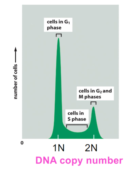
facs
fluorescence activated cell sorting
s phase cells will have more dna than g1 phase
g2/m cells will have twice the dna as s phase
identifaction of cdk mutations
yeast cDNA library → each plasmid has a yeast gene
random uptake of a plasmid
perform rescue experiment at restrictive temp
any colony that can grow has rescued functions
can identify which gene was uptaken
cdc2 and cdc13
mutants delay or fail to enter mitosis
2 encodes cdk1
13 encodes m-cyclin
wee1 mutant
premature mitosis entry
mutant prevents inhibition of cyclin-cdk complex, leads to net activation
cdc25 mutant
delayed mitosis entry
mutant leads to net inhibition of cyclin/cdk complex
positive feedback of m-cycln/cdk
activates cdc25 and inhibits wee1
mitosis challenges
no new protein synthesis after entry
condense chromosomal dna before separation
nuclear envelope disassembly
sister chromatid sorting
mitosis events
6 total
prophase: separation of centrosomes and sister chromatid condensation
prometaphase: nuclear envelope removed, chromosome capture → spindle forms and aligns
metaphase: chromosomes are aligned on plate
anaphase a: sister chromatids move to opposite ends of cell
anaphase b: cell elongates via spindle elongation
telophase: cleavage furrow forms, nuclear envelope reforms
centrioles
one is older than the other
duplication starts in s phase
prophase
disassembly of golgi, mito
dissociation of chromatids
mt dynamics v high
XMAP inhibited, no stabilization
only kinesin-13, which destabilizes mts
metaphase
breakdown of nuclear envelope through lamin phosphorylation
both kinetochores attached, chromosome moves toward spindle
settle at plate
kinetochore
aurora b kinase phosphorylates Ndc80 → loosens mt/kinetochore attachment
must be captured by mt on both sides to prevent aurora b from loosening attachment
thus allows stabilization of interaction
anaphase
separation of sister chromatids
typically held by cohesins
separase can cut cohesins
must have securins ubiquinated and degraded before activation of separase
mediated by apc/c-cdc30
sac is emitted when kinetochores aren’t properly attached, inhibits apc/c-cdc20
thus prevents separase from cutting adhesins, preventing anaphase entry
telophase
nuclear envelope reforms
cytokinesis
cleavage to produce two daughter cells
contractile ring of actin and myosin forms to divide the cytoplasm in two
use ATP to produce contractile force
requires vesicles to maintain membrane homeostasis
contractile ring location mediated by Rho GTPase
cytokinesis midbody
contains molecules that stimulate Rho GTPase to form a contractile ring
recruits GEF
Rho promotes actin formation and myosin
apoptosis
regulated cell death
cell shrinks/bubbles, loses ecm attachment
chromatin condensation → nuclear envelope broken
DNA fragmentation → mediated by endonucleases
ps exposure in outer leaflet → atp dependent flippase usually flips ps into innter leaflet, gets inhibited during apoptosis so ps is always in outer leaflet
dead cells get engulfed
necrosis
cell death caused by trauma or physical damage
cell swells
no chromatin condensation or dna laddering
inflammatory response → immune cells must neutralize the proteins that leak out
apoptosis pathway
intracellular proteolytic cascade mediated by caspases → proteases with cysteine
synthesized as a pro-enzyme, must be activated after cleavage by other activated caspases
two types: intrinsic and extrinsic
bcl2/intrinsic apoptosis pathway
mitochondria
triggered by cell damage of stress
cytochrome c is normally stored within the inner mito membrane
regulated by bcl2 proteins
anti-apoptotic prevents release
pro-apoptotic promotes release
these two can bind and inhibit eo
once released, it binds to Apaf1
activation causes change, leading to multimer formation and binding site exposure
procaspase 9 then binds to form apoptosome
activated caspase9 then activates the caspase cascade
bcl2 specifics
anti-apoptotic proteins prevent aggregation of pro-apoptotic proteins (bax, bak) in outer mito membrane
if apoptotic signal is received, pro-apoptotic proteins from abother faily inhibit anti-apoptotic blc2 proteins
fas/extrinsic apoptosis pathway
receptor on plasma membrane → Fas death receptor
trimeric Fas ligand binds to receptor and causes trimerization within membrane
conformational change recruits FADD protein and procaspase8
procaspase8 cross-cleaves/activates eo
activated caspase8 can then activate the caspase cascade
caspase and dna fragmentation
caspase mediated activation of dna endonucleases lead to the fragmentation observed during apoptosis
apoptosis survival factors
stimulate anti-apoptotic proteins
inhibition of pro-apoptotic bh3 proteins by activation of protein kinases
require constant signals to promote cell survival
cells only survive when and where needed
bc cells compete for resources and stuff