BDS1_BSC1.AHN.08 The cranial fossae and foramina

0.0(0)
Studied by 0 people
call kaiCall Kai
learnLearn
examPractice Test
spaced repetitionSpaced Repetition
heart puzzleMatch
flashcardsFlashcards
GameKnowt Play
Card Sorting

1/47

flashcard set

Earn XP

Description and Tags

o The three cranial fossae and the bones that form them. o Which parts of the brain sit within each fossa. o Which cranial nerves and blood vessels pass through the foramina.

48 Terms

1
New cards

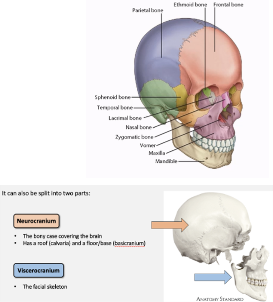
What is the Neurocranium vs viscerocranium? Which bones are in each?

  • Neurocranium: The bony case covering the brain. Has a roof (calvaria) and a floor/base (basicranium)

  • Viscerocranium = the facial skeleton


<ul><li><p><span>Neurocranium: The bony case covering the brain. Has a roof (calvaria) and a floor/base (basicranium)</span></p></li><li><p><span style="font-family: &quot;Times New Roman&quot;; line-height: normal; font-size: 7pt;"> </span><span>Viscerocranium = the facial skeleton</span></p></li></ul><p></p>
2
New cards

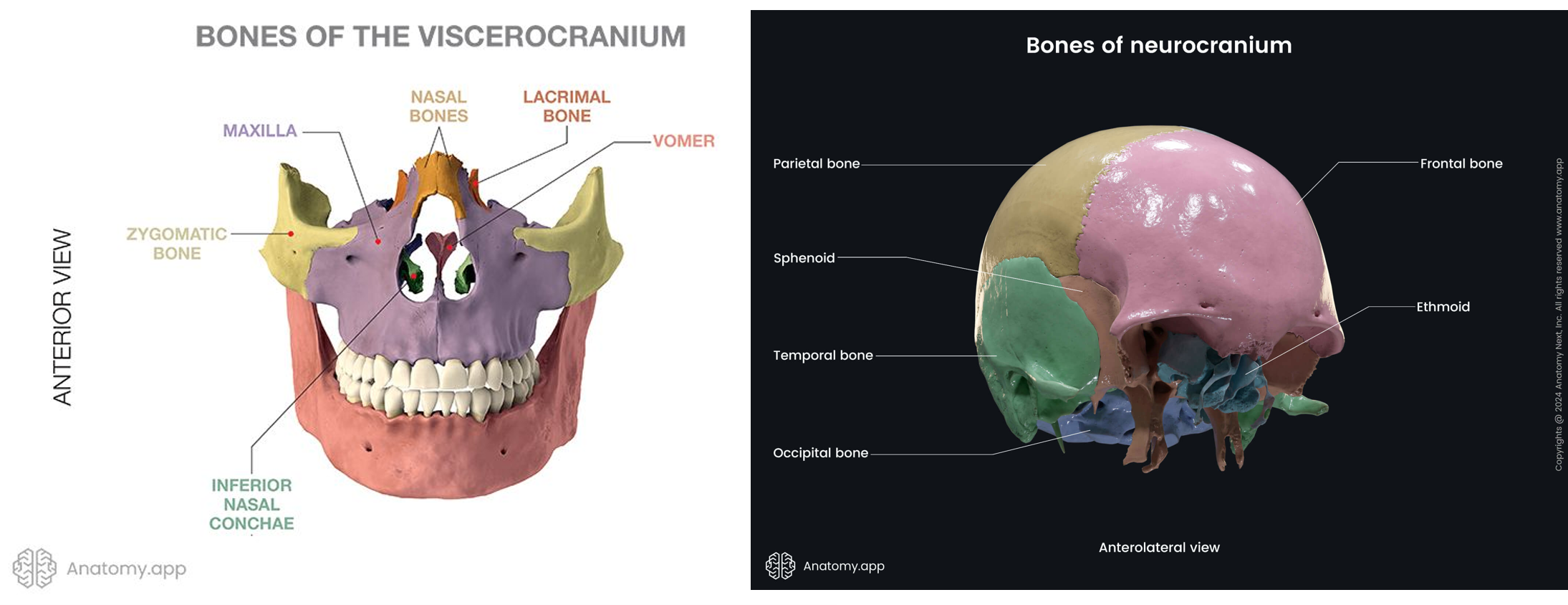
What 5x bones are in the neurocranium and which 10x bones are in the viscerocranium?

  • 5 Neurocranium bones = 2x Occipital, 2x parietal, 2x temporal, 1x frontal and 1x sphenoid.

  • 10x viscerocranium = 2x sphenoid, 2x ethmoid, 2x lacrimal, 2x nasal, 2x zygomatic, 2x maxilla, 1x mandible, 1x vomer, 2x inferior nasal conchae, 2 x palatine,

<ul><li><p><span>5 Neurocranium bones = 2x Occipital, 2x parietal, 2x temporal, 1x frontal and 1x sphenoid.</span></p></li></ul><ul><li><p class="MsoListParagraphCxSpLast"><span>10x viscerocranium = 2x sphenoid, 2x ethmoid, 2x lacrimal, 2x nasal, 2x zygomatic, 2x maxilla, 1x mandible, 1x vomer, 2x inferior nasal conchae, 2 x palatine,</span></p></li></ul><p></p>
3
New cards

 What are the following bony features

Bony prominences

Bony depressions

Bony openings

Skeletal articulations

Process –

Notch –

Foramen –

Joint –

Condyle –

Sulcus –

Fissure -

Suture –

Tuberosity –

Fossa –

Ostium –


Tubercle/eminence –

Fovea –

Canal –


Bony prominences

Bony depressions

Bony openings

Skeletal articulations

Process – general term for any prominence on a bony surface

Notch – indentation at the edge of a bone

Foramen – short window like opening

Joint – articulation between bones in a joint

Condyle – oval prominence usually involved in joints

Sulcus – shallow depression on bony surface, usually marks course of nerve of blood vessel

Fissure -narrow cleft like opening

Suture – union of bones by fibrous tissue 

Tuberosity – large rough prominence on a bony surface

Fossa – deeper depression/concavity on bony surface

Ostium – entrance into a hollow organ or canal


Tubercle or eminence – small, rounded elevation on a bony surface

Fovea – fovea small put or depression

Canal – longer opening tubelike


4
New cards

What are the three cranial fossa and bones that form each?

Cranial fossa: depression in the internal base of the skull that accommodates part of the brain. Anterior —> to posterior is high to low.

  • Anterior cranial fossa: Frontal, ethmoid and sphenoid.

  • Middle cranial fossa: Sphenoid, temporal and parietal

  • Posterior cranial fossa: sphenoid, temporal, parietal and occipital


<p>Cranial fossa: depression in the internal base of the skull that accommodates part of the brain. Anterior —&gt; to posterior is high to low.</p><ul><li><p class="MsoListParagraphCxSpMiddle">Anterior cranial fossa: Frontal, ethmoid and sphenoid.</p></li><li><p class="MsoListParagraphCxSpMiddle"><span style="font-family: &quot;Times New Roman&quot;; line-height: normal; font-size: 7pt;"> </span>Middle cranial fossa: Sphenoid, temporal and parietal</p></li><li><p class="MsoListParagraphCxSpMiddle"><span style="font-family: Aptos, sans-serif;">Posterior cranial fossa: sphenoid, temporal, parietal and occipital</span></p></li></ul><p></p>
5
New cards

What are the positions of and contents of cranial fossa?.

  • Anterior cranial fossa: front/highest contains frontal lobes of brain.

  • Middle cranial fossa: central contains the pituitary gland, the temporal lobes, cavernous sinuses, trigeminal ganglion and important foramina.

  •   Posterior cranial fossa: back/lowest contents the cerebellum and brainstem
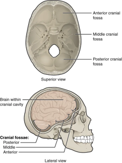

<ul><li><p> Anterior cranial fossa: front/highest contains frontal lobes of brain.</p></li><li><p><span style="font-family: &quot;Courier New&quot;;">Middle cranial fossa: central contains the pituitary gland, the temporal lobes, cavernous sinuses, trigeminal ganglion and important foramina.</span></p></li><li><p class="MsoListParagraphCxSpLast"><span style="font-family: &quot;Times New Roman&quot;; line-height: normal; font-size: 7pt;">&nbsp; </span>Posterior cranial fossa: back/lowest contents the cerebellum and brainstem</p></li></ul><p></p>
6
New cards

What are the important boundaries between the anterior/middle cranial fossa?

Anterior and middle fossae: the lesser wings of the sphenoid laterally, with the prechiasmatic sulcus region marking the boundary in the midline.

<p><span style="font-family: &quot;Times New Roman&quot;; line-height: normal; font-size: 7pt;"> </span>Anterior and middle fossae: the <strong>lesser wings</strong> of the sphenoid laterally, with the<strong> prechiasmatic sulcus </strong>region marking the boundary in the midline.</p>
7
New cards

What are the important boundaries between the middle/posterior cranial fossa?

Middle and posterior fossae: the dorsum sellae medially (sphenoid) and the superior border of the petrous temporal bone laterally.

<p>Middle and posterior fossae: the dorsum sellae medially (sphenoid) and the superior border of the petrous temporal bone laterally.</p>
8
New cards

What bones make up the anterior cranial fossa?

Blue: Orbital plates of the frontal bone.

Yellow: lesser wings of the sphenoid

Purple/pink: Cribriform plate  of the ethmoid bone.
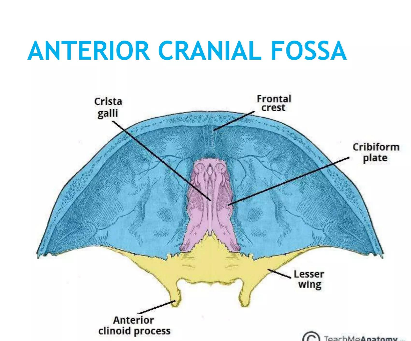
<p class="MsoListParagraphCxSpMiddle">Blue: Orbital plates of the frontal bone.</p><p class="MsoListParagraphCxSpMiddle"><span>Y</span>ellow: lesser wings of the sphenoid</p><p class="MsoListParagraphCxSpMiddle">Purple/pink: Cribriform plate <span>&nbsp;</span>of the ethmoid bone.</p>
9
New cards

What are the 4 contents of the anterior cranial fossa?

  • Frontal lobes of the brain.

  • Crista galli (attachment of the falx cerebri – dural folds separating cerebral hemispheres).

  • Cribriform plate (perforated plate of the ethmoid bone) and olfactory nerve fibres.

  • Foramen caecum (blind-ended midline pit or opening located immediately anterior to the crista galli, at the junction between the frontal bone and the ethmoid bone. During embryonic development it can be patent).


<ul><li><p class="MsoListParagraphCxSpMiddle">Frontal lobes of the brain.</p></li><li><p class="MsoListParagraphCxSpMiddle"><span> </span><strong>Crista galli</strong> (attachment of the falx cerebri – dural folds separating cerebral hemispheres).</p></li><li><p class="MsoListParagraphCxSpMiddle"><span> </span>Cribriform plate (perforated plate of the ethmoid bone) and olfactory nerve fibres.</p></li><li><p class="MsoListParagraphCxSpMiddle">Foramen caecum (blind-ended midline pit or opening located immediately anterior to the crista galli, at the junction between the frontal bone and the ethmoid bone. During embryonic development it can be patent).</p></li></ul><p></p>
10
New cards
<p>What is this? </p>

What is this?

  • Foramen caecum (blind-ended midline pit or opening located immediately anterior to the crista galli, at the junction between the frontal bone and the ethmoid bone. During embryonic development it can be patent).

  • Can transmit the emissary veins.  


11
New cards

What is the crista galli?

Crista Galli in the anterior cranial fossa is a small upward projection of the ethmoid bone. Projects between the two cerebral hemispheres. Provides attachment for the falx cerebri (a fold of dura mater). Essentially a small bony anchor for one of the protective membranes of the brain.

<p><span style="font-family: &quot;Courier New&quot;;">C</span>rista Galli in the anterior cranial fossa is a small upward projection of the ethmoid bone. Projects between the two cerebral hemispheres. Provides attachment for the falx cerebri (a fold of dura mater). Essentially a small bony anchor for one of the protective membranes of the brain.</p>
12
New cards

What is the cribriform plate? What purpose does it serve?

  • The cribriform plate is a perforated part of the ethmoid bone. It has multiple tiny holes (like a sieve).

  • The olfactory nerve fibres (CN I) pass through these holes, travelling from the olfactory receptors in the nasal cavity to the olfactory bulb.

  • Nasal cavity → olfactory nerve fibres → cribriform plate → olfactory bulb → brain.
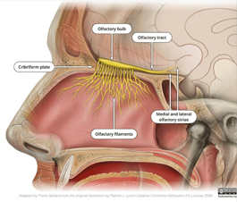

<ul><li><p>The cribriform plate is a perforated part of the ethmoid bone. It has multiple tiny holes (like a sieve).</p></li><li><p>The olfactory nerve fibres (CN I) pass through these holes, travelling from the olfactory receptors in the nasal cavity to the olfactory bulb.</p></li><li><p><span style="font-family: &quot;Times New Roman&quot;; line-height: normal; font-size: 7pt;"> </span>Nasal cavity → olfactory nerve fibres → cribriform plate → olfactory bulb → brain.</p></li></ul><p></p>
13
New cards

What 2 signs and symptoms might a patient develop if they fracture the cribriform plate?

Loss of smell = anosmia.

Leakage of CSF into nose

14
New cards

What and where is the foramen caecum?

  • Located in the anterior cranial fossa. It is often a blind-ended midline pit or opening located immediately anterior to the crista galli, at the junction between the frontal bone and the ethmoid bone. Sometimes has emissary vein connecting the nasal region with the superior sagittal sinus.

  • During embryonic development it can be patent - failure of this region to close properly can be associated with a nasal dermoid cyst.


<ul><li><p>Located in the anterior cranial fossa. It is often a blind-ended midline pit or opening located immediately anterior to the crista galli, at the junction between the frontal bone and the ethmoid bone. <strong>Sometimes has emissary vein connecting the nasal region with the superior sagittal sinus.</strong></p></li><li><p>During embryonic development it can be patent - failure of this region to close properly can be associated with a nasal dermoid cyst.</p></li></ul><p></p>
15
New cards

What are the important boundaries between the anterior/middle and middle/posterior cranial fossa?

  • Anterior and middle fossae: the lesser wings of the sphenoid laterally, with the prechiasmatic sulcus region marking the boundary in the midline.

  • Middle and posterior fossae: the dorsum sellae medially (sphenoid) and the superior border of the petrous temporal bone laterally.
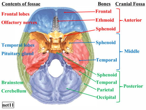

<ul><li><p>Anterior and middle fossae: the <strong>lesser wings of the sphenoid</strong> laterally, with the <strong>prechiasmatic sulcus</strong> region marking the boundary in the midline.</p></li><li><p class="MsoListParagraphCxSpLast">Middle and posterior fossae: the dorsum sellae medially (sphenoid) and the superior border of the petrous temporal bone laterally.</p></li></ul><p></p>
16
New cards

What are the bones forming the middle cranial fossa (2/3) and what is its contents (5)?

  • Bones: greater wing of sphenoid, petrous and squamous portion of the temporal bone.

  • Contents pituitary gland (in hypophysial fossa), the temporal lobes, cavernous sinuses, trigeminal ganglion (and pathway of the three divisions) and several important foramina.


<ul><li><p><span style="font-family: &quot;Times New Roman&quot;; line-height: normal; font-size: 7pt;"> </span>Bones: greater wing of sphenoid, petrous and squamous portion of the temporal bone.</p></li><li><p class="MsoListParagraphCxSpLast"><span style="font-family: &quot;Times New Roman&quot;; line-height: normal; font-size: 7pt;"> </span>Contents pituitary gland (in hypophysial fossa), the temporal lobes, cavernous sinuses, trigeminal ganglion (and pathway of the three divisions) and several important foramina.</p></li></ul><p></p>
17
New cards
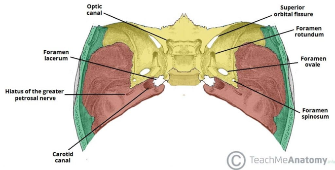
<p><span>Within the middle cranial fossa there are several important foramina. Label the foramina and name the structures passing through them.</span></p>

Within the middle cranial fossa there are several important foramina. Label the foramina and name the structures passing through them.

  • Optic canal, CN II (optic nerve), ophthalmic artery

  • Superior orbital fissure; Oculomotor nerve (CNIII), trochlear nerve (CN IV), ophthalmic division of the trigeminal nerve (CNV1), abducent nerve (CNVI), ophthalmic veins

  • Foramen rotundum, CN V2 (maxillary nerve)

  • Foramen ovale, CN V3 (mandibular nerve), lesser petrosal nerve

  •   Foramen spinosum, Middle meningeal artery

  • Foramen lacerum, No major structure passes completely through it

<ul><li><p>Optic canal, CN II (optic nerve), ophthalmic artery</p></li><li><p class="MsoListParagraphCxSpMiddle">Superior orbital fissure; Oculomotor nerve (CNIII), trochlear nerve (CN IV), ophthalmic division of the trigeminal nerve (CNV1), abducent nerve (CNVI), ophthalmic veins</p></li><li><p class="MsoListParagraphCxSpMiddle">Foramen rotundum, CN V2 (maxillary nerve)</p></li><li><p class="MsoListParagraphCxSpMiddle">Foramen ovale, CN V3 (mandibular nerve), lesser petrosal nerve</p></li><li><p class="MsoListParagraphCxSpMiddle"><span style="font-family: &quot;Times New Roman&quot;; line-height: normal; font-size: 7pt;">&nbsp; </span>Foramen spinosum, Middle meningeal artery</p></li><li><p class="MsoListParagraphCxSpLast">Foramen lacerum, No major structure passes completely through it</p></li></ul><p></p>
18
New cards

What is a sellae and what two sellae are anterior and posterior to the pituitary gland?

  • Sellae is latin for saddle. So describing a saddle structure in the sphenoid bone.

  • Anteriorly: tuberculum sellae

  • In between: hypophysial fossa (also called sella turcia) where pituitary gland sits

  • Posteriorly dorsum sellae


<ul><li><p>Sellae is latin for saddle. So describing a saddle structure in the sphenoid bone.</p></li><li><p class="MsoListParagraphCxSpMiddle">Anteriorly: tuberculum sellae</p></li></ul><ul><li><p class="MsoListParagraphCxSpMiddle">In between: hypophysial fossa (also called sella turcia) where pituitary gland sits</p></li><li><p class="MsoListParagraphCxSpLast">Posteriorly dorsum sellae</p></li></ul><p></p>
19
New cards
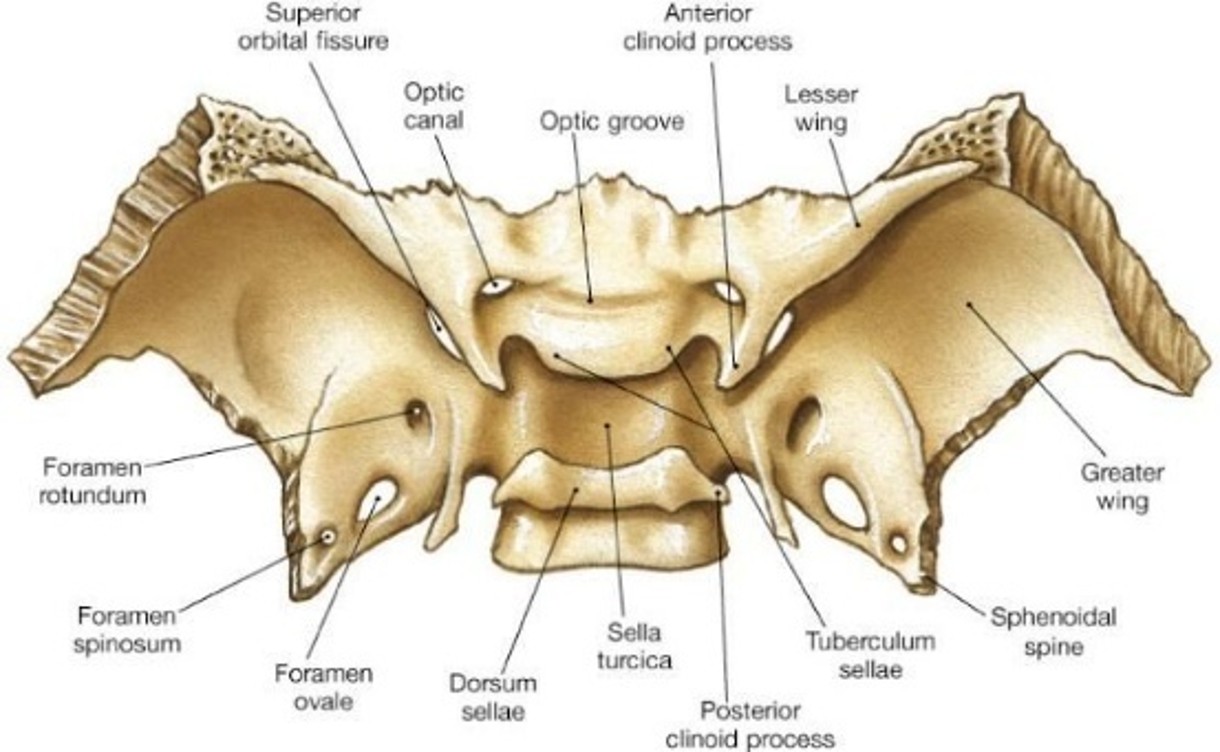
<p>Anterior view of sphenoid. Label the components.</p>

Anterior view of sphenoid. Label the components.

knowt flashcard image
20
New cards
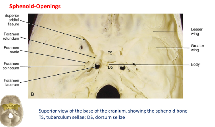
<p>Super view of sphenoid. Label the openings/components</p>

Super view of sphenoid. Label the openings/components
knowt flashcard image
21
New cards

What does foramen rotundum transmit and what is the dental relevance of this?

  • The maxillary division of the trigeminal nerve passes through the foramen rotundum.

  • V2 supplies sensory information from the midface. It provides sensation to the upper teeth, upper gingiva, maxillary sinus and parts of the nasal cavity.


22
New cards

What does foramen ovale transmit and what is the dental relevance of this?

Foramen ovale transmits the mandibular division of the trigeminal nerve. It provides

  • sensory information from the lower teeth and lower jaw.

  • sensation to parts of the tongue (general sensation, not taste)

  • motor innervation to the muscles of mastication.


<p>Foramen ovale transmits the mandibular division of the trigeminal nerve. It provides</p><ul><li><p class="MsoListParagraphCxSpMiddle">sensory information from the lower teeth and lower jaw.</p></li><li><p class="MsoListParagraphCxSpMiddle">sensation to parts of the tongue (general sensation, not taste)</p></li><li><p class="MsoListParagraphCxSpLast">motor innervation to the muscles of mastication.</p></li></ul><p></p>
23
New cards

What does foramen lacerum transmit and what is the dental relevance of this?

  • The foramen lacerum is an opening at the junction of the sphenoid, temporal and occipital bones. Nothing transmits through it. It is FULL of cartilage.

  • The internal carotid artery travels through the carotid canal, It then passes over the superior aspect of the foramen lacerum. It does not pass vertically through the foramen lacerum itself.

  • In addition:  the greater petrosal nerve meets the deep petrosal nerve AND the internal carotid artery passes over the foramen lacerum


<ul><li><p>The foramen lacerum is an opening at the junction of the sphenoid, temporal and occipital bones. Nothing transmits through it. It is FULL of cartilage.</p></li></ul><ul><li><p class="MsoListParagraphCxSpMiddle">The internal carotid artery travels through the carotid canal, It then passes over the superior aspect of the foramen lacerum. It does not pass vertically through the foramen lacerum itself.</p></li></ul><ul><li><p class="MsoListParagraphCxSpLast">In addition: <span>&nbsp;</span>the greater petrosal nerve meets the deep petrosal nerve AND the internal carotid artery passes over the foramen lacerum</p></li></ul><p></p>
24
New cards

Where is the pituitary gland located?

The pituitary gland sits in a depression in the sphenoid bone called the hypophyseal fossa, which is part of the sella turcica. The sella turcica is a saddle-shaped structure.

<p>The pituitary gland sits in a depression in the sphenoid bone called the hypophyseal fossa, which is part of the sella turcica. The sella turcica is a saddle-shaped structure.</p>
25
New cards

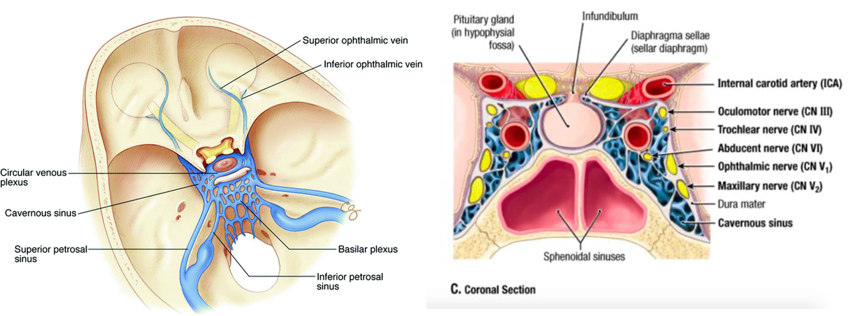
What is the cavernous sinus and what structures are within it?

  • Paired dural venous spaces are located on either side of the sella turcica (contains pituitary gland). They are important because they contain or are closely related to several cranial nerves and the internal carotid artery.

  • Within the sinus: internal carotid artery and abducens nerve (CN6).

  • Within the lateral wall: CN III (oculomotor), CN IV (trochlear), CN V1 (ophthalmic) and CN V2 (maxillary).

<ul><li><p><span>Paired dural venous spaces are located on either side of the sella turcica (contains pituitary gland). </span>They are important because they contain or are closely related to several cranial nerves and the internal carotid artery.</p></li><li><p class="MsoListParagraphCxSpMiddle">Within the sinus: internal carotid artery and abducens nerve (CN6).</p></li><li><p class="MsoListParagraphCxSpMiddle">Within the lateral wall: <span>CN III (oculomotor), CN IV (trochlear), CN V1 (ophthalmic) and CN V2 (maxillary).</span></p></li></ul><p></p>
26
New cards

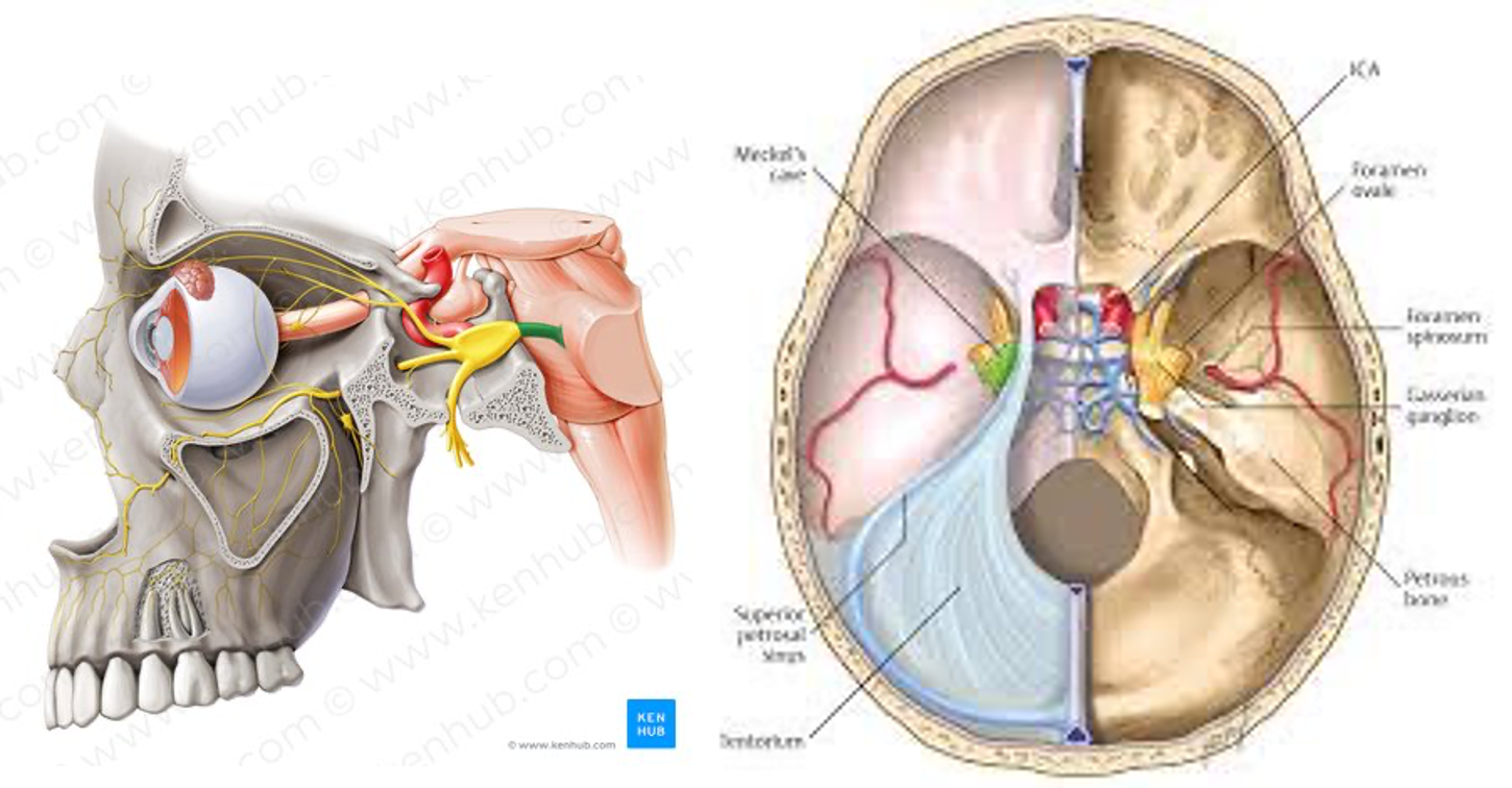
Where is the trigeminal ganglion and what does it produce?

The trigeminal ganglion  is located inside Meckel's cave, a small fluid-filled pouch of dura mater on the floor of the middle cranial fossa (near the apex of the petrous part of the temporal bone, just in front of the ear and near the side of the head)

This is where the sensory cell bodies of the trigeminal nerve are located.

The trigeminal ganglion gives rise to the three divisions:

  • V1 – Ophthalmic.

  • V2 – Maxillary.

  • V3 – Mandibular.


<p><span>The trigeminal ganglion&nbsp; is located inside Meckel's cave, a small fluid-filled pouch of dura mater on the floor of the middle cranial fossa (near the apex of the petrous part of the temporal bone, just in front of the ear and near the side of the head)</span></p><p class="MsoListParagraphCxSpMiddle">This is where the sensory cell bodies of the trigeminal nerve are located.</p><p class="MsoListParagraphCxSpMiddle">The trigeminal ganglion gives rise to the three divisions:</p><ul><li><p class="MsoListParagraphCxSpMiddle">V1 – Ophthalmic.</p></li><li><p class="MsoListParagraphCxSpMiddle">V2 – Maxillary.</p></li><li><p class="MsoListParagraphCxSpMiddle">V3 – Mandibular.</p></li></ul><p></p>
27
New cards

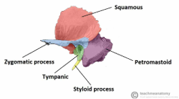
What are the 4 sections of the temporal bone?

  1. Squamous Part (Squama Temporalis): The largest thin/flat and most superiorly positioned part. Forms the lateral wall of the middle cranial fossa and provides an origin point for the temporalis muscle. The zygomatic process projects forward from this part to join the zygomatic bone and form the zygomatic arch (cheekbone).

  2. Petrous Part (Petrous Pyramid): A hard, wedge- or pyramid-shaped region located deep at the base of the skull between the sphenoid and occipital bones. It houses the delicate internal and middle ear structures (including the cochlea, semicircular canals, and bony labyrinth).

  3. Tympanic Part: A small, curved plate situated inferior to the squamous part and anterior to the mastoid part. It surrounds and forms the walls (floor and anterior/posterior walls) of the external acoustic meatus (ear canal).

  4.   Mastoid Part: Located at the posterior (back) region of the temporal bone behind the ear. It contains air-filled spaces called mastoid air cells that help equalize pressure in the middle ear

<ol><li><p>Squamous Part (Squama Temporalis): The largest thin/flat and most superiorly positioned part. Forms the lateral wall of the middle cranial fossa and provides an origin point for the temporalis muscle. <strong>The zygomatic process projects</strong> forward from this part to join the zygomatic bone and form the zygomatic arch (cheekbone).</p></li><li><p><span>Petrous Part (Petrous Pyramid): A hard, wedge- or pyramid-shaped region located deep at the base of the skull between the sphenoid and occipital bones. It houses the delicate <strong>internal and middle ear </strong>structures (including the cochlea, semicircular canals, and bony labyrinth).</span></p></li><li><p><span>Tympanic Part: A small, curved plate situated inferior to the squamous part and anterior to the mastoid part. It surrounds and forms the walls (floor and anterior/posterior walls) of the external acoustic meatus (ear canal).</span></p></li><li><p><span style="font-family: &quot;Times New Roman&quot;; line-height: normal; font-size: 7pt;">&nbsp; </span><span>Mastoid Part: Located at the posterior (back) region of the temporal bone behind the ear. It contains air-filled spaces called mastoid air cells that help equalize pressure in the middle ear</span></p></li></ol><p></p>
28
New cards

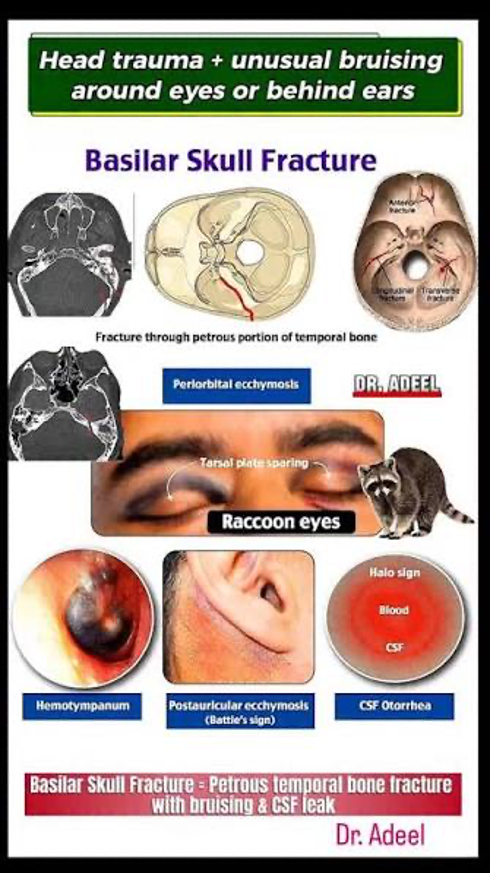
Fracture of what bone is associated with battles sign?

Battles sign = mastoid ecchymosis. Is associated with base of skull fracture most commonly the petrous part of the temporal bone.
<p><span>Battles sign = mastoid ecchym</span>osis. Is associated with base of skull fracture most commonly the petrous part of the temporal bone.</p>
29
New cards

What are the important boundaries between the anterior/middle and middle/posterior cranial fossa?

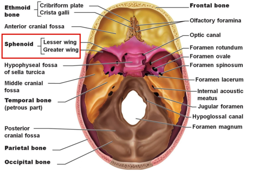
o    Anterior and middle fossae: the lesser wings of the sphenoid laterally, with the prechiasmatic sulcus region marking the boundary in the midline.

o    Middle and posterior fossae: the dorsum sellae medially (sphenoid) and the superior border of the petrous temporal bone laterally.
<p><span style="font-family: &quot;Courier New&quot;;">o</span><span style="font-family: &quot;Times New Roman&quot;; line-height: normal; font-size: 7pt;">&nbsp;&nbsp;&nbsp; </span>Anterior and middle fossae: the lesser wings of the sphenoid laterally, with the prechiasmatic sulcus region marking the boundary in the midline.</p><p class="MsoListParagraphCxSpLast"><span style="font-family: &quot;Courier New&quot;;">o</span><span style="font-family: &quot;Times New Roman&quot;; line-height: normal; font-size: 7pt;">&nbsp;&nbsp;&nbsp; </span>Middle and posterior fossae: the dorsum sellae medially (sphenoid) and the superior border of the petrous temporal bone laterally.</p>
30
New cards

What bones form the posterior cranial fossa (2/3)?  And what are the contents of the posterior cranial fossa (3)?

  • Bones: Occipital PLUS squamous and mastoid parts of the temporal bone.

  • Posterior cranial fossa components: cerebellum, brainstem and occipital lobes lie above the tentorium at the back of the cavity.


<ul><li><p><span style="font-family: &quot;Times New Roman&quot;; line-height: normal; font-size: 7pt;"> </span>Bones: Occipital PLUS squamous and mastoid parts of the temporal bone.</p></li><li><p><span>Posterior cranial fossa components: cerebellum, brainstem and <strong>occipital </strong>lobes lie above the tentorium at the back of the cavity.</span></p></li></ul><p></p>
31
New cards

There are 5 important foramina in the posterior cranial fossa – what are they?

o    Internal acoustic meatus

o    Jugular foramen

o    Hypoglossal canal

o    Foramen magnum

o    Condylar canal

<p class="MsoListParagraphCxSpFirst"><span style="font-family: &quot;Courier New&quot;;">o</span><span style="font-family: &quot;Times New Roman&quot;; line-height: normal; font-size: 7pt;">&nbsp;&nbsp;&nbsp; </span>Internal acoustic meatus</p><p class="MsoListParagraphCxSpMiddle"><span style="font-family: &quot;Courier New&quot;;">o</span><span style="font-family: &quot;Times New Roman&quot;; line-height: normal; font-size: 7pt;">&nbsp;&nbsp;&nbsp; </span>Jugular foramen</p><p class="MsoListParagraphCxSpMiddle"><span style="font-family: &quot;Courier New&quot;;">o</span><span style="font-family: &quot;Times New Roman&quot;; line-height: normal; font-size: 7pt;">&nbsp;&nbsp;&nbsp; </span>Hypoglossal canal</p><p class="MsoListParagraphCxSpMiddle"><span style="font-family: &quot;Courier New&quot;;">o</span><span style="font-family: &quot;Times New Roman&quot;; line-height: normal; font-size: 7pt;">&nbsp;&nbsp;&nbsp; </span>Foramen magnum</p><p class="MsoListParagraphCxSpLast"><span style="font-family: &quot;Courier New&quot;;">o</span><span style="font-family: &quot;Times New Roman&quot;; line-height: normal; font-size: 7pt;">&nbsp;&nbsp;&nbsp; </span>Condylar canal</p>
32
New cards

Where is the internal acoustic meatus and what does it transmit?

The internal acoustic meatus is in the petrous part of the temporal bone within the posterior cranial fossa. The internal acoustic transmits CN7, CN8 and labyrinthine artery. AND vestibular ganglion.

<p>The internal acoustic meatus is in the petrous part of the temporal bone within the posterior cranial fossa. The internal acoustic transmits CN7, CN8 and labyrinthine artery. AND vestibular ganglion.</p>
33
New cards

Where is the jugular foramen and what does it transmit?

The jugular foramen is in the posterior cranial fossa. It is between the petrous part of the temporal bone and the occipital bone. The jugular foramen transmits internal jugular vein, CN9 (glossopharyngeal), CN10 (vagus) and CN11 (accessory)

<p><span>The jugular foramen is in the posterior cranial fossa. It is between the petrous part of the temporal bone and the occipital bone. The jugular foramen transmits internal jugular vein, CN9 (glossopharyngeal), CN10 (vagus) and CN11 (accessory)</span></p>
34
New cards

Where is the hypoglossal canal and what does it transmit?

Hypoglossal canal is in the posterior cranial fossa in the occipital bone of the skull. It transmits the hypoglossal nerve (and the meningeal branch of the ascending pharyngeal artery – which is ECA branch).

<p><span>Hypoglossal canal is in the posterior cranial fossa in the occipital bone of the skull. It transmits the hypoglossal nerve (and the meningeal branch of the ascending pharyngeal artery – which is ECA branch).</span></p>
35
New cards

Where is the foramen magnum and what does it transmit?

Within the occipital bone of the posterior cranial fossa. Transmits brainstem/spinal cord as well as the vertebral arteries, spinal roots of the accessory nerve (CNXI).

<p><span>Within the occipital bone of the posterior cranial fossa. Transmits brainstem/spinal cord as well as the vertebral arteries, spinal roots of the accessory nerve (CNXI).</span></p>
36
New cards

Where is the condylar canal and what does it transmit?

Bony channel located in the condylar fossa of the posterior cranial fossa -- immediately posterior and lateral to the occipital condyle within the lateral part of the occipital bone. Transmits occipital emissary vein.

<p><span style="font-family: &quot;Times New Roman&quot;; line-height: normal; font-size: 7pt;"> </span><span>Bony channel located in the condylar fossa of the posterior cranial fossa -- immediately posterior and lateral to the occipital condyle within the lateral part of the occipital bone. Transmits occipital emissary vein.</span></p>
37
New cards

Skull base foramina/fissures.

Foramina/fissure

Cranial fossa

Contents

Foramen caecum


Olfactory foramina


Optic canal


Superior orbital fissure


Foramen Rotundum


Foramen Ovale


Foramen spinosum


Foramen lacerum


Carotid canal


Internal acoustic foramen


Jugular foramen


Hypoglossal canal


Foramen magnum


Foramina/fissure

Cranial fossa

Contents

Foramen caecum

Anterior cranial fossa

Emissary veins

Olfactory foramina

Anterior cranial fossa

Olfactory nerve

Optic canal

MIDDLE cranial fossa


Optic nerve (CNII), ophthalmic artery, dural sheath of optic nerve

Superior orbital fissure

Middle cranial fossa

Oculomotor nerve (CNIII), trochlear nerve (CN IV), ophthalmic division of the trigeminal nerve (CNV1), abducent nerve (CNVI), ophthalmic veins

Foramen Rotundum

Middle cranial fossa.

Maxillary division of trigeminal n V2.

Foramen Ovale

Middle cranial fossa.

Mandibular division of the trigeminal nerve (CN V3), accessory meningeal branch of maxillary artery, emissary vein and lesser petrosal nerve

Foramen spinosum

Middle cranial fossa.

Middle meningeal artery

Foramen lacerum

Middle cranial fossa.

Nothing transmits through. The greater petrosal nerve meets the deep petrosal nerve AND the internal carotid artery passes over the foramen lacerum.

Carotid canal


Middle cranial fossa.

Internal carotid artery

Internal acoustic foramen

POSTERIOR cranial fossa.

Facial nerve (CNVII), Vestibulocochlear nerve (CNVIII)

Jugular foramen

Posterior cranial fossa.

Glossopharyngeal nerve (CNIX), vagus nerve (CNX), descending portion of the spinal accessory nerve (CNXI), internal jugular vein

Hypoglossal canal

Posterior cranial fossa

Hypoglossal nerve (CNXII)

Foramen magnum

Posterior cranial fossa

Brainstem/spinal cord, vertebral arteries, ascending portion of the spinal accessory nerve (CNXI)


<table style="min-width: 75px;"><colgroup><col style="min-width: 25px;"><col style="min-width: 25px;"><col style="min-width: 25px;"></colgroup><tbody><tr><td colspan="1" rowspan="1" style="width: 150.25pt; border: 1pt solid black; padding: 0cm 5.4pt; height: 15pt;"><p class="MsoNormal"><span>Foramina/fissure</span></p></td><td colspan="1" rowspan="1" style="width: 150.25pt; border-width: 1pt 1pt 1pt medium; border-style: solid solid solid none; border-color: black black black currentcolor; border-image: none; padding: 0cm 5.4pt; height: 15pt;"><p class="MsoNormal"><span>Cranial fossa</span></p></td><td colspan="1" rowspan="1" style="width: 150.25pt; border-width: 1pt 1pt 1pt medium; border-style: solid solid solid none; border-color: black black black currentcolor; border-image: none; padding: 0cm 5.4pt; height: 15pt;"><p class="MsoNormal"><span>Contents</span></p></td></tr><tr><td colspan="1" rowspan="1" style="width: 150.25pt; border-width: medium 1pt 1pt; border-style: none solid solid; border-color: currentcolor black black; border-image: none; padding: 0cm 5.4pt; height: 15pt;"><p class="MsoNormal"><span>Foramen caecum</span></p></td><td colspan="1" rowspan="1" style="width: 150.25pt; border-width: medium 1pt 1pt medium; border-style: none solid solid none; border-color: currentcolor black black currentcolor; padding: 0cm 5.4pt; height: 15pt;"><p class="MsoNormal"><span>Anterior cranial fossa</span></p></td><td colspan="1" rowspan="1" style="width: 150.25pt; border-width: medium 1pt 1pt medium; border-style: none solid solid none; border-color: currentcolor black black currentcolor; padding: 0cm 5.4pt; height: 15pt;"><p class="MsoNormal"><span>Emissary veins</span></p></td></tr><tr><td colspan="1" rowspan="1" style="width: 150.25pt; border-width: medium 1pt 1pt; border-style: none solid solid; border-color: currentcolor black black; border-image: none; padding: 0cm 5.4pt; height: 15pt;"><p class="MsoNormal"><span>Olfactory foramina</span></p></td><td colspan="1" rowspan="1" style="width: 150.25pt; border-width: medium 1pt 1pt medium; border-style: none solid solid none; border-color: currentcolor black black currentcolor; padding: 0cm 5.4pt; height: 15pt;"><p class="MsoNormal"><span>Anterior cranial fossa</span></p></td><td colspan="1" rowspan="1" style="width: 150.25pt; border-width: medium 1pt 1pt medium; border-style: none solid solid none; border-color: currentcolor black black currentcolor; padding: 0cm 5.4pt; height: 15pt;"><p class="MsoNormal"><span>Olfactory nerve</span></p></td></tr><tr><td colspan="1" rowspan="1" style="width: 150.25pt; border-width: medium 1pt 1pt; border-style: none solid solid; border-color: currentcolor black black; border-image: none; padding: 0cm 5.4pt; height: 15pt;"><p class="MsoNormal"><span>Optic canal</span></p></td><td colspan="1" rowspan="1" style="width: 150.25pt; border-width: medium 1pt 1pt medium; border-style: none solid solid none; border-color: currentcolor black black currentcolor; padding: 0cm 5.4pt; height: 15pt;"><p class="MsoNormal"><span>MIDDLE cranial fossa</span></p><p class="MsoNormal"><span>&nbsp;</span></p></td><td colspan="1" rowspan="1" style="width: 150.25pt; border-width: medium 1pt 1pt medium; border-style: none solid solid none; border-color: currentcolor black black currentcolor; padding: 0cm 5.4pt; height: 15pt;"><p class="MsoNormal"><span><strong>Optic nerve (CNII), ophthalmic artery,</strong> dural sheath of optic nerve</span></p></td></tr><tr><td colspan="1" rowspan="1" style="width: 150.25pt; border-width: medium 1pt 1pt; border-style: none solid solid; border-color: currentcolor black black; border-image: none; padding: 0cm 5.4pt; height: 15pt;"><p class="MsoNormal"><span>Superior orbital fissure</span></p></td><td colspan="1" rowspan="1" style="width: 150.25pt; border-width: medium 1pt 1pt medium; border-style: none solid solid none; border-color: currentcolor black black currentcolor; padding: 0cm 5.4pt; height: 15pt;"><p class="MsoNormal"><span>Middle cranial fossa</span></p></td><td colspan="1" rowspan="1" style="width: 150.25pt; border-width: medium 1pt 1pt medium; border-style: none solid solid none; border-color: currentcolor black black currentcolor; padding: 0cm 5.4pt; height: 15pt;"><p class="MsoNormal"><span>Oculomotor nerve (CNIII), trochlear nerve (CN IV), ophthalmic division of the trigeminal nerve (CN<strong>V1</strong>), abducent nerve (CNVI), <strong>ophthalmic veins</strong></span></p></td></tr><tr><td colspan="1" rowspan="1" style="width: 150.25pt; border-width: medium 1pt 1pt; border-style: none solid solid; border-color: currentcolor black black; border-image: none; padding: 0cm 5.4pt; height: 15pt;"><p class="MsoNormal"><span>Foramen Rotundum</span></p></td><td colspan="1" rowspan="1" style="width: 150.25pt; border-width: medium 1pt 1pt medium; border-style: none solid solid none; border-color: currentcolor black black currentcolor; padding: 0cm 5.4pt; height: 15pt;"><p class="MsoNormal"><span>Middle cranial fossa.</span></p></td><td colspan="1" rowspan="1" style="width: 150.25pt; border-width: medium 1pt 1pt medium; border-style: none solid solid none; border-color: currentcolor black black currentcolor; padding: 0cm 5.4pt; height: 15pt;"><p class="MsoNormal"><span>Maxillary division of trigeminal n V2.</span></p></td></tr><tr><td colspan="1" rowspan="1" style="width: 150.25pt; border-width: medium 1pt 1pt; border-style: none solid solid; border-color: currentcolor black black; border-image: none; padding: 0cm 5.4pt; height: 15pt;"><p class="MsoNormal"><span>Foramen Ovale</span></p></td><td colspan="1" rowspan="1" style="width: 150.25pt; border-width: medium 1pt 1pt medium; border-style: none solid solid none; border-color: currentcolor black black currentcolor; padding: 0cm 5.4pt; height: 15pt;"><p class="MsoNormal"><span>Middle cranial fossa.</span></p></td><td colspan="1" rowspan="1" style="width: 150.25pt; border-width: medium 1pt 1pt medium; border-style: none solid solid none; border-color: currentcolor black black currentcolor; padding: 0cm 5.4pt; height: 15pt;"><p class="MsoNormal"><span>Mandibular division of the trigeminal nerve (CN <strong>V3</strong>), accessory meningeal branch of maxillary artery, emissary vein and<strong> lesser petrosal nerve</strong></span></p></td></tr><tr><td colspan="1" rowspan="1" style="width: 150.25pt; border-width: medium 1pt 1pt; border-style: none solid solid; border-color: currentcolor black black; border-image: none; padding: 0cm 5.4pt; height: 15.75pt;"><p class="MsoNormal"><span>Foramen spinosum</span></p></td><td colspan="1" rowspan="1" style="width: 150.25pt; border-width: medium 1pt 1pt medium; border-style: none solid solid none; border-color: currentcolor black black currentcolor; padding: 0cm 5.4pt; height: 15.75pt;"><p class="MsoNormal"><span>Middle cranial fossa.</span></p></td><td colspan="1" rowspan="1" style="width: 150.25pt; border-width: medium 1pt 1pt medium; border-style: none solid solid none; border-color: currentcolor black black currentcolor; padding: 0cm 5.4pt; height: 15.75pt;"><p class="MsoNormal"><span>Middle meningeal artery</span></p></td></tr><tr><td colspan="1" rowspan="1" style="width: 150.25pt; border-width: medium 1pt 1pt; border-style: none solid solid; border-color: currentcolor black black; border-image: none; padding: 0cm 5.4pt; height: 15pt;"><p class="MsoNormal"><span>Foramen lacerum</span></p></td><td colspan="1" rowspan="1" style="width: 150.25pt; border-width: medium 1pt 1pt medium; border-style: none solid solid none; border-color: currentcolor black black currentcolor; padding: 0cm 5.4pt; height: 15pt;"><p class="MsoNormal"><span>Middle cranial fossa.</span></p></td><td colspan="1" rowspan="1" style="width: 150.25pt; border-width: medium 1pt 1pt medium; border-style: none solid solid none; border-color: currentcolor black black currentcolor; padding: 0cm 5.4pt; height: 15pt;"><p class="MsoNormal"><span>Nothing transmits through. The greater petrosal nerve meets the deep petrosal nerve AND the internal carotid artery passes over the foramen lacerum.</span></p></td></tr><tr><td colspan="1" rowspan="1" style="width: 150.25pt; border-width: medium 1pt 1pt; border-style: none solid solid; border-color: currentcolor black black; border-image: none; padding: 0cm 5.4pt; height: 15pt;"><p class="MsoNormal"><span>Carotid canal</span></p><p class="MsoNormal"><span>&nbsp;</span></p></td><td colspan="1" rowspan="1" style="width: 150.25pt; border-width: medium 1pt 1pt medium; border-style: none solid solid none; border-color: currentcolor black black currentcolor; padding: 0cm 5.4pt; height: 15pt;"><p class="MsoNormal"><span>Middle cranial fossa.</span></p></td><td colspan="1" rowspan="1" style="width: 150.25pt; border-width: medium 1pt 1pt medium; border-style: none solid solid none; border-color: currentcolor black black currentcolor; padding: 0cm 5.4pt; height: 15pt;"><p class="MsoNormal"><span>Internal carotid artery</span></p></td></tr><tr><td colspan="1" rowspan="1" style="width: 150.25pt; border-width: medium 1pt 1pt; border-style: none solid solid; border-color: currentcolor black black; border-image: none; padding: 0cm 5.4pt; height: 15pt;"><p class="MsoNormal"><span>Internal acoustic foramen</span></p></td><td colspan="1" rowspan="1" style="width: 150.25pt; border-width: medium 1pt 1pt medium; border-style: none solid solid none; border-color: currentcolor black black currentcolor; padding: 0cm 5.4pt; height: 15pt;"><p class="MsoNormal"><span>POSTERIOR cranial fossa.</span></p></td><td colspan="1" rowspan="1" style="width: 150.25pt; border-width: medium 1pt 1pt medium; border-style: none solid solid none; border-color: currentcolor black black currentcolor; padding: 0cm 5.4pt; height: 15pt;"><p class="MsoNormal"><span>Facial nerve (CNVII), Vestibulocochlear nerve (CNVIII)</span></p></td></tr><tr><td colspan="1" rowspan="1" style="width: 150.25pt; border-width: medium 1pt 1pt; border-style: none solid solid; border-color: currentcolor black black; border-image: none; padding: 0cm 5.4pt; height: 15pt;"><p class="MsoNormal"><span>Jugular foramen</span></p></td><td colspan="1" rowspan="1" style="width: 150.25pt; border-width: medium 1pt 1pt medium; border-style: none solid solid none; border-color: currentcolor black black currentcolor; padding: 0cm 5.4pt; height: 15pt;"><p class="MsoNormal"><span>Posterior cranial fossa.</span></p></td><td colspan="1" rowspan="1" style="width: 150.25pt; border-width: medium 1pt 1pt medium; border-style: none solid solid none; border-color: currentcolor black black currentcolor; padding: 0cm 5.4pt; height: 15pt;"><p class="MsoNormal"><span>Glossopharyngeal nerve (CNIX), vagus nerve (CNX), descending portion of the spinal accessory nerve (CNXI), internal jugular vein</span></p></td></tr><tr><td colspan="1" rowspan="1" style="width: 150.25pt; border-width: medium 1pt 1pt; border-style: none solid solid; border-color: currentcolor black black; border-image: none; padding: 0cm 5.4pt; height: 15pt;"><p class="MsoNormal"><span>Hypoglossal canal</span></p></td><td colspan="1" rowspan="1" style="width: 150.25pt; border-width: medium 1pt 1pt medium; border-style: none solid solid none; border-color: currentcolor black black currentcolor; padding: 0cm 5.4pt; height: 15pt;"><p class="MsoNormal"><span>Posterior cranial fossa</span></p></td><td colspan="1" rowspan="1" style="width: 150.25pt; border-width: medium 1pt 1pt medium; border-style: none solid solid none; border-color: currentcolor black black currentcolor; padding: 0cm 5.4pt; height: 15pt;"><p class="MsoNormal"><span>Hypoglossal nerve (CNXII)</span></p></td></tr><tr><td colspan="1" rowspan="1" style="width: 150.25pt; border-width: medium 1pt 1pt; border-style: none solid solid; border-color: currentcolor black black; border-image: none; padding: 0cm 5.4pt; height: 15pt;"><p class="MsoNormal"><span>Foramen magnum</span></p></td><td colspan="1" rowspan="1" style="width: 150.25pt; border-width: medium 1pt 1pt medium; border-style: none solid solid none; border-color: currentcolor black black currentcolor; padding: 0cm 5.4pt; height: 15pt;"><p class="MsoNormal"><span>Posterior cranial fossa</span></p></td><td colspan="1" rowspan="1" style="width: 150.25pt; border-width: medium 1pt 1pt medium; border-style: none solid solid none; border-color: currentcolor black black currentcolor; padding: 0cm 5.4pt; height: 15pt;"><p class="MsoNormal"><span>Brainstem/spinal cord, vertebral arteries, ascending portion of the spinal accessory nerve (CNXI)</span></p><p class="MsoNormal"><span>&nbsp;</span></p></td></tr></tbody></table><p></p>
38
New cards
term image
knowt flashcard image
39
New cards
term image
  • Optic canal, CN II (optic nerve), ophthalmic artery

  • Superior orbital fissure; Oculomotor nerve (CNIII), trochlear nerve (CN IV), ophthalmic division of the trigeminal nerve (CNV1), abducent nerve (CNVI), ophthalmic veins

  • Foramen rotundum, CN V2 (maxillary nerve)

  • Foramen ovale, CN V3 (mandibular nerve), lesser petrosal nerve

  • Foramen spinosum, Middle meningeal artery

  • The sella turcica is also known as the hypophyseal fossa, pituitary fossa, or Turkish saddle


<ul><li><p>Optic canal, CN II (optic nerve), ophthalmic artery</p></li><li><p>Superior orbital fissure; Oculomotor nerve (CNIII), trochlear nerve (CN IV), ophthalmic division of the trigeminal nerve (CNV1), abducent nerve (CNVI), ophthalmic veins</p></li><li><p>Foramen rotundum, CN V2 (maxillary nerve)</p></li><li><p>Foramen ovale, CN V3 (mandibular nerve), lesser petrosal nerve</p></li><li><p>Foramen spinosum, Middle meningeal artery</p></li><li><p><span>The sella turcica is also known as the hypophyseal fossa, pituitary fossa, or Turkish saddle</span></p></li></ul><p></p>
40
New cards

Which foramen does the Optic nerve travel thorough?

Optic nerve CN2 travels through optic canal with ophthalmic artery.

41
New cards

Which foramen does mandibular branch of the trigeminal nerve travel through?

o    Foramen ovale, CN V3 (mandibular nerve) AND lesser petrosal nerve

42
New cards

Which foramen does the oculomotor nerve travel thorough?

Oculomotor nerve (CN3) travels through Superior orbital fissure not foramen. And travels with trochlear nerve (CN IV), ophthalmic division of the trigeminal nerve (CNV1), abducent nerve (CNVI), ophthalmic veins

43
New cards

Which foramen does the middle meningeal artery pass through?

Foramen spinosum

44
New cards

Which foramen does the CN V1 nerve travel thorough?

Ophthalmic division of trigeminal nerve (CN5) passes through Superior orbital fissure not foramen along with Oculomotor nerve (CN3), trochlear nerve (CN4), abducent nerve (CNVI), ophthalmic veins

45
New cards

Which foramen does the maxillary branch of the trigeminal nerve travel through?

Foramen rotundum, CN V2 (maxillary nerve)

46
New cards

  Which foramen does abducent nerve travel through?

Abducens nerve (CN6) travels through the superior orbital fissure not foramen. Also travelling through the superior orbital fissure is oculomotor nerve (CN3), trochlear nerve (CN4), V1 of trigeminal nerve AND the ophthalmic vein (artery travels through the optic canal with the optic nerve).

47
New cards

Which foramen does the lesser petrosal nerve travel through?

Lesser petrosal nerve is parasympathetic branch of the glossopharyngeal nerve   it passes through foramen ovale with CN V3 (mandibular nerve)

48
New cards

Which foramen does the trochlear nerve travel thorough?

Trochlear nerve CN4 travels through Superior orbital fissure Superior orbital fissure not foramen. And travels with Oculomotor nerve (CN3), ophthalmic division of the trigeminal nerve (CNV1), abducent nerve (CNVI), ophthalmic veins