Physiology Block 3
 Call Kai
 Learn
 Practice Test
 Spaced Repetition
 Match
 Flashcards
 Knowt Play
1/49

There's no tags or description
Looks like no tags are added yet.
50 Terms

Structure of
the Heart
• Four chambers, Right atrium does what?
receives
deoxygenated blood
from the body

Structure of
the Heart
• Four chambers, left atrium does what?
receives
oxygenated blood
from the lungs

Structure of
the Heart
• Four chambers, right ventricle does what?
pumps
deoxygenated blood
to the lungs

Structure of
the Heart
• Four chambers, left ventricle does what?
pumps oxygenated
blood to the body

Pulmonary: between
heart and lungs
Blood pumps to lungs via
pulmonary _____.
Blood returns to heart via
pulmonary _____ .
Blood pumps to lungs via
pulmonary arteries.
Blood returns to heart via
pulmonary veins.

Systemic: between heart
and body tissues
Blood pumps to body
tissues via _____
Blood returns to heart via
________
Blood pumps to body
tissues via aorta.
Blood returns to heart via
superior and inferior
venae cava

• Rearrange these structures in the correct sequence that a red blood cell passes through, starting immediately after the Right Ventricle:
• A) Left Atrium
• B) Systemic Capillaries
• C) Pulmonary Artery
• D) Right Atrium
• E) Aorta
• F) Left Ventricle
• G) Pulmonary Veins
• H) Vena Cava
C (Pulmonary Artery)
G (Pulmonary Veins)
A (Left Atrium)
F (Left Ventricle)
E (Aorta)
B (Systemic Capillaries)
H (Vena Cava)
D (Right Atrium)
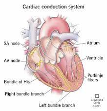
• Put the components of the cardiac electrical conduction pathway in order, starting from the generation of the pacemaker potential:
• A) Purkinje fibers
• B) SA Node
• C) AV Node
• D) Bundle of His (AV bundle)
• E) Bundle Branches
1. B (SA Node) → Generates excitation
2. C (AV Node) → Creates a brief delay for atrial filling
3. D (Bundle of His) → Travels down the septum
4. E (Bundle Branches) → Splits toward the apex
5. A (Purkinje fibers) → Spreads upward through ventricular myocytes

• Arrange the 4 main mechanical phases of the left ventricle in order, starting immediately after the mitral (AV) valve closes:
• A) Isovolumetric Relaxation
• B) Ventricular Filling
• C) Isovolumetric Contraction
• D) Ventricular Ejection
1. C (Isovolumetric Contraction) → Pressure builds, all valves shut.
2. D (Ventricular Ejection) → Aortic valve opens, volume drops.
3. A (Isovolumetric Relaxation) → Aortic valve closes, pressure drops.
4. B (Ventricular Filling) → Mitral valve opens, volume rises

Order the steps of the homeostatic reflex loop that occurs immediately after a sudden drop in blood pressure (e.g., standing up too fast)
• A) Increase in Sympathetic output / Decrease in Parasympathetic output
• B) Decreased stretch and reduced firing rate of baroreceptors
• C) Vasoconstriction (increased TPR) and increased Cardiac Output
• D) Blood pressure returns toward normal
• E) Cardiovascular control center in the brain processes reduced signal
B → E → A → C → D

• An ECG records a QRS complex.
1. What electrical event just finished?
2. What mechanical phase immediately follows?
1. Atrial repolarization and Ventricular depolarization.
2. Isovolumetric contraction of the ventricles

During Ventricular Ejection, the semilunar (aortic/pulmonary) valves are open. What is the exact sequence of valve movements that allows the heart to transition into Isovolumetric Relaxation?
1. Ventricular pressure drops below arterial pressure.
2. Semilunar valves snap shut (causing the second heart sound).
3. Note: AV valves remain closed during this transition until ventricular pressure falls below atrial pressure.

As blood moves from the arterial end of a capillary bed to the venous end, how does the sequence of Starling forces change, and what is the net direction of fluid movement?
• Arterial End: Hydrostatic pressure is high → exceeds Oncotic pressure → Net Filtration (fluid leaves capillary).
• Venous End: Hydrostatic pressure drops → Oncotic pressure exceeds it → Net Absorption (fluid re-enters capillary).

If the Left Ventricle fails to contract effectively, trace the backward sequence of pressure backups that leads to breathing difficulties.
• Left Ventricle fails to empty → Blood backs up into Left Atrium → Backs up into Pulmonary Veins → Increases hydrostatic pressure in Pulmonary Capillaries → Fluid filters into lungs (Pulmonary Edema).

• Both large arteries and large veins are reservoirs, but they serve completely different purposes.
1. Which one is the pressure reservoir, and why?
2. Which one is the volume reservoir, and why?
1. Large Arteries = Pressure Reservoir. They have thick, highly elastic walls. They expand during systole and recoil during diastole to maintain a constant pressure gradient and keep blood flowing while the heart relaxes.
2. Large Veins = Volume Reservoir. They have highly compliant (stretchy) walls and large diameters, allowing them to hold the highest percentage of the body's total blood volume at low pressure.

What are the two major cell types in the heart, and what is the single most important voltage-gated channel difference that prevents contractile cardiac muscle from undergoing tetany (sustained contraction)?
1. Pacemaker (Autorhythmic) cells (initiate/conduct electrical signals) and Contractile cells (mechanical pumping).
2. Voltage-gated Ca2+ channels. These channels open during the contractile action potential, creating a prolonged plateau phase. This lengthens the absolute refractory period, ensuring the heart muscle relaxes and fills with blood (a twitch) rather than locking up (tetany).

Capillaries are uniquely optimized for fluid and gas exchange. What universal structural feature do all blood vessels share, and how are capillaries structurally stripped down to facilitate rapid diffusion?
• All vessels share a thin inner lining of endothelial cells (endothelium).
• Capillaries lack smooth muscle and connective tissue entirely. They consist only of a single layer of endothelial cells and a basement membrane, minimizing the diffusion distance to maximize simple diffusion.
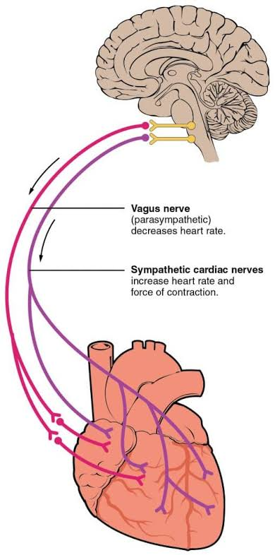
• How do Sympathetic and Parasympathetic stimulation physically alter the pacemaker potential (the phase before threshold) to change your heart rate?
• Sympathetic stimulation: Increases the rate of the slow Na+ leak. This makes the slope steeper, causing the cell to reach threshold faster and increasing heart rate.
• Parasympathetic stimulation: Decreases the rate of the slow Na+ leak (and opens K+ channels). This flattens the slope, taking longer to reach threshold and decreasing heart rate.

• Arterioles are primarily regulated by the sympathetic nervous system.
1. What is meant by "sympathetic tone"?
2. What neurotransmitter is released, and how do you achieve vasodilation if the parasympathetic nervous system does not innervate most systemic blood vessels?
1. Sympathetic tone is a continuous, baseline level of sympathetic nerve firing that keeps blood vessels partially constricted.
2. The neurotransmitter is Norepinephrine (NE).
3. Vasodilation is achieved simply by decreasing the sympathetic firing rate below baseline. (More sympathetic firing = vasoconstriction; less sympathetic firing = vasodilation).

If an arteriole undergoes sudden vasoconstriction, what happens to the blood pressure upstream from that point versus downstream from that point?
• Upstream pressure increases (blood backs up against the narrow resistance point, increasing systemic arterial blood pressure).
• Downstream pressure decreases (less blood is able to squeeze through the constricted vessel into the capillaries).
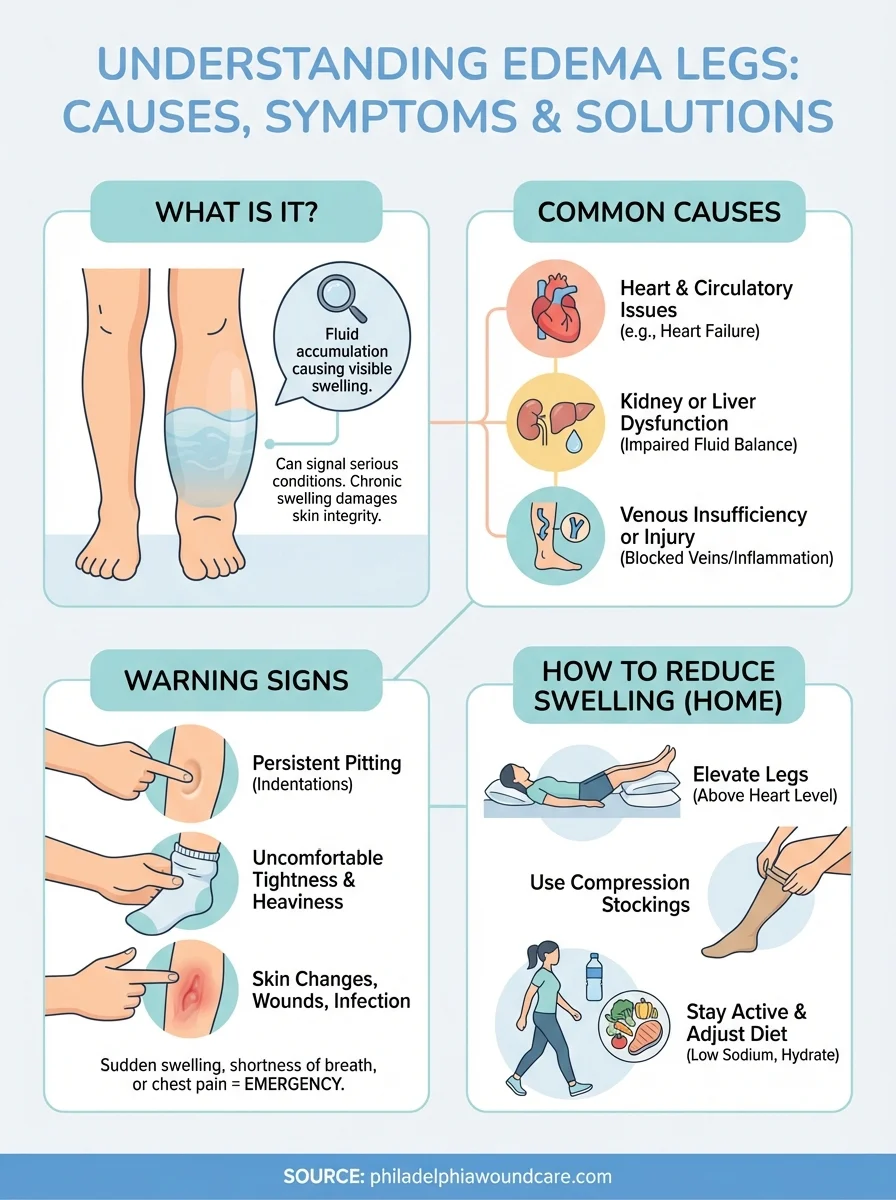
Define edema. Based on Starling forces, name three distinct physiological imbalances that could theoretically cause fluid to accumulate in tissues.
• Edema is swelling caused by excess fluid accumulating in the interstitial fluid (ISF).
• Three potential causes:
1. Increased capillary hydrostatic pressure (e.g., high blood pressure or venous backup forcing fluid out).
2. Decreased plasma oncotic pressure (e.g., severe liver disease or malnutrition causing a drop in plasma proteins like albumin).
3. Lymphatic blockage (preventing the lymphatic system from returning the naturally filtered excess fluid back to the blood).
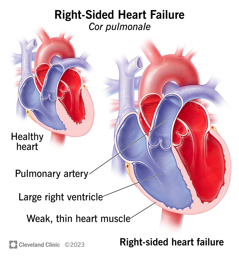
If a patient has a defect that prevents their Right Ventricle from contracting effectively, why does this lead to swelling in their feet and ankles (peripheral edema)?
The weak right ventricle fails to pump blood forward efficiently into the lungs Blood backs up into the Right Atrium Backs up into the Vena Cava Backs up into the systemic veins and capillaries. This dramatic rise in systemic capillary hydrostatic pressure forces excess fluid out into the lower body tissues.

Why do patients with long-term, untreated high blood pressure (hypertension) develop a thickened left ventricular wall (hypertrophy)?
Chronic high blood pressure means afterload is permanently elevated (the aortic pressure the ventricle must overcome to open the valve is too high). To pump blood against this extreme resistance, the left ventricular muscle must work harder with every single beat. Just like skeletal muscle exposed to heavy weightlifting, the cardiac myocytes undergo hypertrophy (grow thicker) to cope with the chronic workload.

What is the exact step-by-step path of blood flow through the heart, starting from its return from the body? (12 steps)
1. Venae Cava (Superior & Inferior)
2. Right Atrium
3. Tricuspid Valve
4. Right Ventricle
5. Pulmonary Valve
6. Pulmonary Arteries → Lungs (to get O₂)
7. Pulmonary Veins
8. Left Atrium
9. Mitral/Bicuspid Valve
10. Left Ventricle
11. Aortic Valve
12. Aorta → Body Tissues

How do the Pulmonary and Systemic circulation loops compare in terms of their start, end, and oxygen states?
Pulmonary Loop: Right Ventricle → Pulmonary Arteries (Low O₂) → Lungs → Pulmonary Veins (High O₂) → Left Atrium.
Systemic Loop: Left Ventricle → Aorta (High O₂) → Body Tissues → Venae Cavae (Low O₂) → Right Atrium.
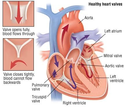
What underlying physical mechanic causes the heart valves to open and close?
Pressure differentials. The valves are strictly one-way doors:
Open: When pressure is higher on the leading side (upstream).
Close: When pressure is higher on the trailing side (downstream), forcing the valve shut to prevent backflow.

Exactly when do the "Lub" and "Dub" sounds happen, and which valves cause them?
"Lub" (1st Sound): Caused by the closure of the AV valves (Tricuspid & Mitral). It marks the start of ventricular contraction (systole).
"Dub" (2nd Sound): Caused by the closure of the Semilunar valves (Aortic & Pulmonic). It marks the start of ventricular relaxation (diastole).

What is the correct chronological order of the 4 main phases of the cardiac cycle, starting with relaxation?
1. Ventricular Filling (Diastole)
2. Isovolumetric Contraction (Start of Systole)
3. Ventricular Ejection (Systole)
4. Isovolumetric Relaxation (Start of Diastole)

What happens to Ventricular Pressure and Volume during each of the 4 cycle phases?
1. Filling: Pressure is very low; Volume increases to its highest.
2. Isovolumetric Contraction: Pressure shoots up rapidly (developing pressure); Volume stays exactly the same because all valves are closed.
3. Ejection: Pressure remains high; Volume decreases rapidly as blood pumps out.
4. Isovolumetric Relaxation: Pressure drops rapidly; Volume stays exactly the same because all valves are closed again.

On a Left Ventricular Pressure-Volume (PV) Loop, what specific sequential cardiac milestones occur at points A, B, C, and D?
Point A: Mitral (AV) valve opens, kicking off ventricular filling.
Point B: Mitral (AV) valve closes because the ventricle starts contracting.
Point C: Aortic valve opens once ventricular pressure beats aortic pressure, starting ejection.
Point D: Aortic valve closes because the ventricle finishes contracting and begins relaxing.

What is the state of the heart valves during the vertical lines vs. the horizontal lines of the PV Loop?
Vertical Lines (Isovolumetric phases): All valves are closed. Blood cannot move, so volume is locked while pressure changes rapidly (Upwards line = Contraction; Downwards line = Relaxation).
Horizontal Lines (Flow phases): One valve is open. Blood is moving actively (Bottom right-to-left line = Filling via open Mitral valve; Top left-to-right curve = Ejection via open Aortic valve).

In the sequence of a heartbeat, when do End-Diastolic Volume (EDV) and End-Systolic Volume (ESV) occur?
End-Diastolic Volume (EDV): The total volume of blood sitting in the ventricle at the very end of filling (right before contraction begins). This is the maximum volume of the loop.
End-Systolic Volume (ESV): The leftover blood left behind in the ventricle at the very end of ejection (right before relaxation begins). This is the minimum volume of the loop.

What is Stroke Volume (SV), and how is it calculated using your volume points?
Stroke Volume is the exact amount of blood physically pumped out of the ventricle during a single beat.
Formula: Stroke Volume = EDV - ESV
On the loop: It is represented visually by the horizontal width of the PV loop box.
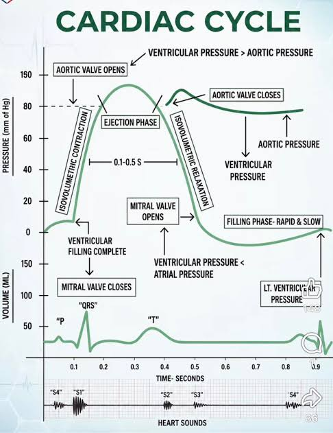
What is phase 4+ (Late Filling), and what mechanical event happens here?
Atrial contraction (atrial systole). While the ventricle is already mostly filled passively, the atrium squeezes at the very end of diastole to force the final remaining volume of blood down into the ventricle right before the AV valve snaps shut

How do cardiac muscle cells communicate electrically, and what is a functional syncytium?
Cells are interconnected by gap junctions located at intercalated discs. These allow the action potential (AP) to flow freely from cell to cell.
A functional syncytium means an entire region of cells contracts together as a single unit. The heart has two separate units: the atria contract as one unit, and the ventricles contract as another. They are separated by a non-conductive fibrous skeleton.

What is the exact chronological pathway an electrical impulse follows through the heart's conduction network?
1. Sinoatrial (SA) Node (The primary pacemaker where depolarization begins)
2. Atrial Myocytes (Depolarization spreads across the atria)
3. Atrioventricular (AV) Node (Conduction slows down intentionally here)
4. Bundle of His (AV Bundle in the interventricular septum)
5. Right and Left Bundle Branches (Carries signal down toward the apex)
6. Purkinje Fibers (Distributes signal upward through the walls)
7. Ventricular Myocytes (Triggers coordinated ventricular contraction)

How do pacemaker cells depolarize spontaneously without a resting membrane potential?
• Through HCN channels (funny Na+ channels).
• Following hyperpolarization, these channels open and cause a slow, spontaneous inward influx of sodium Na+ called the pacemaker potential.
• Once this slow climb reaches -40mV voltage-gated calcium Ca 2+) channels open to rapidly drive the true action potential.

How do the Sympathetic and Parasympathetic nervous systems modify pacemaker cell activity to alter heart rate?
By changing the slope of the pacemaker potential:
Sympathetic (Epinephrine/NE): Binds to beta 1 receptors → opens more HCN channels → increases Na+ influx → steeper slope to threshold → faster heart rate.
• Parasympathetic (Acetylcholine): Binds to muscarinic receptors → opens K+ channels → K+ efflux fights against the Na+ influx → shallower slope to threshold → slower heart rate.

What ionic movements create the depolarization, plateau, and repolarization phases in contractile cardiac myocytes?
Phase 0 (Depolarization): Rapid influx of sodium Na+ through voltage-gated channels.
Phase 2 (Plateau Phase): Slow influx of calcium Ca 2+ into the cell. This prolongs the action potential and refractory period to prevent tetanus (sustained cramping), ensuring the heart can relax and fill between beats.
Phase 3 (Repolarization): Rapid efflux of potassium K+ out of the cell, bringing the potential back down.

hat electrical events do the P wave, QRS complex, and T wave represent on an EKG?
P wave: Atrial depolarization (which initiates atrial contraction).
PR/PQ segment: The flat line where the electrical signal is traveling slowly through the AV Node and conduction fibers.
QRS complex: Ventricular depolarization (which initiates ventricular contraction). (Atrial repolarization is also buried here).
T wave: Ventricular repolarization (which signals the ventricles to relax).

Blood flow and valves: • "Try Before you Buy"
• Meaning:
Blood passes through the Tricuspid valve on the right side before it passes through the Bicuspid (mitral) valve on the left side.
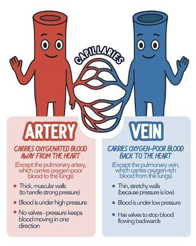
Blood flow and valves: "Arteries Away, Veins Visit"
• Meaning:
Arteries always carry blood Away from the heart; Veins always return to Visit the heart

Blood flow and valves • "Lub is Left-in, Dub is Departed"
• Meaning:
The 1st heart sound (Lub) is the closure of the AV valves trapping blood inside the ventricles; the 2nd sound (Dub) is the closure of the semilunar valves as blood departs the heart

PV loops: • "Ventricles Fill at the Floor, and Shoot up the Elevator Door"
• Meaning:
On the PV Loop graph, filling happens at the very bottom line. Point B shuts the door (Mitral closes), and Isovolumetric Contraction shoots straight up like an elevator to point C without changing volume.

PV Loops: • "Ventricles Fill at the Floor, and Shoot up the Elevator Door"
• Meaning:
On the PV Loop graph, filling happens at the very bottom line. Point B shuts the door (Mitral closes), and Isovolumetric Contraction shoots straight up like an elevator to point C without changing volume

PV Loops • "Width is the Widthdrawal"
• Meaning:
The horizontal width of your PV loop box is exactly your Stroke Volume (the amount of blood withdrawn/pumped out during that beat)
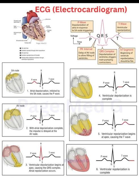
Electrical Activity & EKG
• "Send Another Awesome Box of Purple Veggies
Conduction Pathway Order: SA node → Atria → AV node → Bundle of His → Purkinje fibers → Ventricles.

Electrical activity and EKG: • "Funny Sodium makes the heart beat fun, Calcium sh*ots the loaded g*n"
• Meaning:
Pacemaker cells rely on Funny Sodium Na+ channels to climb slowly to threshold, but it's the Calcium Ca 2+ influx that fires the actual action potential

Electrical activity and EKG: • "A Calcium Plateau keeps the heart on track, it can't cramp up or it won't pump back"
• Meaning:
Calcium influx creates the long plateau phase in myocytes, preventing a sustained contraction (tetanus) so the heart is forced to relax and pump cyclically

Electrical activity and EKG: • "P for Pumping Atria, QRS for Squeezing Ventricles, T for Time to Rest"
• Meaning:
P wave = atrial depolarization; QRS complex = massive ventricular squeeze; T wave = ventricular repolarization and relaxation.