Musculoskeletal & Integumentary System Disorders & Medications
 Call Kai
 Learn
 Practice Test
 Spaced Repetition
 Match
 Flashcards
 Knowt Play
1/73

Earn XP
Description and Tags
Module 7
Name | Mastery | Learn | Test | Matching | Spaced | Call with Kai | Chat |
|---|
No analytics yet
Send a link to your students to track their progress
74 Terms
Calcium Homeostasis & remodeling
dynamic balance between resorption & deposition
constant turnover
skeleton acts as calcium reservoir regulated by endocrine feedback loops
cellular players
osteoblasts build bone matrix while osteoclasts resorb mineralized tissue
Blasts are the builders
Clasts are the cleavers
Primary Hormones
parathyroid hormone, calcitonin, and active vitamin D control flux
Parathyroid hormone (PTH)
Secreted by parathyroid glands in response to low serum calcium
parathyroid functions
stimulates osteoclasts
activates bone resorption to releases stored calcium into bloodstream
renal conservation
increases calcium reabsorption in kidney tubules, reducing urinary loss
activates vitamin D
stimulates renal conversion of calcidiol to calcitrol for gut absorption

Calcitonin
secreted by thyroid parafollicular cells in response to high serum calcium
calcitonin functions
inhibits osteoclasts
rapidly slows bone breakdown to prevent hypercalcemia
promotes deposition
encourages calcium incorporation into bone matrix
counter-regulatory role
acts as a physiological antagonist to PTH to protect skeletal mass

Vitamin D (Calcitriol)
essential steroid hormone governing mineral absorption & bone mineralization
Vitamin D (Calcitriol) functions
intestinal absorption
directly enhances dietary calcium & phosphate absorption in gut
bone mineralization
ensures adequate mineral ions available for hydroxyapatite crystal formation
deficiency impact
leads to osteomalacia or secondary hyperparathyroidism

bone remodeling process
continuous lifelong process: old bone is replaced by new bone
balance is key
healthy bone = balanced activity of osteoclasts and osteoblasts
bone strength depends on this equilibrium
osteoclasts
break down/resorb bone

osteoblasts
build new bone (deposition)

when osteoclast activity > osteoblasts activity
net bone loss occurs
leads to decreased bone mass & weakening of bone structure

osteopenia
is bone loss but not severe T-score
(-1 to -2.5)
osteoporosis
bone loss but substantial T-score
(-2.5 or lower)
What is a T-score?
A comparison of a person's bone mineral density to that of a healthy young adult.
Dual-Energy X-ray Absorptiometry (Bone mineral Density DEXA)
calculates T-score
sends to types of X-ray beams
One absorbed by soft tissue, the other bone
by subtracting the difference, tells you how dense

DEXA recommendations
no widely accepted recommendations on how often
65 yo should obtain baseline (some sources)
regular repeat scans are controversial
high risk patients may need earlier & often
Osteopenia (low bone mass)
a condition where bone mineral density is lower than normal but not low enough to be classified as osteoporosis
consider a precursor
T-score between -1.0 and -2.5
moderate risk of fracture; higher than normal but lower than osteoporosis
focus on lifestyle changes:
weight bearing exercise, adequate calcium and vitamin D intake, and monitoring
Osteoporosis (severe bone loss)
A progressive disease characterized by significant bone mineral density reduction and deterioration of bone structure, leading to fragility
T-score of -2.5 or lower
high risk of fracture, particularly in the hip, spine, and wrist, often from minor trauma
requires lifestyle changes PLUS medical intervention (medications to strengthen bones, fall prevention, and monitoring
osteopenia & osteoporosis risk factors
age: increased risk with age (especially >50 years)
sex: female (especially post menopausal
family history: genetic predisposition
low body weight/ BMI
sedentary lifestyle: lack of weight bearing exercise
smoking & excessive alcohol
medications: steroids, anticonvulsants, proton pump inhibitors
hormonal imbalances
decreased estrogen/ testosterone (postmenopause, hypogonadism)
increased parathyroid hormone (hyperparathyroidism)
nutritional deficiencies: calcium & vitamin D deficiency
chronic diseases: rheumatoid athritis, chronic kidney disease, malabsorption
osteoporosis treatment
proper nutrition
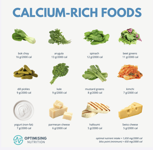
vitamin D & calcium rich diet
lifestyle changes
ETOH & smoking cessation
weight bearing exercises
fall prevention to help prevent fractures
osteomalacia/rickets
characterized by an abnormally high ratio of osteoid (inadequately mineralized bone matrix) to mineralized bone
bone formation progresses to osteoid formation but calcification does not occur; the result is soft bones
pain, bone fractures, vertebral collapse, bone malformation
Osteomalacia etiology
two main causes:
vitamin D metabolism
Deficiency of vitamin D lowers the absorption of calcium from the intestines
lack of utilization of calcium, not lack of calcium
could also be just a lack of dietary calcium
renal tubular phosphate loss
hereditary defect of the renal tubules, leading to decreased reabsorption of phosphate & therefore reduced serum phosphate levels
Osteomalacia treatment
oral supplements: vitamin D, calcium or phosphate supplements, depending on the individual case
osteomalacia symptom management
wearing braces to reduce or prevent bone irregularities
surgery to correct bone deformities (in severe cases)
adequate exposure to sunlight
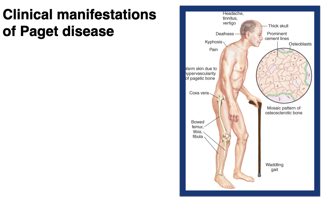
paget disease
“bowing of legs”
radiograph of the fibula & tibia of a patient demonstrating the effect of advanced proliferate paget’s disease
pagets disease pathology
chronic condition of bone turnover characterized by disorder of the normal bone remodeling process
excessive breakdown of bone tissue followed by abnormal bone formation
normal bone marrow is replaced by vascular, fibrous, connective tissue that leads to formation of larger, disorganized, & weaker bone tissue
most commonly affecting: skull, femur, tibia, pelvic bones, & vertebrae
Pagets disease symptoms
bone pain → fractures
skeletal deformities: bowed legs, fronto-occiptial skull enlargement
nerve damage: hearing loss, visual abnormalities
cardiac abnormalities: shortness of breath
pagets disease treatment
meds
bisphospnates work by controlling the excessive breakdown and formation of bone that happens in pagets disease
calcitonin- promotes the formation of new bone
exercise: to building bones, prevent wt gain, keep joints mobile
surgery:
reposition of bones that are fractured
joint replacement
osteomyelitis patho
infection of the bone; acute or chronic
2/2 direct penetration, skin infections, bloodstream infection (primarily in children) or contagious spread from adjacent wound
osteomyelitis s/sx
chills, fever, malaise, bone pain, poor incisional would healing and closure, drainage, localized tenderness, redness & swelling
osteomyelitis diagnosis
I.D. causitive organism via blood cx, bone aspiration (typically s.aureus); may be visualized with routine debridement; CT scan, MRI
osteomyelitis treatment
difficult to treat
IV abx (long course) followed by PO
debridement, amputation with clean margins
activity intolerance
the amount of physical activity a person can perform without injury or excessive exertion
no enough physical or psychological energy reserves to endure to complete ADL
assessment:
have patient recount normal activites & level of tolerance of fatigue
formal assessment tool like Humana activity profile
fatigue
the sensation that comes with having exhausted those energy reserves
it is a state experienced by everyone at some time in life. it is subjective
can be acute or chronic
treatment is symptomatic
Osteoarthritis
Degeneration and loss of articular cartilage, sclerosis of bone underneath cartilage, and formation of bone spurs (osteophytes)
Also referred to as degenerative joint disease
Incidence increases with age
Primary disease is idiopathic
Characterized by local areas of damage and loss of articular cartilage, new bone formation of joint margins, subchondral bone changes, and variable degrees of mild synovitis and thickening of the joint capsule
Manifestations
Pain, stiffness, enlargement of the joint, tenderness, limited motion, and deformity
osteoarthritis treatment
meds
NSAIDS
acetaminophen
Duloxetine (Cymbalta)
topicals like biofreeze, diclofenac, etc.
therapy; PT/OT assistive devices
low impact exercises
surgery & procedures
joint injections- corticosteroids (reduce inflammation) or hyaluronic acid (lubrication)
total joint replacement - removes the damaged parts of joint and inserts a prosthesis made of metal and plastic
Rheumatoid Arthritis
chronic autoimmune systemic disease
peak incidence 40-50 years of age
thought to be genetic
70-80% have an antibody called Rheumatoid Factor (RF)
Manifestations are joint pain, swelling, redness, warmth, deformity, loss of function, and joint stiffness
usually bilateral & symmetrical
nodules in hands and feet in later stages
RA treatment
therapy: PT/OT assistive devices
low impact exercises
surgery & procedures: synovectomy, tendon repair, joint fusions, total joint replacement
meds
Synovectomy
surgery to remove the imflamed lining of the joint (synovium) can help reduce pain and improve the joints flexibility
tendon repair
inflammation and joint damage may cause tendons around joint to loosen or rupture
joint fusion
surgically fusing a joint may be recommended to stabilize or realign a joint and for pain relief when a joint replacement isn’t a option
total joint replacement
removed the damaged parts of joint and inserts a prosthesis made of metal and plastic
Drugs for treating Rheumatoid Arthritis (RA)
NSAIDS
glucocorticoids
disease modifying antirheumataic drugs DMARDS
conventional synthetic nonbiologic (traditional) DMARDS
biologic DMARDS
Target synthetic DMARDS
Gout
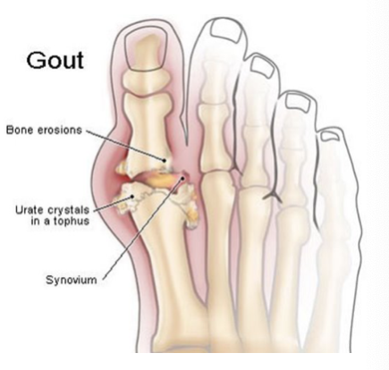
metabolic disorder characterized by elevated uric acid levels in blood resulting in deposistion of irate crystals in synovial fluid and joint tissues
high serum: > 8.5mg/dL Uric Acid
mostly seen in males, particularly AA
Gout Symptoms
intense pain comes on suddenly - often in the middle of the night or upon arising
inflammation: redness, swelling, and warmth over the joints
Gout Treatment
anti-inflammatory
NSAIDs
Colchicine
corticosteroids
Manage uric acid:
block uric acid production - allopurinol
improve uric acid removal - probenecid
Gout treatment - dietary recommendations
cherry consumption is associated with lower risk of gout attacks
Dietary restrictions
Purine restricted diet
avoid foods & beverages w/high levels of purines
organ meats (kidney, liver, etc)
seafood includes anchovies, sardines, mussels, scallops, trout &tuna
red wine, beer, & red meat
sugary drinks
low impact exercises
Muscular Dystrophy
multiple types but most common is Duchenne’s or DMD
Genetic disorder - produce progressive deterioration of skeletal muscle because of mixed muscle cell hypertrophy, atrophy, and necrosis
progressive, usually manifest in early childhood as weakness, abnormal posture or joint immobility
no cure, treatment is supportive
therapy, braces, breathing assistive devices, mobility aids
death normally occurs from respiratory or cardiac involvement in young adulthood
Soft tissue injury
include injury to muscle, ligaments & tendons
trauma
contusion
hematoma
laceration
management
contusion - elevation & ice; if hematoma forms - may need aspiration, pain management
laceration - cleanse area and close wound (suture, staples derma bond), apply sterile dressing, assess need for tetanus immunization
Arthropod Infestation: Scabies
A mite, Sarcoptes scabiei, burrows into the epidermis
transmitted person to person, includes sexual contact
healthcare facilities via sheets (can live up to 2 days on sheets)
affected areas: interdigital web of the finger, flexor surface of the wrist, inner surface of the elbow, axilla, nipple, penis, belt line, and gluteal crease
main symptom pruritus (itching of the skin) & accompanying bites on skin
Diagnosis - skin scraping showing + mites, ova, or feces
treatment - topical medication (permethrin, malathion) applied on entire body after bathing. leave on for 12 hours

Arthropod Infestation: Lice
lice - Pediculosis , insects that ive off of human blood
cannot survive outside the host
types:
Pediculus humanus corporis (body lice), Phthirus pubis (pubic lice), and Pediculus humanus capitis (head lice)
Spread via close personal contact
Arthropod Infestation: Lice diagnosis/treatment
presence of lice or nits, pruritus, papules, and wheals
treatment
eradicate louse & nits on body/clothing/bedding
shampoos or topical preparations of malathion or other pediculicides
may require multiple treatments
discard hairbrush, combs, etc
allergic contact dermatitis
inflammation of the skin
cell-mediated type IV hypersensitivity, such as to poison ivy
usually, mild erythema but can be vesicle or bullae
location & pattern can help identify cause
irritant contact dermatitis
caused by chemicals irritating skin
sopas fragrances, harsh chemicals etc
can be classified as subjective, acute, chronic, and chemical burns
characterized by dryness & erythema or edema, inflammation & vesicles
contact dermatitis treatment
patch test
remove allergen, treat symptoms; if systemic effects may need corticosteroids or antihistamines
Atopic dermatitis (aka atopic eczema)
itchy, inflammatory skin disorder
poorly defined erythema with edema, vesicles & weeping at the acute stage and skin thickening (lichenification) in the chronic stage
although often described as an immunoglobulin IgE-mediated hypersensitivity (atopic) disease, allergic causation is difficult to document
viewed as a skin disease that predisposes to allergies
Nummular Eczema
Idiopathic but common to have a history of allergy-related disorders
Lesions are coin shaped, papulovesicular patches mainly involving the arms and legs.
Lichenification and secondary bacterial infections are common.
Mostly chronic, with weeks to years between exacerbations.
Exacerbations are more frequent in the cold winter months.

Eczema treatment
remove allergen if indicated
anti-pruritic creams/lotions
topical emollients
topical cortiocosteroids
may need systemic meds like corticosteroids or antihistamines
may need immunosuppressive agents in severe cases (cyclosporine, tacrolimus, and azathioprine
phototherapy (use of UV light for flare ups, itching, redness)
Acne Vulgaris background information
Issue with pilosebaceous unit, which comprises a tiny vellus hair, hair follicle, and sebaceous gland
Comprised of non-inflammatory and inflammatory lesions
Non-Inflammatory – comedones (whiteheads and blackheads)
Inflammatory – Papules, pustules, and nodules
Acne vulgaris is most common type
Acne Vulgaris Etiology
Cause unknown, possible genetic
Possible contributing factors include: ↑ sebum production, ↑ proliferation of the keratinizing epidermal cells forming sebaceous cells, colonization/proliferation of Propionibacterium acnes, and inflammation.
Diagnosed as mild, moderate, or moderately severe
Acne Vulgaris treatment
Treatment
Mild - Topical retinoids, benzoyl peroxide, and azelaic acid
Moderate – same as mild + systemic antibiotics
Rosacea background information
Chronic inflammatory process that occurs in adults between the ages of 30 and 50 years
Fair skinned people, most common in white women >30 y/o
Rosacea Etiology
Unknown, but believed to be an inflammatory process accompanied by vascular instability with leakage of fluid and inflammatory mediators into the dermis
what are the 4 types of rosacea
Erythematotelangiectatic (flushing and persistent central facial erythema)
Papulopustular (inflammatory)
Phymatous (thickening of the skin with irregular surface nodularities and enlargement)
Ocular (involving the eyes)
starts as “blushing” evolves into permanent dark red erythema of nose and cheeks
rosacea treatment
Topical metronidazole and azelaic acid, topical and systemic abx
If rhinophyma is present can be treated using surgical methods, including electrosurgery, laser ablation, dermabrasion, cryosurgery, and excision
Psoriasis background information
Common, chronic inflammatory skin disease characterized by circumscribed, red, thickened plaques with an overlying silvery-white scale.
Incidence lower in warmer, sunnier climates

Psoriasis Etiology
Genetic component; apparent association of arthritis and psoriasis
Activated T lymphocytes produce chemical messengers that stimulate abnormal growth of keratinocytes and dermal blood vessels. Accompanying inflammatory changes are caused by infiltration of neutrophils and monocytes.
Köbner reaction
Psoriasis Patho
Increased epidermal cell turnover with marked epidermal thickening (hyperkeratosis)
Permanent damage to capillary beds even in the presence of remission or resolution
Psoriasis Types
Plaque-type psoriasis (most common)
Guttate psoriasis
Pustular psoriasis (localized and generalized)
Erythrodermic psoriasis
Psoriasis assessment
Primarily located on elbows, knees, and scalp
Primary lesions are sharply demarcated, thick, red plaques with a silvery scale, vary in size/shape. In darker skinned people, plaques may appear purple. Lesions may have excoriation, thickening, or oozing.
Auspitz sign
Psoriasis Treatment
NO CURE
Goal: Sign and symptom management and suppression
Topical: Emollients, keratolytic agents, coal tar products, anthralin, corticosteroids, and calcipotriene.
Systemic: phototherapy, photochemotherapy, methotrexate, retinoids, corticosteroids, and cyclosporine