Introduction to Radiography
 Call Kai
 Learn
 Practice Test
 Spaced Repetition
 Match
 Flashcards
 Knowt Play
1/90

There's no tags or description
Looks like no tags are added yet.
91 Terms
Radiograph
Image produced on a sensitive plate or film by X-rays, gamma rays, or similar radiation, typically used in medical examinations.
Radiography
Art and Science
The process or occupation of taking radiographs to assist in medical examination.
Basic X-ray Concepts (Include)
X-ray tube
Central ray (CR)
Collimation
Image Receptor
Patient + Part
X-ray Tube
Creates/emits radiation
Where handling takes place
Central Ray (CR)
Comes from focal point of x-ray tube
Angled out causes langevity
Collimation
Field of view
Acts as a lead shutter and is able to decrease/increase field of view
Knobs move this
Image Receptor (IR)
Helps produce image (radiation interacts with receptor)

X-ray Tube

Center Ray (CR)

Image Receptor (IR)

Collimation
Anatomic Position
Reference position (standard)
Facing forward, palms out
Types of planes in the body
Sagitial
Oblique
Horizontal (axial)
Coronal
What is the surfaces of the foot
Bottom: planter surface (posterior surface)
Top: dorsal surface (Anterior surface)
Directional terms
Superior
Inferior
Medial
Lateral
Proximal
Distal
Superificial
Deep
Ipsilateral
Contralateral
Superior
Higher
Inferior
Lower
Medial
Closer to the middle
Lateral
Close to the edge
Proximal
Close to point of attachment
Distal
Further to the point of attachment
Superificial
Close to the surface
Deep
Far away from the skin (deep)
Ipsilateral
Same side of the body
Contralateral
Opposite side of the body
Projection
Path of the x-ray beam as it passes through the patient

Posterior to Anterior

Anterior to Posterior
What type of lateral projection
Mediolateral
Lateromedial

A lateral projection
Mediolateral

Lateral
Lateromedial
what type of oblique projections
AP medial oblique
PA lateral Oblique

AP Medial Oblique (lateral projection)

PA lateral Oblique (lateral projection)
Axial (types of projections)
Cephalad (toward the head)
Caudad (away from the head)
Inferosuperior
Superoinderior
Tangential
Plantodorsal
Dorsoplantar
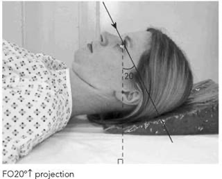
Cephalad (toward the head)(axial projection)

Caudad (away from the head)(axial projection)

Infersuperior (Axial projection)

Superoinferior (axial projections)

Tangential (axial projection)
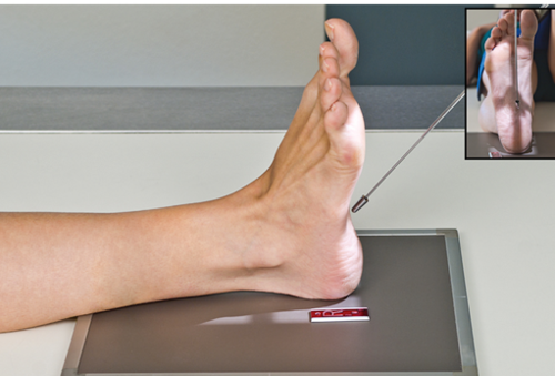
Plantodorsal (axial projection)

Dorsoplantar (axial projection)
Position
Descrides how patient or part of interest is situated/located during an X-ray
Types of positions
Erect/upright
Recumbent
Supine
Prone
Trendelburg
Fowlers
Sims
Lithotomy position

Erect/upright (position)
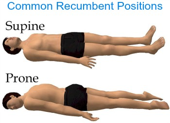
Recumbent (position)(Laying down in any position)

Supine (position)

Prone (position)
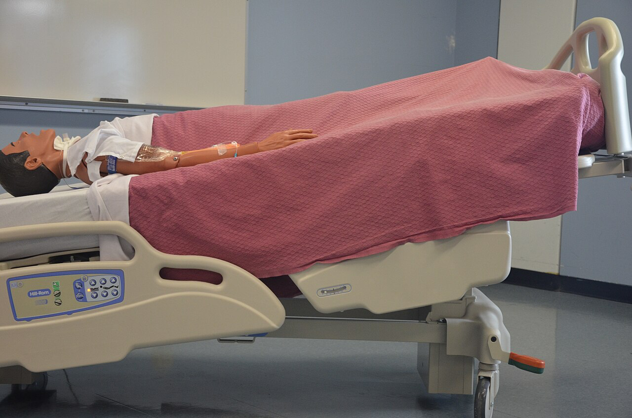
Trendelenburg (position)
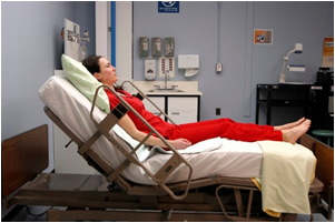
Fowler’s (position)(can be called reverse Tendelenburg)

Sim’s (position)

Lithotomy (position)
Lateral (how to classify)
As right or left (whatever is touching the IR)
Lateral: coronal plane, perpendicular to IR OR 90 degrees from AP or PA.
Types of lateral Position
Right, lateral
Left lateral recumbent
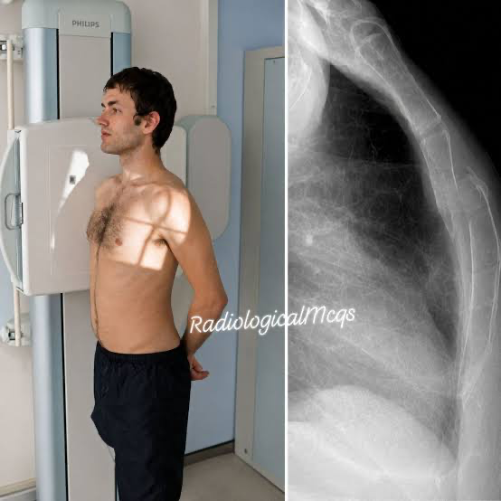
Right, lateral (upright)

Left lateral recumbent
Oblique
Neither perpendicular nor parallel
Left or right (which side the body is angled) Anterior or posterior (which side IR is touching)
Oblique (types of positions)
Left posterior oblique
Left anterior oblique
Right posterior oblique
Right anterior oblique
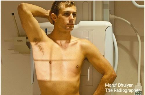
Left posterior oblique (position)

Right anterior oblique (position)

Top: Right anterior oblique (position)
Bottom: Left anterior obique (position)
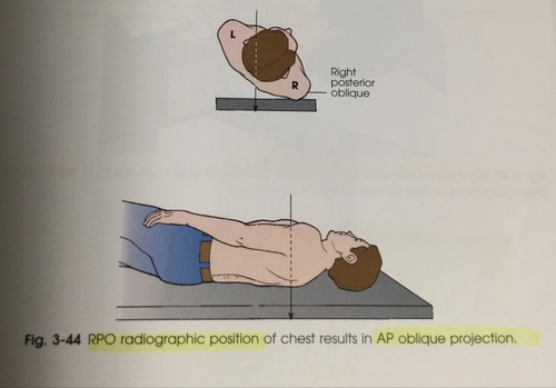
Right posterior oblique (Position)
Decubitus
A position assumed in lying down where the central ray is parallel to the floor (horizontal way)
Decubitus (types)
Left lateral decubitus
Right lateral decubitus
(left) dorsal decubitus
(right) ventral decubitus
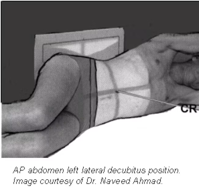
Left lateral decubitus (position)

Right lateral decubitus (position)

(right) dorsal decubitus (position)
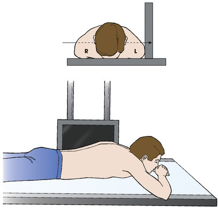
(left) ventral decubitus (position)
Movement Terms
Flexion
Extension
Ulnar deviation
Radial deviation
Plantar flexion
Dorsiflexion
Inversion (varus)
Eversion (valgus)
Medial Rotation
Lateral Rotation
Supination
Pronation
Abduction
Adduction
Elevation
Depression
Circumduction
Tilting
Rotation
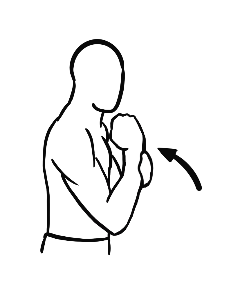
Flexion

Extension

Ulnar Deviation
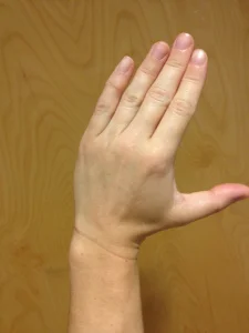
Radial Deviation

Top: Dorsiflexion
Bottom: Plantar flexion
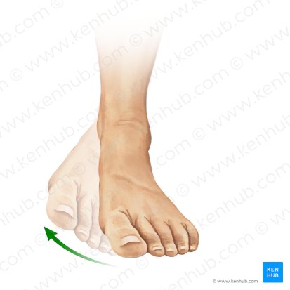
Inversion of the foot (leads to Bow legs)(varus)

Eversion (valgus)(Leads to knock knees)

Lateral Rotation (palm outside)

Medial Rotation

Supination

Pronation

Abduction

Adduction

Elevation

Depression

Circumduction (Not all joints can do this)(Ex: wrist, shoulders, fingers, ankles)(circular motion)

Tilting

Rotation
N.O.D
N: Name
O: Occupation
D: Doing
A.C.E
A: Announce
C: Communication
Explain
Radiographs are evaluated on 3 main criteria
1) Anatomy
2) Position
3) Exposure
How much projections
Minimum two (90 degrees from both)
Joins: take three (AP OR PA, lateral, and oblique)