Week 2: Bounce/Nine Lives
 Call Kai
 Learn
Learn Practice Test
 Spaced Repetition
 Match
Match Flashcards
 Knowt Play
1/49

There's no tags or description
Looks like no tags are added yet.
Name | Mastery | Learn | Test | Matching | Spaced | Call with Kai | Chat |
|---|
No analytics yet
Send a link to your students to track their progress
50 Terms
superficial tangential layer
a:

intermediate transitional layer
b:

deep radial layer
c:

calcified cartilage; tide mark between c & d
d:

subchondral compact bone
e:

medullary bone
f:
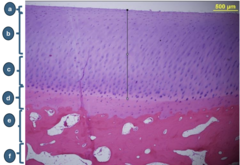
superficial tangential layer
a:

intermediate transitional layer
b:

deep radial layer
c:

calcified cartilage
d:

subchondral compact bone
e:

medullary bone
f:

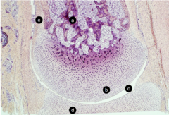
up
growth plate. which direction is the bone growing - up or down?

resting
growth plate, b: zone

proliferative
growth plate, c: zone

hypertrophic cartilage
growth plate, d:

calcified
growth plate, e: zone
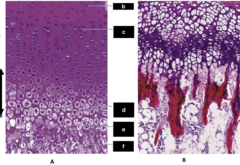
ossification
growth plate, f: zone

zone of hypertrophy: chondrocytes are hypertrophying, and then degrade and become calcified
zone of resorption: medullary capillary loops invade the calcified cartilage along with osteoprogenitor cells which form longitudinal spicules on the cartilage
zone of ossification: osteoprogenitor cells differentiate into osteoblasts and deposit bone on the spicules
what is happening in image b?

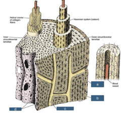
volkmann's canal
a: structure
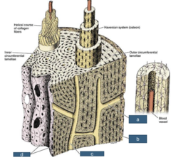
periosteum
b: outermost layer
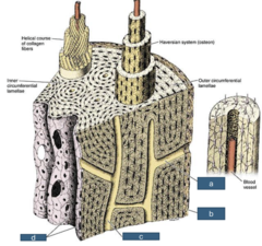
haversian canal
c: structure
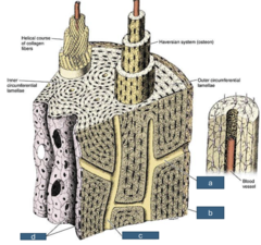
endosteum
d: innermost layer
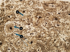
haversian canal
a: space

osteocytes
b: cell type

osteocyte processes
c: structures

compact bone
specific tissue type

osteon/haversian system
b: structure
haversian canal
c: structure

osteocytes
d: cells

trabecular bone, spongy bone
a: specific tissue type

hyaline cartilage, articular cartilage
b: tissue type

joint cavity
c: space

hyaline cartilage, articular cartilage
d: tissue type

mesenchymal cells/connective tissue
e: tissue type
tropocollagen
a:

myofibril
b:

fibril
c:

fibroblast
d: cell that produces tropocollagen

crimp structure
e:

endotenon
f:

epitenon
g:

skeletal muscle
specific tissue type

peripheral nucleus, striations
why is this skeletal muscle?

endomysium
c: tissue at the end of the arrows

motor end plate
a: entire functional structure

axon terminals
b: rounded structures

axon
c: single line
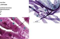
nerve bundle
d: structure

myofiber
e: structure
