Exam 2 -- Antigen Recognition by BCRs and TCRs
 Call Kai
 Learn
 Practice Test
 Spaced Repetition
 Match
 Flashcards
 Knowt Play
1/87

There's no tags or description
Looks like no tags are added yet.
88 Terms
TRUE or FALSE — Innate and Adaptive Immunities are very different
FALSE !! — They are extremely related and intertwined systems
How are antibodies shaped?
Y-Shape!
TRUE or FALSE — BCRs and TCRs bind and recognize antibodies?
FALSE — Antigens
What’s the chain situation on antibodies?
2 Heavy and 2 light chains
What are the 5 Ig classes?
IgG, IgM, IgD, IgA, IgE
Structure of Antibody 1
1
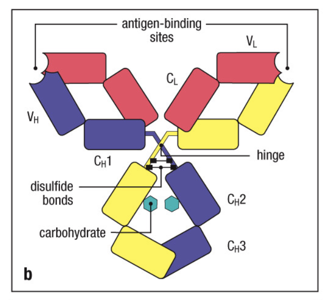
Structure of Antibody 2
2

What is the N-Terminus of an antibody responsible for?
Antigen binding
What is the C-Terminus of an Antibody responsible for?
Effector Functions
What are the two kinds of ligtht chains?
Lambda ( λ ) and Kappa ( κ )
What are the five kinds of heavy chains?
IgM / Mu (μ) — IgD / Delta (δ) — IgG / Gamma (γ) — IgA / Alpha (α) — IgE / Epsilon (ε)
How long is the total IgG?
Heavy Chain = 50 kDa // Light Chain = 25 kDa
Total chain length = 150 kDa
TRUE or FALSE — Antibodies can exist with both a Lambda ( λ ) and Kappa ( κ ) light chain
FALSE !!!!! ONLY ONE!!!!!!!
TRUE or FALSE — Antibodies can exist with multiple types of heavy chains
FALSE !!!!! ONLY ONE!!!!!!!
How many Disulfide bonds connect an Antibody?
4 - 2 connecting heavy chains and 2 connecting light chains

:: 4 Disulfide bonds connect the antibody chains
Antibodies are very stable!
Into what two pieces can Papain proteylytic cleavage break antibodies?
Fab and Fc

What is the Fab Fragment?
Fragment Antigen Binding
What is the Fc Fragment?
Fragment Crystallization - Effector Domain / Constant Region / C-Terminus
What’s the difference between the Fab and Fc ?
Fab - Many different antigen binding spots, high variability, non-crystalizable
Fc - All the same region, crystalizable
Why is Fab non-crystalizable?
High-variability
Into what two pieces can Pepsin cleave antibodies?
F(ab’)2 and pFc’

What is F(ab’)2 ?
Two Fab’s bound/cleaved together
What is pFc’?
Pieces of Fc
Are antibodies cleaved by Pepsin crystallizable?
NO!!!!!
Are Polyclonal Antibodies derived from ONE or MULTIPLE B-Cells?
Multiple
Are Monoclonal Antibodies derived from ONE or MULTIPLE B-Cells?
One
:: Polyclonal Antibodies are derived from multiple B-Cells
Polyclonal antibodies are not genetically identical, and can recognize many epitopes
:: Monoclonal antibodies are derived from one B-Cell
Monoclonal antibodies have the exact same amino acid sequence, are crystalizable, and recognize the exact same epitope
TRUE or FALSE — Polyclonal antibodies are crystalizable
FALSE !!
TRUE or FALSE — ALL PARTS of monoclonal antibodies are crystalizable
TRUE ! - They are genetically identical - even monoclonal-derived p(Fab’)2
Monoclonal Antibody shorthand
mAb
What is Recombinant Antibody Technology?
A means of genetically altering antibodies to combine variable regions, allowing for rapid production of Abs
What parts are included in a recombinant antibody?
Just the heavy and light variable chain regions (Ab-binding sites)
TRUE or FALSE — Recombinant antibodies are still able to bind antigens
TRUE !
Benefits of Recombinant Antibody Technology
Quick/easier creation of antibodies
Therapeutic benefits (smaller, easier entry into tissue, more specific targeting, tumor therapy against custom (cancer, toxin, protein) antigens)
TRUE or FALSE — Antibody arms are extremely rigid
FALSE — Ab arms are joined by a flexible hinge region

Picture of the Immunoglobin Domain
Picture of the Immunoglobin Domain
How is the immunoglobin domain shaped?
2 anti-parallel beta sheets held together by disulfide bonds
Forming a Beta Barrel
“Immunoglobin Fold”
What part of an antibody is the immunoglobin domain?
The heavy and light variable chains
When dealing with Antigen-Antibody binding, what is the framework region?
AA sequences that form the boarder structural framework of the domain
AA portions that make up the bulk of the immunoglobin domain - Beta barrels
Pretty constant amongst Ab’s
When dealing with Antigen-Antibody binding, what is the Hypervariable region?
Highly variable AA regions that allow Abs to bind a multitude of Ags
How are Hypervariable regions organized?
3 Discrete loops in the folded structure of an antibody
What’s another name for the Hypervariable Reason?
The Complementary Determining Region (CDR)
What are CDR’s (Complementary Determining Regions) ?
More specific hypervariable regions that complement the antigen that they bind
Still more variable than Framework regions, but more specific than hypervariable regions
How many CDR’s (Complementary Determining Regions) are contained within an antigen binding site?
6
How many CDR’s (Complementary Determining Regions) are contained within an antibody?
12
How many CDR’s (Complementary Determining Regions) are contained within a heavy chain?
3
How many CDR’s (Complementary Determining Regions) are contained within a light chain?
3
How do antibodies bind antigens?
Through tons of noncovalent, reversible interactions
TRUE or FALSE — Antibodies bind antigens reversibly
TRUE !
What’s unique about camelid and shark Abs?
All heavy chains
Camelid - Smaller (2 Fc, 1 Fab)
Shark - Bigger (5 Fc, 1 Fab)
How do BCRs differ from Igs?
Surface bound - short transmembrane domain
Igβ and Igα related subunits help with signaling
How do TCRs differ from Igs
Resembles a membrane-bound Fab fragment
Cα, Vα, Vβ, and Cβ
What are the two kinds of TCR heterodimers?
α:β and γ:δ
Which TCR heterodimer is more common?
α:β
What’s special about γ:δ TCR heterodimers (prevalence)
Much more uncomming (5-15% of all T-Cells)
What makes up an α:β TCR’s antigen-binding site?
CDRs 1, 2, and 3
Their disulfide bonds
TRUE or FALSE — For proper presentation, TCR epitopes must be present on a protein’s surface
FALSE !!
TRUE or FALSE — TCR epitopes must be linear/continuous peptide sequences
TRUE !
Why do TCR epitopes need to be linear, but do not need to be present on protein surfaces?
Because proteins are chopped up prior to presentation, and small fragments are what’s presented in MHC molecules
What are the two ways antibody epitopes can appear
Linear/Continuous and Conformational/Discontinuous
What kind of antibody epitope conformation is ALWAYS recognized by TCRs?
Linear/Continuous ONLY
What kind of antibody epitope conformation is ALWAYS recognized by BCRs/Abs/Igs?
Conformational or Discontinuous (typically, not a requirement)
In what form do TCRs recognize Antigens?
In the form of a peptide-MHC molecule complex
TRUE or FALSE — Abs/Igs have a wide range of epitopes
TRUE ! Their epitopes are not always proteins!
What are the domains of an MHC I protein? (# / makeup)
3 Domains
α chain, β-2 Microglobin, Peptide-Binding cleft
1 Transmembrane domain (MHC I)
What are the domains of an MHC II protein? (# / makeup)
4 Domains
2 α chains, 2 β chains, and the peptide binding cleft
2 Transmembrane domains (MHC II)
Why is MHC turnover important?
Recency of peptides displayed allows immune cells to recognize more recent pathogen infections
Describe the MHC I peptide binding cleft
Closed, holds shorter peptides
Stabilizing noncovalent interactions in the middle + held in on the ends
Presents cytosolic/endogenous peptides

Describe the MHC II peptide binding cleft
Open on the ends, holds longer peptides
Stabilizing noncovalent interactions in the MIDDLE of the peptide
Presents exogenous peptides
:: MHC I molecules have closed sides of the the presentation cleft, and many stabilizing interactions at the sides of their binding cleft
:: MHC I molecules bind smaller amino acids (~9 sequences) and have higher specificity
What are anchor residues?
Structurally related amino acids in a MHC I molecule’s binding cleft
Typically Phenylalanine (Phe/F) and Tyrosine (Tyr/Y)
What do anchor residues do? (MHC I)
Proteins bind anchor residues at their amino and carboxyl ends
They bind at the amino/carboxyl ends because MHC I molecules are closed on the ends
:: MHC II presentation clefts are open on the sides, and have many stabilizing interactions in the middle of their binding cleft
:: MHC II molecules can bind longer amino acids (~13-17 sequences) and have higher specificity
TRUE or FALSE — There’s a lot of variation within MHC binding clefts
FALSE !! — Anchor residues correspond with common core sequences present within bound peptides
What specific sequences do peptides contain to mediate MHC II binding
Core sequences
In the middle of the protein, noncovalently binds the middle of the binding cleft
How do TCR’s bind the MHC I complex?
CDR (Complement Determining Region) 3α and 3β bind the center of the peptide
CDR 1 and 2 α (of the TCR) contact the Amino terminus
CDR 1 and 2 β (of the TCR) contact the Carboxylic Acid terminus
In what context do TCR's bind the MHC II peptide?
TCR’s understand the protein in the context of the MHC II molecule
Presentation of peptide on the MHC molecule matches to the cognate/matching TCR
:: MHC molecules are highly polymorphic, there are hundreds of alleles of MHC class 1 and 2 genes in humans
:: MHC molecules can bind a vast array of proteins!
(Due to the differences in their protein binding clefts)

The CD4+ T-Cell Co-Receptor has 4 Ig-Like domains
The CD4+ T-Cell Co-Receptor has 4 Ig-Like domains
Where are the main polymorphic areas in MHC molecules?
The peptide binding cleft!

The CD8+ T-Cell Co-Receptor is a dimer
The CD8+ T-Cell Co-Receptor is a dimer
MHC I :
CD8+ - Cytotoxic T-Lymphocytes (CTLs)
MHC II :
CD4+ - Regulatory / Helper T-Cells (Tregs/Th)
What cells express MHC I cells?
All nucleated cells
NOT RED BLOOD CELLS!!!! No nucleus
What cells express MHC II cells?
Antigen-presenting cells
Dendritic Cells, Macrophages, B-Cells
Epithelial cells of the thymus

Where do T-Cell CD8+ and CD4+ co-receptors sit when interacting with MHC molecules?
Next to the MHC molecule