Summarised Lower Limb Ligaments
 Call Kai
 Learn
 Practice Test
 Spaced Repetition
 Match
 Flashcards
 Knowt Play
1/24

Earn XP
Description and Tags
Comprehensive vocabulary flashcards detailing the anatomical origins, insertions, and movement limitations of the hip, knee, and ankle ligaments based on the lower limb lecture summary.
25 Terms

Iliofemoral Ligament
An anterior hip ligament originating from the anterior inferior iliac spine (AIIS) of the innominate bone and inserting into the intertrochanteric line of the femur; limits hip extension, adduction, and lateral rotation.

Pubofemoral Ligament
An anterior hip ligament originating from the iliopubic eminence and superior pubic ramus of the pubic bone and inserting into the lower part of the intertrochanteric line of the femur; limits hip extension, abduction, and lateral rotation.

Ischiofemoral Ligament
A hip ligament originating from the body of the ischium behind and below the acetabulum of the innominate bone and inserting into the superior part of the neck and root of the greater trochanter of the femur (wrapping posterior to anterior); limits hip extension, abduction, and medial rotation.

Transverse Ligament of the Acetabulum
A ligament spanning across the deficient inferior acetabular rim between the ischial and pubic sides of the acetabular notch; leaves a gap for blood vessels to enter the hip and provides a bloodstream to the femur in children.

Ligamentum Teres (HOF Ligament)
A hip ligament originating from the margins of the acetabular notch and inserting into the fovea capitis on the head of the femur; it is taut in flexion and adduction, and carries the obturator artery to supply blood to the femoral head and hip joint.
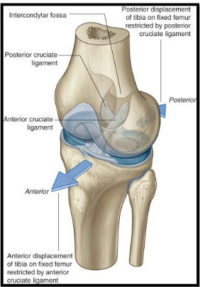
Anterior Cruciate Ligament (ACL)
A knee ligament originating from the anterolateral aspect of the tibia anterior to the tibial spines and inserting into the posterior part of the medial surface of the lateral femoral condyle; limits anterior translation of the tibia on the femur.
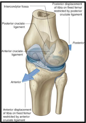
Posterior Cruciate Ligament (PCL)
A knee ligament originating from a depression in the posterior intercondylar area of the tibial plateaux and inserting into the lateral surface of the medial condyle of the femur; limits posterior translation of the tibia on the femur.

Medial Collateral Ligament (Tibial)
A knee ligament originating from the medial epicondyle of the femur and inserting into the medial condyle of the tibia; limits valgus forces of the knee (tilting inwards).

Lateral Collateral Ligament (Fibular)
A knee ligament originating from the lateral epicondyle of the femur and inserting into the lateral surface of the head of the fibula; limits varus forces of the knee.

Patellar Ligament
A structure extending from the apex of the patella into the tibial tuberosity; transmits quadriceps muscle force to enable knee extension, stabilizes the knee, and maintains patellar alignment.
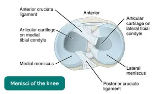
Menisci
Two crescent-shaped cartilages that sit atop the medial and lateral condyles of the tibia; act as shock absorbers and weight bearers, and aid joint lubrication.
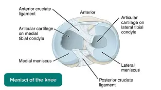
Lateral Meniscus
A crescent-shaped cartilage of the knee whose anterior horn attaches in front of the intercondylar eminence and whose posterior horn attaches behind it.
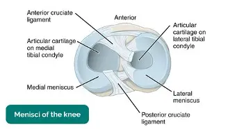
Medial Meniscus
A crescent-shaped cartilage of the knee whose anterior horn attaches to the intercondylar area in front of the ACL and whose posterior horn attaches to the posterior intercondylar area.

Oblique Popliteal Ligament
A capsular strengthening ligament of the knee that is an expansion of the semimembranosus tendon; passes upward and laterally to the intercondylar line of the femur.

Arcuate Popliteal Ligament
A capsular strengthening ligament that passes in two bands from the fibular head: one to the posterior intercondylar area of the tibia and the other to the back of the lateral femoral condyle.
Deltoid Ligament
A triangular medial ankle ligament complex made up of four bands: deep parts (anterior and posterior tibiotalar bands) and superficial parts (tibionavicular and tibiocalcaneal bands).
Anterior Tibiotalar Band
A deep component of the deltoid ligament originating from the tip of the medial malleolus and inserting into the medial part of the neck of the talus; resists forward displacement of the foot.
Posterior Tibiotalar Band
A deep component of the deltoid ligament originating from the tip of the medial malleolus and inserting into the medial neck and medial tubercle of the talus; resists backward displacement of the foot.
Tibionavicular Band
A superficial component of the deltoid ligament originating from the tip of the medial malleolus and inserting into the tuberosity on the navicular; resists eversion forces.
Tibiocalcaneal Band
A superficial component of the deltoid ligament originating from the tip of the medial malleolus and inserting into the sustentaculum tali of the calcaneus bone; resists backward displacement of the foot.
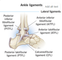
Anterior Talofibular Ligament (ATFL)
A lateral ankle ligament originating from the tip/anterior margin of the lateral malleolus and inserting into the neck of the talus (in front of the lateral articular facet); resists forward displacement of the foot (anterior talar translation), inversion, and plantarflexion.
Posterior Talofibular Ligament (PTFL)
A lateral ankle ligament originating from the tip/medial surface of the lateral malleolus (malleolar fossa) and inserting into the lateral tubercle of the posterior process of the talus; resists inversion forces, backwards displacement of the foot, and extreme dorsiflexion.
Calcaneofibular Ligament (CFL)
A lateral ankle ligament originating from the tip/anterior border of the lateral malleolus and inserting into the peroneal tubercle on the lateral surface of the calcaneus; resists backwards displacement of the foot and inversion in dorsiflexion.
Anterior Ligament (Ankle)
An ankle ligament originating from the anterior margin of the lower end of the tibia and inserting into the anterior part of the neck of the talus; resists forward displacement of the foot.
Posterior Ligament (Ankle)
An ankle ligament originating from the posterior aspects of the tibia and fibula and inserting into the medial tubercle of the posterior surface of the talus; resists backward displacement of the foot.