A&P : Practical #1 (mitra)
 Call Kai
 Learn
 Practice Test
 Spaced Repetition
 Match
 Flashcards
 Knowt Play
1/524

There's no tags or description
Looks like no tags are added yet.
525 Terms
synarthrosis
immovable joint
amphiarthrosis
slightly movable joint
diarthrosis
freely movable joint
fibrous
solid mass of connective tissue between bones
cartilaginous
cartilage between bones
synovial
joint incorporates lubricated cavity
hinge joint
synovial joint; uniaxial ; ex: elbow joint
pivot joint
synovial joint; uniaxial; ex: atlanto-axial joint
condyloid or elliposidal joint
synovial joint; bilateral ; ex: radiocarpal, metacarpophalangeal
glenohumeral joint
shoulder joint
glenohumeral ligaments of glenohumeral joint
What is number 6 ? (superior, middle, and inferior bands forming the anterior joint capsule)
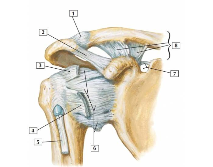
Coracoacromial ligament of glenohumeral joint
What is number 2 ? (connects the coracoid process to the acromion)

Coracohumeral ligament of glenohumeral joint
What is number 3 ? Circled in red (connects the coracoid process to the greater tubercle of the humerus)

acromioclavicular ligament of glenohumeral joint
What is number 1 ? (connects the acromion of the scapula to the clavicle)

coracoclavicular ligament of glenohumeral joint
What is number 8 ? (composed of the trapezoid ligament laterally and the conoid ligament medially)

ulnar collateral ligament
What is number 2 ?

radial collateral ligament
What is number 1 ?
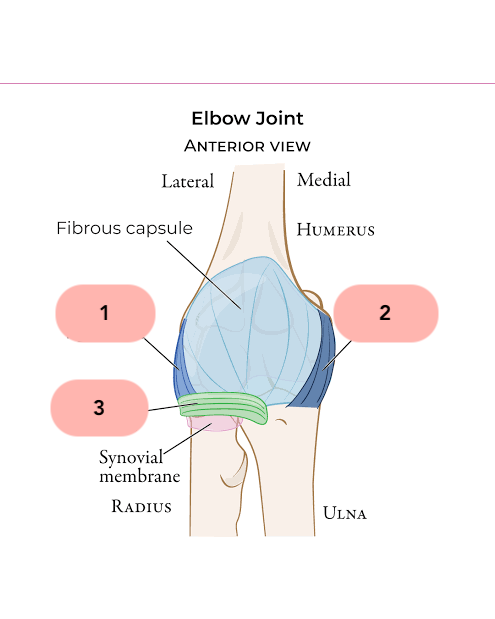
annular ligament of radius
What is number 3 ?

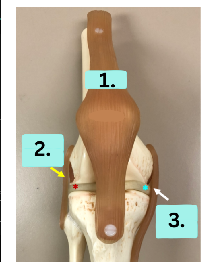
patellar ligament of tibiofemoral joint
What is number 1 ?

fibular collateral ligament of tibiofemoral joint
What is number 2 ?

tibial collateral ligament of tibiofemoral joint
What is number 3 ?
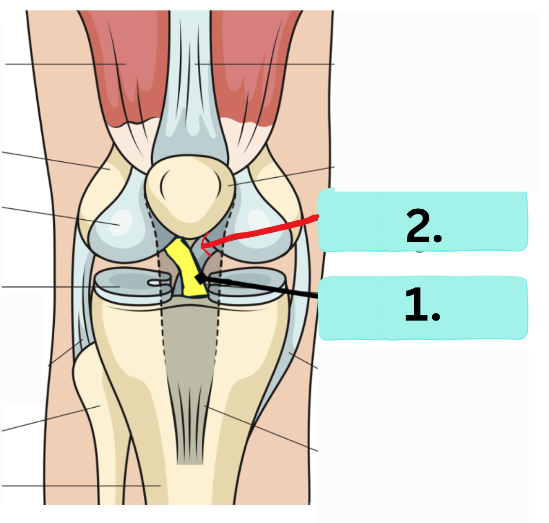
anterior cruciate ligament of tibiofemoral joint
What is number 1 ? (the yellow area w black arrow)

posterior cruciate ligament of tibiofemoral joint
What is number 2 ? (the blue area w red arrow)
iliofemoral ligament of coxal joint
What is number 2 ? (attaches from femur to ilium)

pubofemoral ligament of coxal joint
What is number 3 ? (attaches from femur to pubis)
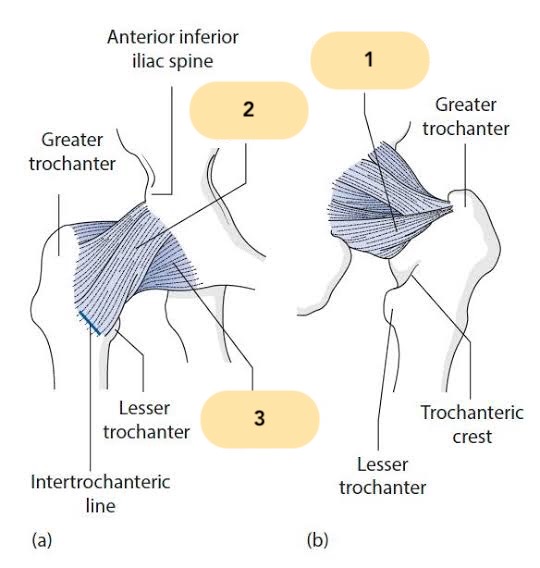
ischiofemoral ligament of coxal joint
What is number 1 ? (attaches femur to ischium)

transverse acetabular ligament
What is number 2 ? (completes bowl of acetabulum)

ligamentum teres
What is number 1 ?

plane joint
Non-axial or gliding - Intercarpal & Intertarsal joints
saddle joint
Biaxial - Carpometacarpal joint of digit 1
hinge joint
Uniaxial or monoaxial - Humeroulnar joint
ball and socket joint
Multiaxial or triaxial - Glenohumeral & Acetabulofemoral joint
synarthrosis
suture (fibrous joint)
suture
coronal suture
syndesmosis
amphiarthrosis ; distal tibiofibular joint
Gomphosis
Synarthrosis ; Tooth-Alveolar socket
synchondrosis
synarthrosis ; Epiphyseal plate
Symphysis
Amphiarthrosis ; Intervertebral discs
Angular movements
Sagittal & Frontal plane
Sagittal plane
Flexion , Extension , Hyperextension , Dorsiflexion (ankle flexion) , Plantarflexion (ankle extension)
Frontal plane
Abduction , Adduction , Inversion , Eversion , Lateral flexion
Rotation
Lateral , Medial , Right , or Left rotation ; Supination , pronation
Special movements
Opposition , Retraction , Protraction , Depression , Elevation
Circumduction
One of the movements allowed by synovial joints
Lateral meniscus
What is red asterisk ?

Medial meniscus
What is blue asterisk ?

iliac crest (ilium)
what is 1?; superior ridge of the ilium; site to obtain donated bone marrow

ala (ilium)
what is 2?; (also called the wing of the ilium) is the large, upper, fan-shaped portion of the hip bone that forms the lateral wall of the false pelvis
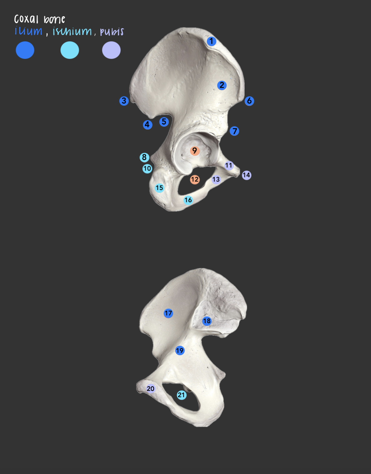
posterior superior iliac spine
what is 3?; posterior end of the iliac crest

posterior inferior iliac spine
what is 4?; projection below the PSIS on the posterior ilium
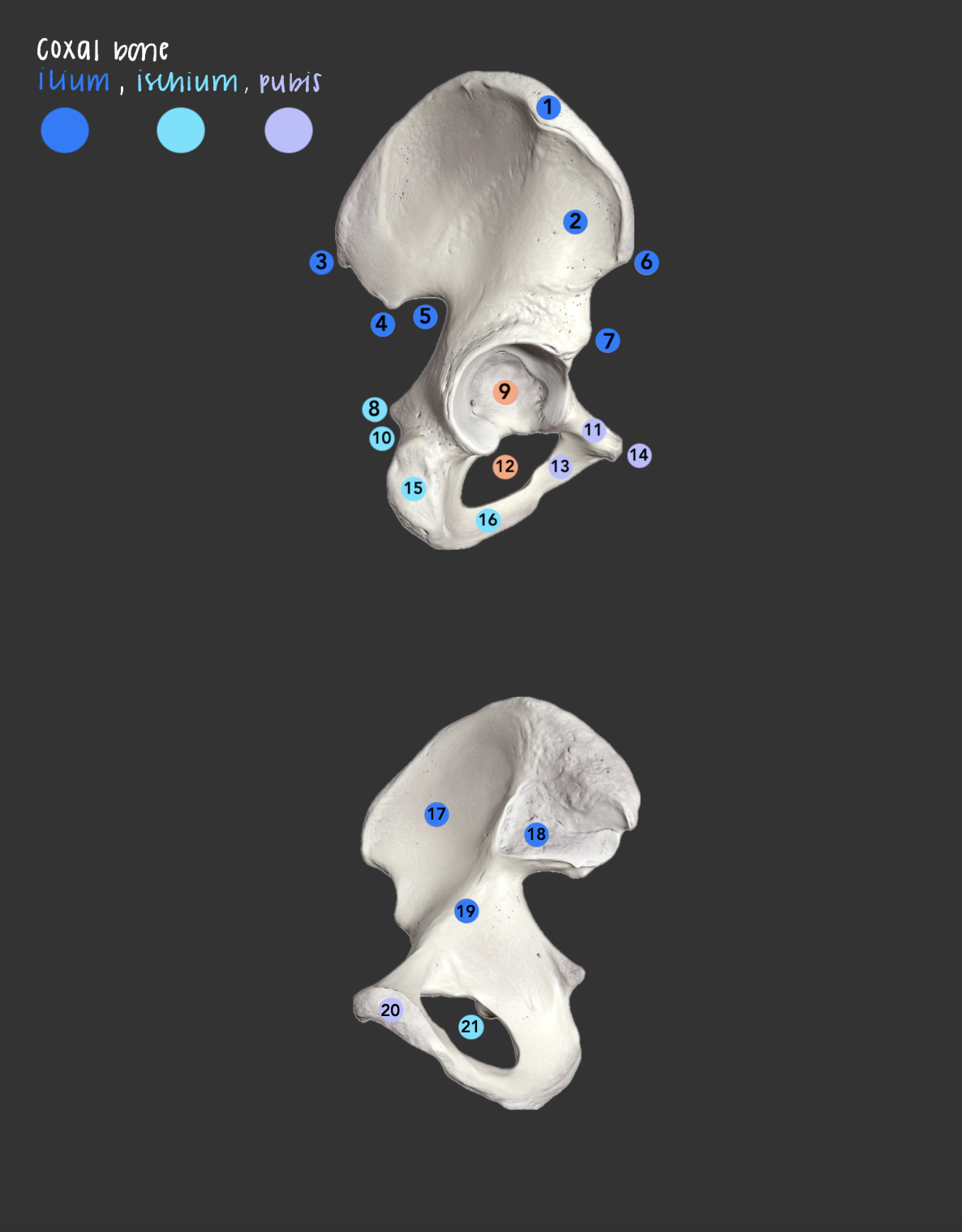
greater sciatic notch (ilium)
what is 5?; sciatic nerve passes through

anterior superior iliac spine
what is 6?; anterior end of the iliac crest

anterior inferior iliac spine
what is 7?; projection below the ASIS on the anterior ilium

ischial spine
what is 8?; a pointed bony projection on the back border of the ischium bone in the pelvis, obstetric landmark

acetabulum of coxal bone
what is 9?; socket formed by all three coxal parts; head of the femur articulates here

lesser sciatic notch (ischium)
what is 10?; notch below the ischial spine

superior ramus (pubis)
what is 11?; superior branch of pubis
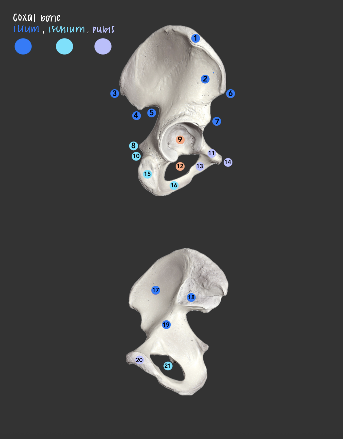
obturator foramen (ischium & pubis)
what is 12 & 21 (same structure)?; large opening formed by only the pubis and ischium
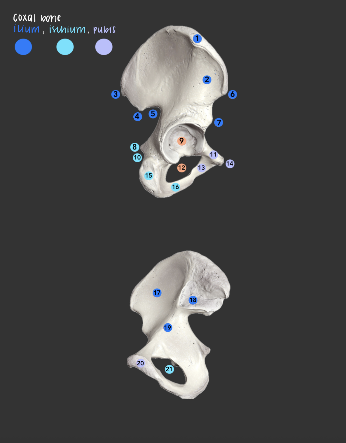
inferior ramus (pubis)
what is 13?; inferior branch of the pubis

pubic tubercle
what is 14?; small bump on the superior ramus near the pubic symphysis

ischial tuberosity
what is 15?; obstetric landmark, a pointed bony projection on the back border of the ischium bone in the pelvis

ischial ramus
what is 16?; part of the ischium that joins the inferior ramus of the pubis
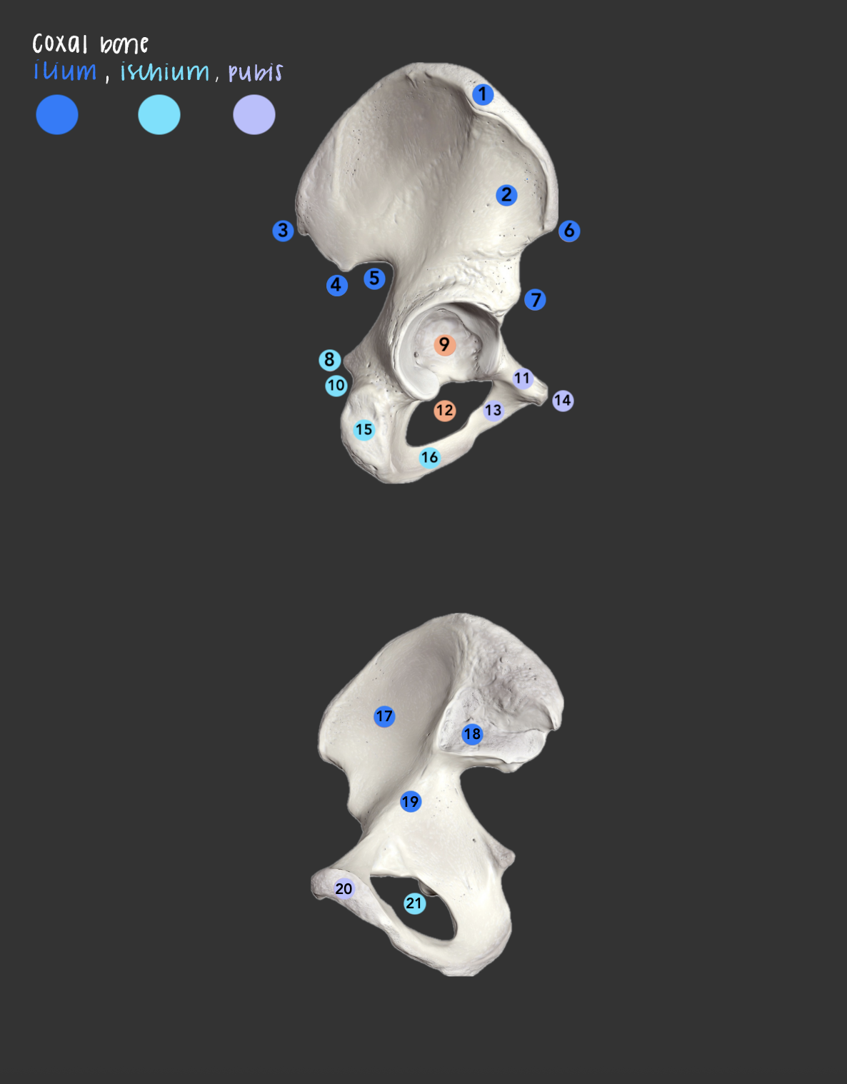
iliac fossa
what is 17?; a large, smooth, and concave depression on the inner surface of the hip bone
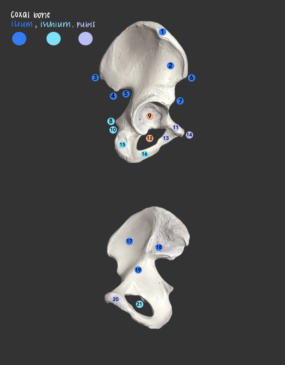
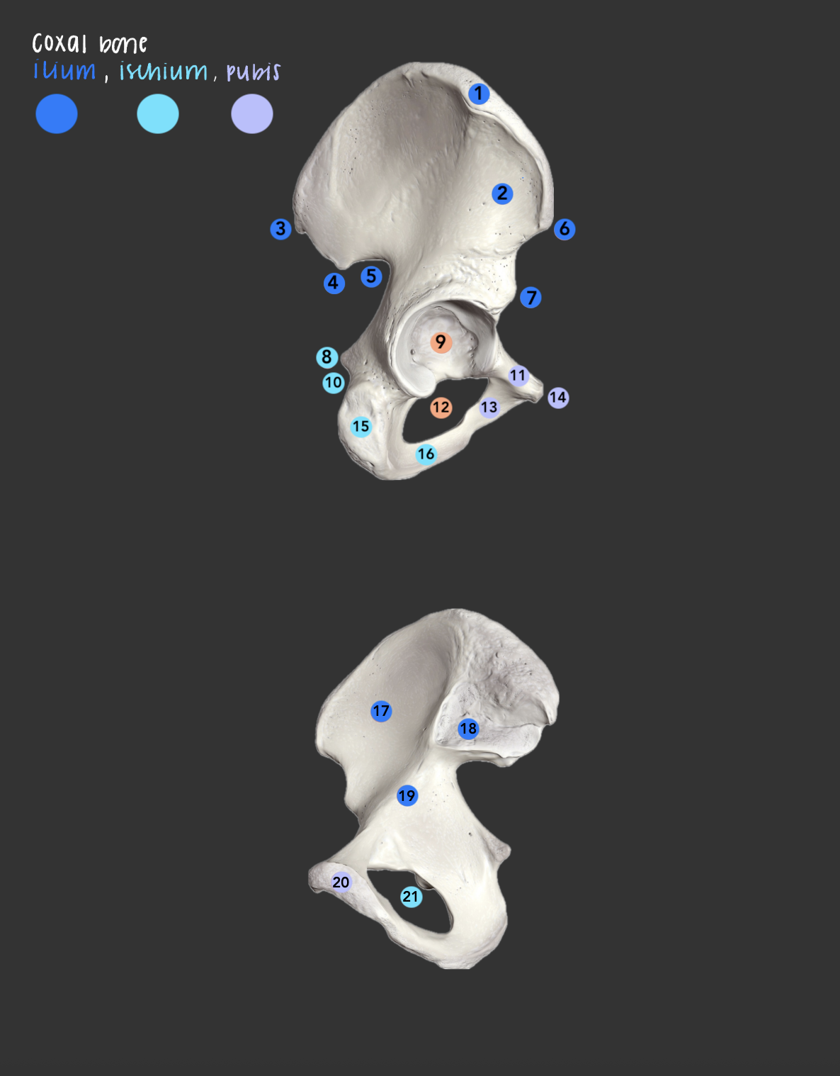
auricular surface (ilium)
what is 18?; ear-shaped articular area on the inner (medial) surface of the hip bone that connects with the sacrum to form the sacroiliac joint

arcuate line (ilium)
what is 19?; ridge on the inner ilium forming part of the pelvic brim

articular surface (pubic symphysis)
what is 20?; where the two pubic bones meet at the midline
frontal bone
makes up the forehead and upper part of eye sockets

supraorbital margin
superior rim of orbit (eyes)

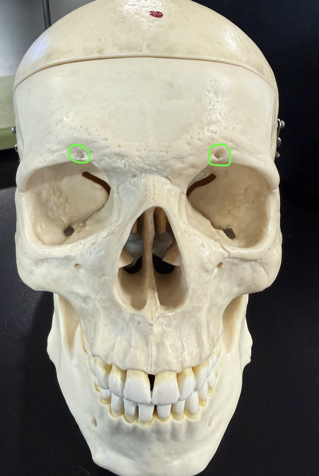
supraorbital foramen (notch)
opening in the supraorbital margin for the supraorbital nerves & vessels
glabella
smooth area b/w the eyebrows

frontal sinus
What is the Blue region ? (cavity in frontal bone above eyes)

parietal bone
top of the head

coronal suture
joins frontal & parietal bones

squamous suture
joins parietal & temporal bones

lambdoid suture
joins parietal & occipital bones

sagittal suture

occipital bone
back of head

external occipital protuberance
midline bump on the back of the skull

external occipital crest
midline ridge running from protuberance toward the foramen magnum

superior nuchal line
ridge extending laterally from protuberance (attachment for neck muscle)

inferior nuchal line
ridge below the superior nuchal line (attachment for neck muscle)

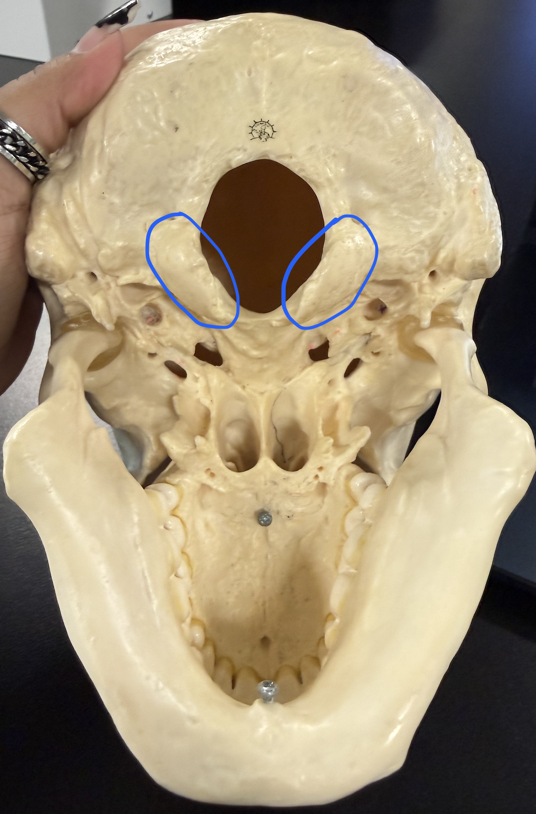
occipital condyle
articulates w/ C1 (atlas)
foramen magnum
large opening where the spinal cord leaves the skull

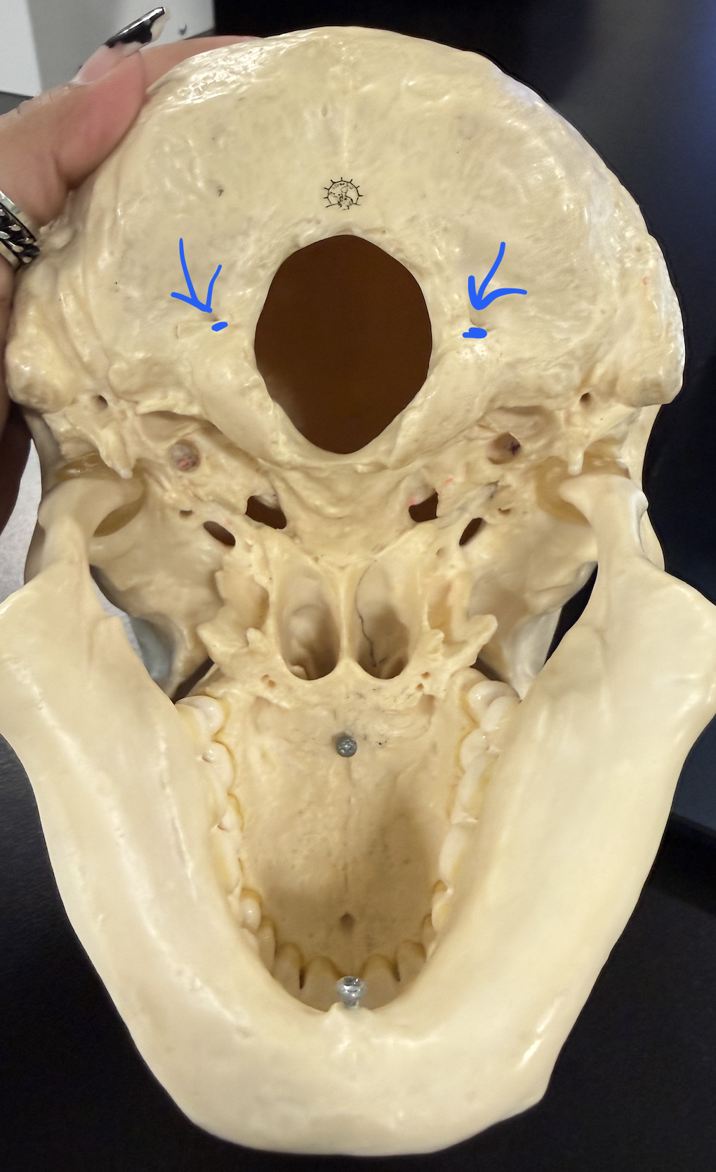
hypoglossal canal
under occipital condyles (need to tilt to be visible)
temporal bone
sides of head

squamous region
thin, flat, platelike part of the temporal bone

zygomatic process
projects forward to help form zygomatic arch
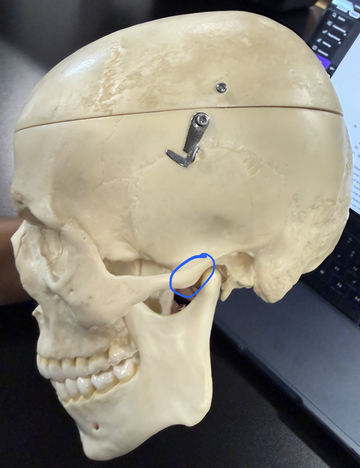
mandibular fossa
articulates w/ mandibular condyle of the mandible

external acoustic meatus
ear canal opening

styloid process
slender spike below the ear canal

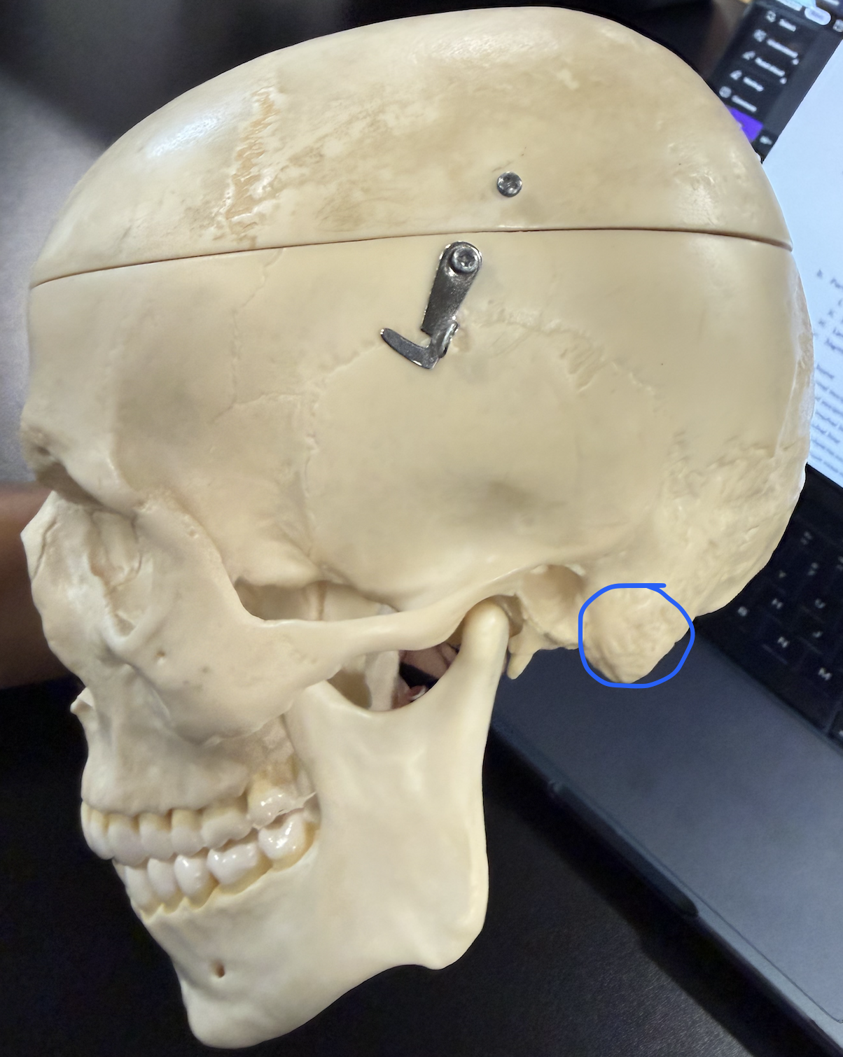
mastoid process
middle ear infection can spread here (mastoiditis), then to meninges (meningitis); sternocleidomastoid attaches here
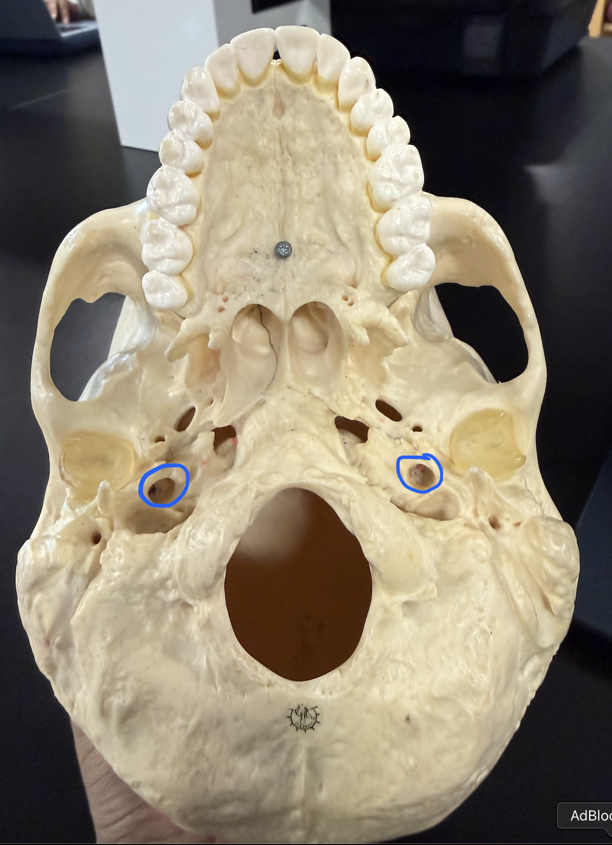
jugular foramen
internal jugular vein, CN XI, CN X, CN IX

internal acoustic meatus
CN VIII , CN VII

foramen lacerum
jagged opening at base of the skull b/w the temporal, sphenoid and occipital bones

carotid canal
sphenoid bone
complex, butterfly-shaped bone located at the middle base of the skull

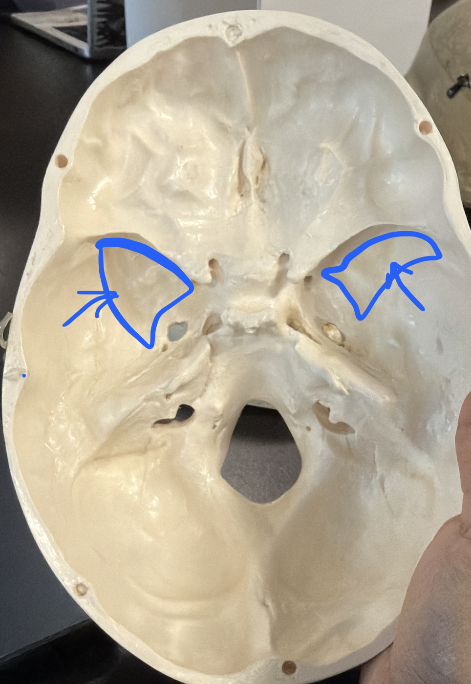
lesser wings
thin wings forming part of the anterior cranial floor and eyes

greater wings
large lateral wings forming part of the cranial floor and eyes
optic canal

superior orbital fissure
CN III, CN IV, CN VI, ophthalmic branch of CN V
