L10: Respiratory I
 Call Kai
 Learn
Learn Practice Test
 Spaced Repetition
 Match
Match Flashcards
 Knowt Play
1/44

There's no tags or description
Looks like no tags are added yet.
Name | Mastery | Learn | Test | Matching | Spaced | Call with Kai | Chat |
|---|
No analytics yet
Send a link to your students to track their progress
45 Terms
Respiratory functions of the respiratory system
Gas transport for metabolism
Move oxygen from the air into pulmonary blood
Clearance of carbon dioxide
Non-respiratory functions of the respiratory system
Lungs receive 100% of the cardiac output from the right heart.
Filter blood, chemical processing, maintenance and defenses (part of the first line of defense)
Facilitate venous return (respiratory pump)
Define respiration
Interchanges of gases between the atmosphere and the cells of the body
Define ventilation (breathing)
Air transported through the airways to and from the atmosphere to the respiratory zone of the lungs
Define gas exchange
O2/CO2 exchange between the air in the lungs and cells in the body
Define cellular respiration
oxidation of cellular molecules that produces CO2, water and ATP
Overall gas transport includes:
Ventilation: movement of bulk airflow, delivering air to the respiratory zone where gas exchange occurs
Gas exchange/Lung diffusion: gas exchange between respiratory zone and blood - O2 moves across to blood and red blood cells; reverse process for CO2
Circulation/Transport (Blood ⇔ tissues): Requires adequate function of the pulmonary and systemic circulations
Tissue diffusion: Erythrocyte/plasma ⇔ tissue cells = Passive diffusion
5. Internal respiration: metabolism using O2 and producing CO2
Airways for ventilation include:
System of tubular structures:
Nasal and oral cavities
Pharynx and larynx
Trachea
Bronchi
Bronchioles
*Bronchioles connect to alveoli where gas exchange occurs

Main functions of airways
-Delivering gas to the respiratory zone (alveoli)
-Conditioning of the inhaled air:
Air warmed to core body temperature (prevents temperature choc in the alveoli)
Gas humidification: saturation with vapor to prevent dehydration of the respiratory epithelium in alveoli
Filtration, cleansing: prevents foreign objects and microorganisms to enter the lungs (reduces risks of injury and infection)
Structure of nasal/oral cavities
Inner surface = mucous membrane that warms and humidifies air
Some species = hair in nostrils act as first filter
Epithelium contains ciliated cells and mucus cells (goblet): trap foreign objects, move the mucus towards the pharynx
Structure of pharynx
Connection between nasal/oral cavity and the larynx
Structure of larynx
Connects pharynx and the trachea: glottis and epiglottis = cartilage that prevents food to enter the trachea
Contains the vocal cords
Structure of trachea
Flexible tube kept open by cartilage rings
Inner surface lined with ciliated and mucus cells
Mucus traps particles, and coordinated cilia movements push the trash back toward the pharynx
Structure of bronchi (in the thoracic cavity)
Possess cartilage plates to maintain the shape
Starts with 1 tube per lung = primary bronchi
Branches off to narrower tubes with less cartilage
possess ciliated and mucus producing epithelial cells
Structure of bronchioles
Lack cartilage = depend upon lung recoil to maintain potency. Possess smooth muscle
possess ciliated and mucus producing epithelial cells

Airway cross-sectional area increases DRAMATICALLY moving from:
Trachea to respiratory zone:
Geometric increase in number of small airways
Reduces velocity of airflow to virtually ZERO
Movement of gas in respiratory zone by DIFFUSION only
Velocity (cm/sec) = flow (cm3/sec)/cross-section area (cm2)
Airway clearance
Cilia and globlet cells work to move thin sheet of mucus from lower parts of the lungs to the throat region
Defensins: Airway “Lysol” – destroy bacteria; first line of defense (2nd line is immune system proper)
Endoscopic View: Trachea
Accumulation of mucus and pus in the trachea associated with inflammation of the lower airways. Turbulence is create, and more resistance to air.
Role of the environment

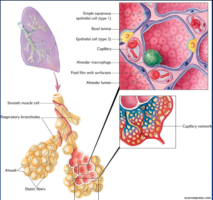
Alveoli characteristics
Respiratory zone
Clusters around terminal bronchioles
Formed by a single layer of epithelial cells
Adult human: covers 75-80 m2
Directly surrounded by a capillary network
Air separated from blood by 2 layers of cells (epithelium and endothelium): best for gas exchange
Some epithelial cells (type 2) produce fluid = surfactant which reduces surface tension, by breaking h-bonds
If small particles reach alveoli = “phagocytized” by macrophages (immune defense)
Thoracic cavity characteristics
Space within the thoracic cage between:
Thoracic vertebras
Ribs and intercostal muscles
Sternum
Includes the Diaphragm and Mediastinum

Diaphragm characteristics
separate, thoracic and abdominal cavity
sheet of skeletal muscles and tendon
Mediastinum characteristics
Divide thoracic cage in 2 halves (from spine to sternum)
Connective tissue containing vessels, nerves, trachea, esophagus and the heart
Each lung fills 1 half
Pleural membranes and characteristics
wet epithelial surfaces, attached to and covering the lungs
parietal membrane is connected to the diaphragm which distends the lungs and also anchored around the cage
Intrapleural space (between visceral and parietal pleura) filled with small amount of fluid = lubrication for friction free movements

Ventilation characteristics
Follows the laws of physic: flow from high to low pressure = depends on ΔP
Resistance to flow (R) due to friction of air particles with each others and with the ducts
Air flow concept similar to blood flow (F = ΔP/R)
Resistance in airways normally small → ΔP major factor
Blood flow
F (flow) = ΔP/R
If R ↑, ΔP must ↑ to maintain air flow
If ΔP ↓, R must ↓ to maintain air flow
What controls ΔP
Compression/expansion of the lungs by respiratory muscles
Ventilation Pump (mechanics): Inspiration
Active mechanism
Diaphragm contracts → expands thoracic volume, creates negative pressure → lung expands → increase in alveolar volume → negative pressure gradient facilitates flow down airways from outside towards the respiratory zone
at rest diaphragm most important muscle
P alveolar < P atmos = propels air through the airway until P alveolar = P atmos
Ventilation Pump (mechanics): Expiration
Passive mechanism (at rest)
End of inspiration – inspiratory muscles relax, allows lung to spontaneously recoil → increase pressure in alveoli → create pressure gradient from alveoli to atmosphere
P alveolar > P atmos = airflow outward until equilibrium reached
Boyle’s law of gases
For a gas at a set temperature, pressure and volume are linked: P1V1 = P2V2
If volume (V) ↑ = pressure (P) ↓
If volume (V) ↓ = pressure (P) ↑

Special case in horses
End of expiration active → beginning of inspiration passive (recoil)
Special case: Locomotion (walking, running)
More than just the diaphragm and intercostal muscles involved
Muscles take active part in expiration (speeds it up)
Galloping: synchronization with breathing
Special case: Diving mammals
Voluntary apnea (up to 1 h)
Strong conducting airways
When under water, compressed gas pushed towards the air ways (no gas exchange) = prevents entry of gas in blood
Generally, do not dive with full lungs
Factors influencing ventilation
Air ways resistance:
Mostly in nasal cavity (diseases, obstruction)
During exercise, animals mainly use mouth to reduce resistance (except horses: flare nostrils)
Degree of turbulence: more turbulence = more resistance
Diameter: reduced with branching = increase resistance. Parallel branches results in increased cross/section (surface area) which compensates for increased resistance
Autonomic nervous system controls smooth muscle in bronchiole: Sympathetic relaxes cells = increased diameter, Parasympathetic contracts cells = reduces diameter
Lung compliance in ventilation
Ability of the lung to distend followed by the ability to recoil:
Elastic fibers in the lung
Muscle tissue in the intercostal muscles
Depends on the elasticity of the tissues in the lung and the thoracic cage
Depends on the surface tension in the alveoli
What causes alveolar surface tension
Due to hydrogen bonds in water molecules when on contact with air (gives water drops their shape)
Alveolar surface tension causes:
Tends to reduce the surface area
Since alveoli lined with moisture, it creates a surface tension against distension
Alveolar surface tension is reduced by the presence of..
SURFACTANT
Mixture of phospholipids, Ca2+ and proteins
Phopholipids reduce the surface tension between hydrogen molecules
Barker syndrome in piglets
inadequate production of surfactant = ↑ surface tension = ↑ resistance to distension = ↓ ventilation.
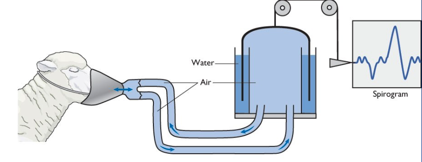
Pulmonary volumes: Spirometer
measure volumes of air inhaled and exhaled (Spirogram = recording)
Tidal Volume (CVT)
Volume flowing through airways

Inspiratory Reserve Volume (IRV)
It is possible to further force more air during inspiration by maximum contraction of respiratory muscles

Expiratory Reserve Volume (ERV)
After normal expiration, it is possible to force more air out
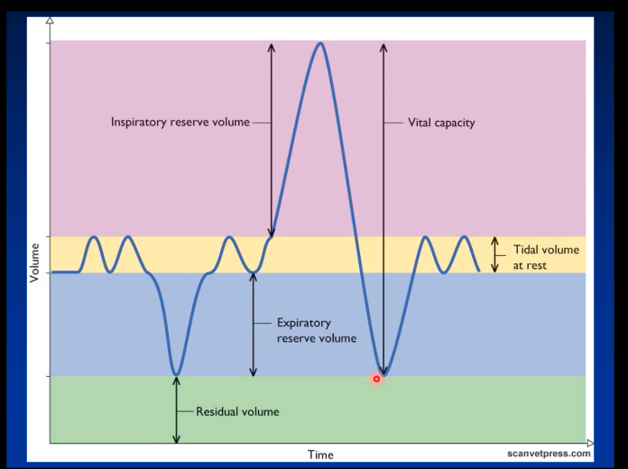
Residual Volume (RV)
After ERV forced out, there always remains air in the lungs

Vital capacity (VT)
is the maximum air that can be inhaled and exhaled

Total lung capacity =
Residual Volume (RV) + Vital capacity (VT)