2) B-Scan/A-scan Ultrasound, Retinal Evaluation with 3-Mirror Lens
 Call Kai
 Learn
 Practice Test
 Spaced Repetition
 Match
 Flashcards
 Knowt Play
1/76

There's no tags or description
Looks like no tags are added yet.
77 Terms
→ focused, high frequency, short- wavelength sound waves
Frequency = 10–15 MHz
tissues will reflect, refract, and/or scatter these sound waves
acoustic waves are generated by vibrations of crystals in the probe and move toward the eye
reflected waves are received by the probe, amplified, filtered, and displayed on the screen as a 2D image (echogram)
focused beam’s reflection is represented as a dot on the screen with its strength indicated by the dot’s brightness
↑ freq = ↓ λ = ↓ penetration = better resolution
(we don’t need deep penetration for ocular structures, but we need more resolution/detail)
sound wave reflected back to the probe
What is acoustic impedance? Give examples.
→ medium's resistance to ultrasound transmission
↑ dense medium = slower sound goes through it (weaker echo) = more light reflected back = brighter it appears on B-scan
Buried optic nerve head drusen
Choroidal osteoma
Retinoblastoma
↓ dense medium = stronger echo = darker it appears on B-scan
Cysts
Retinoschisis

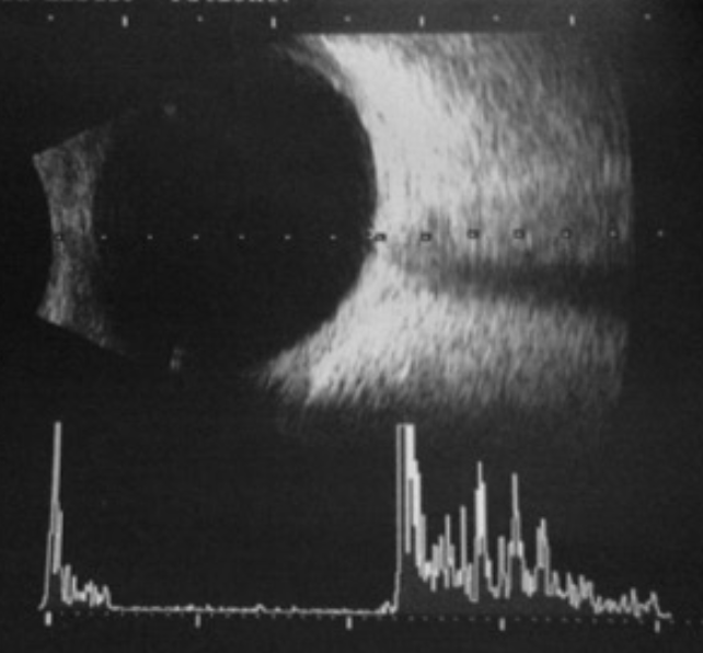
What is a A-scan (Amplitude scan)?
→ 1D amplitude display (sends just one beam of ultrasound)
returning echoes are displayed as spikes from a set baseline
taller spike = ↑ echo strength
commonly used to measure axial length to calculate IOL power

What happens to A-scan spikes when tissue interfaces differ greatly vs if its similar?
Interface differs = spikes become taller
Interface similar = spikes become shorter

What is a B-scan (Brightness scan)?
→ 2D brightness display (sends various waves of the ultrasound)
returning echoes are displayed as dots of varying brightness
brighter dot = ↑ echo strength
What is gain in B-scan ultrasound?
→ amplification of all displayed echoes w/out changing the probe's emitted energy
What’s the difference b/w a high gain vs low gain?
High gain
useful for gross retinal lesions or vitreous findings
(+) weak echoes become more visible (e.g., vitreous)
(+) makes image brighter & visualization depth deeper
(-) resolution decreases + image may wash out
Low gain
→ useful for subtle fundus elevations, choroid, sclera, orbital structures & optic nerve drusen
weak echoes are filtered out
(+) improves resolution of strong echoes (e.g., choroid)
(-) ↓ depth of penetration

Why should both high and low gain be used?
High gain - reveals weak signals
Low gain - provides better detail of strong signals
What is the 2 main uses of B-scans?
Detect and assess ocular and orbital disorders
Monitor and manage existing lesions (i.e., suspicious choroidal nevus)
List the indications for B-Scan Ultrasound.
Evaluating the posterior segment when direct visualization is difficult or abnormalities are suspected:
media opacification (very dense cataract, vitreous heme, dense vitritis, hyphema, dense corneal opacities, miotic pupil)
differentiate PVD vs RD vs choroidal detachment
Dislocated lens
Optic disc drusen vs. papilledema
Choroidal melanoma vs metastasis vs choroidal hemangioma
IOFB vs orbital FB
EOM disease (EOMs inflammation)
Posterior staphyloma
Posterior scleritis
What are the contraindications to B-scan?
Recent ocular surgery
Perforating injury
Scleral laceration
Compare the advantages vs disadvantages of B-scan.

What does the horizontal axis vs vertical axis represent on a normal B-scan?
Horizontal = Tissue depth from anterior to posterior
Vertical = scanned segment of the globe or orbit
Label the following letters.

A – probe/corneal surface
B – posterior lens
C – vitreous
D – retina
E – sclera (very hyper)
F – optic nerve
G – orbital fat and tissue
No - they usually appear together
they’re only separated during pathology (e.g., RD)
Label B-scan ultrasound (axial scan).

Cornea – most superficial echogenic, curved line
A/C – shows no echo (dark)
Iris
Lens – notice how the lens shows no echo, but its edges do
Vitreous – usually echo-free but with aging may show some
Retina/choroid
Optic nerve – hypoechogenic surrounded by echogenic fat
Retrobulbar fat
Optic disc – this is where you’d look for
Why can EOMs appear dark on B-scan?
→ their uniform muscle bellies produce relatively weak echoes
BUT tendon surrounding EOMs and any orbital fat around it will be hyper-reflective


What are the 2 B-scan documentation modes?
Dynamic
Static
What is a dynamic B-scan?
a moving/kinetic examination showing how structures move
Where can a B-scan probe be placed?
Directly on an anesthetized eye or over closed eyelids

List the disadvantages of scanning over closed eyelids.
Waves are attenuated so the echo differentiation is reduced
Hard to tell exactly where on the eye the probe is with the eyelids closed
What coupling agents may be used for B-scan?
Celluvisc
Refresh gel
Ocular ultrasound gel
Where should the patient look during a B-scan?
Toward the meridian being evaluated
What 3 types of B-scans are performed? Mention what they’re performed for.
Axial (front to back) – horizontal and vertical at the optic axis (line of sight)
good for imaging the posterior pole (optic nerve and macula)
determine axial length, evaluate any central axis lesion (of optic nerve and macula)
Transverse – circumferential scan in 8 meridians, scanning multiple clock hours
good for screening a broad retinal area
determines the circumferential and lateral extents of a RD tumor
Longitudinal (1 clock hour at a time)– radial, to isolate a lesion in 1 clock hours
good for measuring a lesion's anterior-to-posterior length & height (cornea to optic nerve)

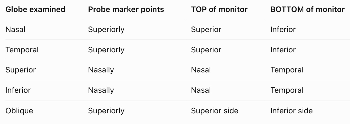
What are the B-scan types and their corresponding probe marker orientations?

Where would you put the probe marker for a Transverse?
Transverse = | | to limbus
180o away from lesion but| | to limbus
⭐ pt looks in the direction to be evaluated (lesion at 6 o’clock, pt look at 6 o’clock)

How do you scan from posterior to anterior with a transverse probe?
Move the probe from the limbus toward the fornix (horizontal scan) or canthus (vertical scan)

What does the top of a B-scan image correspond to?

side where the probe marker is located
If the marker is nasal, what appears at the top of the scan?

nasal side of the globe
If the marker is nasal, what appears at the bottom of the scan?
temporal side of the globe
Which structures are examined relative to the probe tip?
Ocular structures directly opposite from the the probe tip are what is examined

Where would you put probe for a longitidunal scan?
Longitudinal scan = ⊥ to limbus


How does a longitudinal scan appear on the monitor?
Top = Anterior structures
Bottom = Posterior structures (optic nerve)
Where would you put probe for a Axial scan?
Axial = at the cornea/visual axis


How can transverse and longitudinal scans be combined to measure a lesion?
Use transverse for lateral diameter and perpendicular longitudinal for anterior-posterior extent and apical height
How does tissue density affect reflectivity and brightness on B-scan, and what are examples?
Denser tissue → Stronger echo → Brighter(high reflectivity)
e.g., buried optic nerve head drusen, retinoblastoma
Less dense tissue → Weaker echo → Darker(low reflectivity)
e.g., fluid-filled cysts, retinoschisis
Which is more reflective on B-scan: choroid or vitreous?
Choroid
How is reflectivity assessed using B-scan?
→ evaluated by looking at the spike height on an A-scan vector overlay and signal brightness of B-scan
e.g., lesions - membranes, opacities, tumors, FB
What normal ocular structure has the highest reflectivity?
Sclera (100%)
How is the internal structure on B-scan? How is a regular vs irregular internal structure represented?
→ evaluated by looking at the difference in height and length of the A-scan spikes and differences in echo densities on B-scan
Regular internal structure = little or no variation in A-scan spikes and uniform appearance of echoes on B-scan
indicates a homogenous internal structure within a lesion
Irregular internal structure = significant differences in A-scan spikes and echo densities
indicates a heterogenous internal structure

What is acoustic attenuation? List what material can produce attenuation.
→ dark area behind a structure that blocks or attenuates sound waves (dark due to loss of ultrasound energy)
wave energy can decrease due to absorption, reflection or scattering of the sound waves while moving through tissue
e.g., Calcification, FB, bones
What is after-movement on kinetic B-scan?
→ movement of lesion echoes after the eye stops moving
3 types = good, restricted, and none
Which has greater after-movement: PVD or RD?
PVD
RD = restricted after-movement
What ultrasound finding suggests a metallic intraocular foreign body?
highly reflective object with acoustic shadowing

How does the vitreous appear in a young, healthy eye?
Relatively echolucent/dark
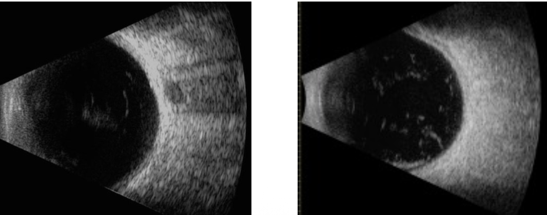
How does a PVD behave during kinetic B-scan?
highly mobile
How does fresh vitreous hemorrhage appear on B-scan?
Low-reflectivity, mobile clump-like or membrane-like echoes
How can fresh vitreous hemorrhage be visualized better?
↑ gain
How does a Retinoschisis appear?
→ won’t move much with kinetic B-scan
clear space below it
sensory retina is split in the OPL
more rigid, dome shaped, surface is highly reflective, and underlying fluid is anechoic (appearing black)

How does a Retinal tear appear?
→ will look like a “flap” on longitudinal scans
flap may appear attached to vitreous still
with the retinal edges folded posteriorly is connected to folded retina
How does a RD appear?
→ should be viewed dynamically/kinetic
moves quite a bit
if total RD, will look funnel shaped b/c still attached to ora and optic nerve
What is this an image of?

Total RD on axial scan – highly reflective, stay anchored to the nerve and ora, funnel-shaped, limited mobility
What is this an image of?

RD on transverse scan – you will get a different view, if not visualizing the anchoring to the nerve
What retinal layer does the lecture identify as the site of splitting in retinoschisis?
OPL
What does a transverse RD scan show differently from an axial RD scan?
different cross-sectional view that may not show the optic nerve attachment
How do vitreous echoes compare with retinal echoes?
Vitreous echoes are generally less bright and have shorter A-scan spikes

How does a PVD attachment near the optic disc differ from an RD attachment?
PVD attachment may be eccentric
RD attaches directly at the disc

How can A-scan spike heights distinguish PVD from RD?
Retinal spikes remain consistently high
PVD spikes decrease in some areas
How do optic nerve head drusen appear on B-scan?
→ Highly reflective calcific nodules at or within the ONH
bilateral, asymmetric
bilateral edema is assoc with ↑ ICP (papilledema) & HTN crisis
nerves look edematous when burried

What is another name for ONH drusen when they mimic swelling?
Pseudopapilledema
What is posterior staphyloma? What is it indicative of? Who is it found in?
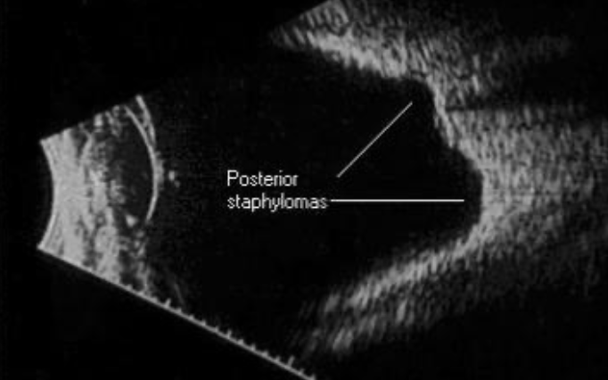
→ localized area of scleral thinning and outward bulging
indicative of degenerative myopia
found in pt with CT disorders
What is this an image of?
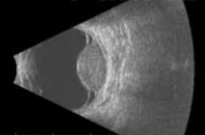
Choroidal melanoma with surrounding serous retinal detachments
What is this an image of?

Choroidal detachment (choroidal effusion)
Which probe placement gives better resolution: conjunctiva or eyelid?
Directly on the conj
How should the probe be oriented relative to the tissue for better resolution?
⟂ to the tissue
Where is B-scan resolution generally best?
At the center of the scan
What is the purpose of the 3-mirror retinal examination?
→ stereoscopic examination of the vitreous and retina using a slit lamp
⭐ not a practical technique to screen the peripheral retina due to small FOV
What is the rule for mirror size and retinal location? Mention what each mirror views.
smaller the mirror, the more peripheral the view
Central circle = posterior pole (better than 78/90D) + vitreous
macula - macular edema, holes & AMD
optic nerve
Trapezoid = equatorial fundus
Square = anterior equator to posterior ora serrata
Thumbnail = ora serrata/pars plana

Rank the peripheral mirrors from least to most anterior.
Anterior = peripheral

What type of view does the central lens provide?
Direct, upright, magnified
Where should the peripheral mirror be positioned relative to the retinal area being examined?
180° opposite the retinal area
to observe nasal retina → mirror should be temporal
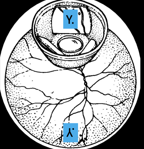
What happens as the angle of a peripheral mirror becomes smaller?
view becomes more anterior/peripheral, closer to the ora
How can you extend the view more anteriorly vs posterioly?
Anteriorly = Tilt the lens slightly toward the mirror
Posteriorly = Tilt or rock the lens away from the mirror
What gaze direction can help obtain a more anterior retinal view?
Ask the patient to look opposite the mirror