practical 3
 Call Kai
 Learn
 Practice Test
 Spaced Repetition
 Match
 Flashcards
 Knowt Play
1/48

There's no tags or description
Looks like no tags are added yet.
49 Terms
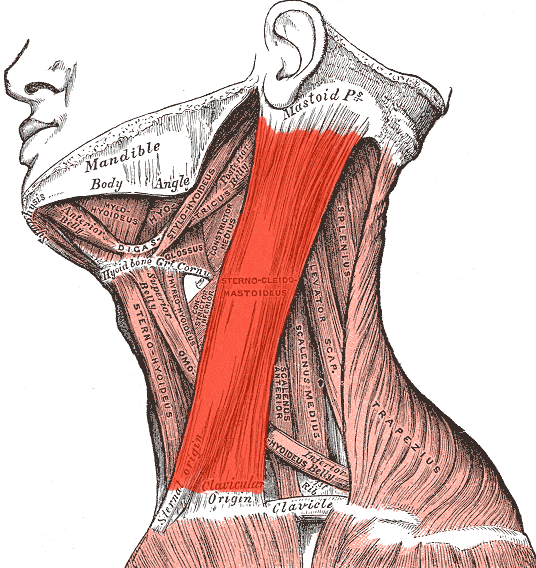
Sternocleidomastoid
Two-headed neck muscle running diagonally from the manubrium and medial clavicle to the mastoid process; laterally flexes the neck and rotates the head to the opposite side.

Trapezius
Large, superficial diamond-shaped muscle of the posterior neck and upper back that extends from the occiput down to the lower thoracic vertebrae; elevates, retracts, and depresses the shoulder blade.

Occipitofrontalis
Epicranial muscle with anterior and posterior bellies linked by a central sheet-like tendon; wrinkles the forehead, raises the eyebrows, and draws the scalp posteriorly.

Orbicularis oculi
Sphincter muscle arranged in concentric rings around each orbital margin; closes eyelids during winking, squinting, and sleeping.

Orbicularis oris
Complex circular sphincter muscle enclosing the mouth opening; compresses, purses, and protrudes the lips.

Zygomaticus major and minor
Pair of diagonal cheek muscles extending from the malar bone downward to the corner of the mouth; elevates the oral angle laterally to produce a smile.

Mentalis
Small V-shaped muscle pair located on the apex of the chin; elevates and wrinkles chin skin while pushing the lower lip outward.

Masseter
Thick, rectangular muscle of mastication covering the lateral mandibular ramus; elevates the jaw to close the mouth during chewing.
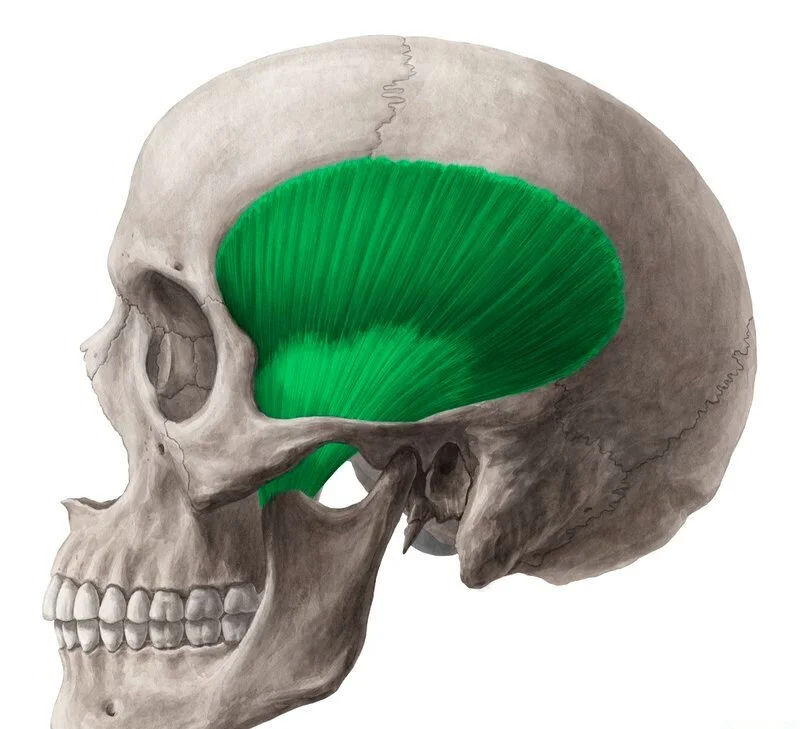
Temporalis
Fan-shaped muscle filling the temporal depression on the lateral skull that passes beneath the zygomatic arch to the coronoid process; elevates and retracts the mandible.

Suprahyoid muscles
Cervical muscle group located superior to the hyoid bone that elevates the hyoid and larynx to support swallowing and vocalization.

Infrahyoid muscles
Ribbon-like strap muscles positioned below the hyoid bone that depress the hyoid and thyroid cartilage following deglutition.

External intercostals
Superficial intercostal layer with fibers angled inferomedially ('hands in pockets'); raises the ribs to expand the chest cavity during inspiration.

Pectoralis major
Broad, fan-shaped chest muscle arising from the sternum and clavicle and inserting into the upper humerus; adducts, flexes, and medially rotates the arm.

Diaphragm
Dome-shaped musculotendinous internal divider between thoracic and abdominal compartments; flattens downward to drive negative-pressure breathing.

Rectus abdominis
Pair of vertical abdominal straps bound within an aponeurotic sheath and divided by horizontal tendinous intersections; flexes the trunk and compresses viscera.

External abdominal oblique
Outermost lateral abdominal wall muscle whose fibers run obliquely downward and inward; compresses the abdomen and rotates the trunk.

Internal abdominal oblique
Intermediate abdominal layer with fibers oriented upward and medially; flexes and rotates the vertebral column while compressing abdominal organs.

Transversus abdominis
Deepest abdominal wall muscle characterized by horizontally traveling fibers; constricts abdominal contents to stabilize core posture.

Rhomboids (major and minor)
Deep back muscles positioned between the medial scapular border and the upper thoracic spine; retract and downwardly rotate the shoulder blade.

Serratus anterior
Saw-toothed muscle slips lining the lateral rib cage originating on ribs 1-8; protracts the scapula forward around the thorax ('boxer muscle').

Pectoralis minor
Deep triangular anterior thoracic muscle originating from ribs 3-5 and attaching to the coracoid process; tilts the scapula anteriorly and downward.

Supraspinatus
Superior rotator cuff muscle occupying the fossa above the scapular spine, inserting onto the humeral greater tubercle; initiates arm abduction.

Infraspinatus
Rotator cuff muscle positioned in the broad fossa below the scapular spine, inserting on the greater tubercle; laterally rotates the arm.

Teres minor
Narrow cylindrical rotator cuff muscle positioned directly beneath the infraspinatus along the lateral border; externally rotates and stabilizes the shoulder joint.

Subscapularis
Anterior rotator cuff muscle filling the ventral concavity of the shoulder blade and inserting on the lesser tubercle; medially rotates the arm.

Brachialis
Deep anterior arm muscle underlying the superficial flexor, inserting on the ulnar coronoid process; chief flexor of the elbow.

Biceps brachii
Two-headed anterior arm muscle originating from the scapular coracoid process and supraglenoid tubercle, inserting onto the radial tuberosity; flexes and supinates forearm.

Brachioradialis
Superficial lateral forearm muscle spanning from the lateral supracondylar ridge to the radial styloid process; flexes elbow in mid-pronation.

Triceps brachii
Large three-headed posterior arm muscle inserting into the olecranon of the ulna; primary extensor of the forearm at the elbow.

Extensor digitorum
Superficial posterior forearm muscle that splits into four long tendons inserting into phalanges 2-5; extends the fingers and wrist.

Palmaris longus
Slender superficial forearm muscle bearing a long cord-like tendon that inserts directly into the palmar aponeurosis; tenses palmar fascia and weakly flexes wrist.

Flexor carpi ulnaris
Medial-most superficial forearm flexor inserting onto the pisiform and fifth metacarpal; flexes and ulnar adducts the wrist.

Iliacus
Fan-shaped muscle filling the internal basin of the ilium that joins the lumbar spine flexor to insert on the lesser trochanter; flexes the hip.

Psoas major
Long, cylindrical muscle arising from the lumbar vertebrae and descending under the inguinal ligament to the lesser trochanter; flexes hip and lumbar spine.
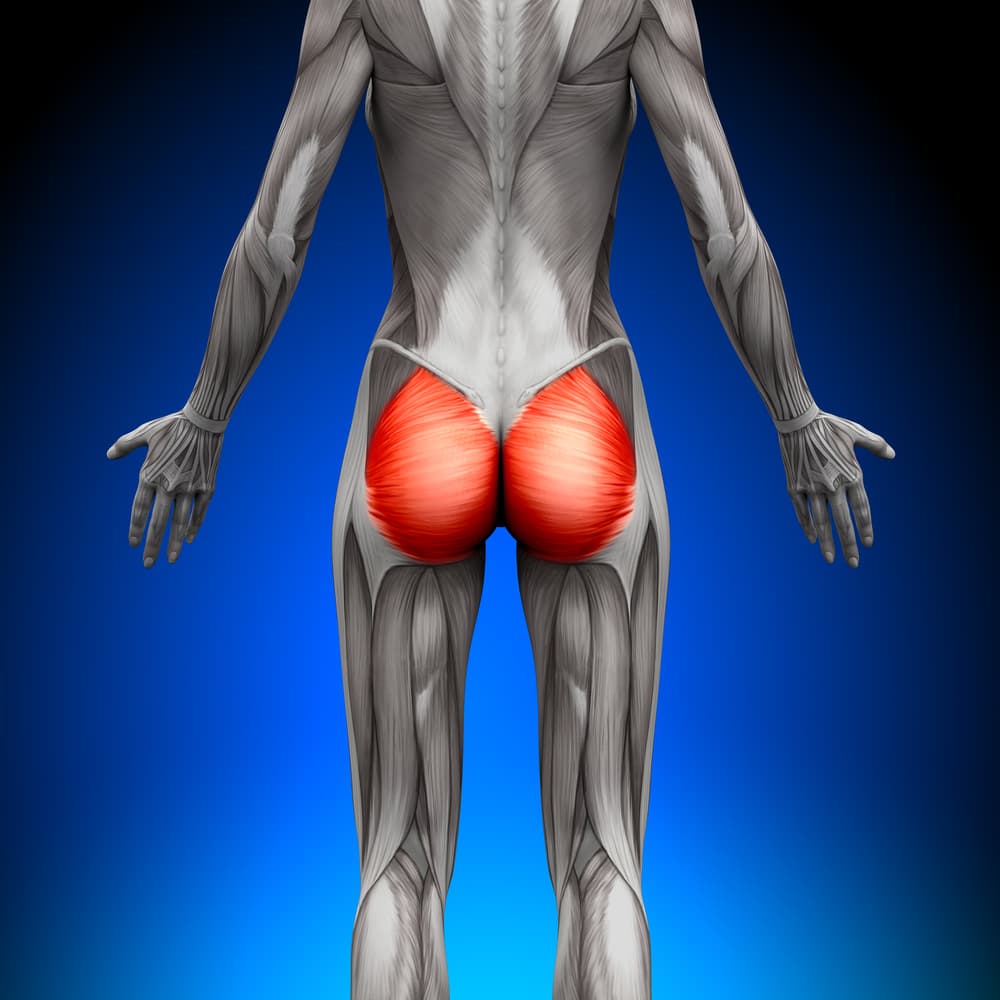
Gluteus maximus
Bulky superficial buttock muscle originating from the ilium and sacrum and inserting on the femur and iliotibial band; powerfully extends and laterally rotates the thigh.

Gluteus medius
Fan-shaped lateral hip muscle underlying the largest gluteal muscle and inserting on the greater trochanter; abducts the thigh and keeps the pelvis level when walking.

Gluteus minimus
Deepest and smallest of the buttock muscle group, sitting directly beneath the middle gluteal muscle; abducts and medially rotates the thigh.

Rectus femoris
Central superficial quadriceps muscle arising from the anterior inferior iliac spine and crossing two joints; extends the knee and flexes the hip.
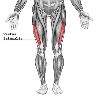
Vastus lateralis
Largest component of the quadriceps group situated on the lateral aspect of the thigh; extends the knee joint.

Vastus medialis
Teardrop-shaped quadriceps muscle situated on the anteromedial distal thigh; extends knee and stabilizes patellar tracking.

Vastus intermedius
Deep quadriceps muscle lying directly underneath the central superficial flexor on the anterior femoral shaft; extends the knee.

Sartorius
Long, narrow strap muscle crossing diagonally from the anterior superior iliac spine to the medial proximal tibia; flexes, abducts, and laterally rotates the thigh.

Biceps femoris
Lateral hamstring muscle featuring long and short heads inserting on the fibular head and lateral tibial condyle; extends the thigh and flexes the knee.

Semitendinosus
Medial hamstring muscle with a long round distal tendon attaching to the medial proximal tibia; extends thigh and flexes knee.

Semimembranosus
Deep medial hamstring muscle marked by a broad membranous proximal attachment that inserts onto the posterior medial tibial condyle; extends thigh and flexes knee.

Tibialis anterior
Superficial muscle lying directly lateral to the anterior shin crest, inserting into the first metatarsal and cuneiform; dorsiflexes and inverts the foot.

Gastrocnemius
Two-bellied superficial posterior calf muscle spanning from the femoral condyles to the calcaneus via the Achilles tendon; plantarflexes the foot and flexes the knee.

Soleus
Broad, flat calf muscle situated deep to the gastrocnemius heads that inserts onto the calcaneus; primary plantarflexor used for standing posture and walking.

Flexor carpi radialis
A muscle located in the forearm that flexes and abducts the wrist. It originates from the medial epicondyle of the humerus and inserts on the bases of the second and third metacarpal bones.