Surface Properties and Analysis (Protein Adsorption)
 Call Kai
 Learn
Learn Practice Test
 Spaced Repetition
 Match
Match Flashcards
 Knowt Play
1/99

There's no tags or description
Looks like no tags are added yet.
Name | Mastery | Learn | Test | Matching | Spaced | Call with Kai | Chat |
|---|
No analytics yet
Send a link to your students to track their progress
100 Terms
Why focusing on the surface?
first contact w/ biological system
different from the bulk of a material
easily contaminated
structure is often mobile (atoms/groups of atoms move according to the environment)
Surface Picture

Surface Parameters to be Measures
wettability (contact angle)
chem. composition (IR, XPS, SIMS)
topography/roughness (SEM, AFM)
surface mobility
crystallinity
heterogeneity
Wettability
parameter of the surface; how easy it is for water/aqueous soln. to adhere or spread across the material; diff. for hydrophobic and hydrophilic surfaces
Wettability - Hydrophobic Surfaces
have low surface energy and water beads at the surface; ex. PE and PTFE (np/hydrophobic groups)
Wettability - Hydrophilic Surfaces
have high surface energy and water wets the surface; ex. polar/hydrophilic groups: -COO-, -NH4+, -OH, -COOH, -C=O, -NH2 (can H-bond with water and more charged = more hydrophilic)
Surface Structure Mobility
hydrophilic domains/groups may rearrange to face the surface in an aqueous environment

Contact Angle - Wettability
describes the shape of a liquid drop resting on a soid surface; a measure of the liquid wetting of a surface, an estimate of surface energy, which in turn, is a characteristic of chemical bonding
higher angle = lower surface E/tension → hydrophobic
lower angle = higher surface E/tension → hydrophilic
Contact Angle Picture

WCA
water contact angle
Contact Angle Measurement Concerns/Errors
operator dependent (use of software helps)
surface roughness and heterogeniety (could vary across surface)
liquid contamination (same syringe. etc.)
liquid changes the surface (reorientation, absorption, swelling, or dissolving)
Typical Water Contact Angle Values
skin = 90o → slighty hydrophobic bc of lipids
PE and PP = 87o
PTFE = 112o → low surface E; allows it is be “anti-stick”
polyamide (Nylon) = 73o → slightly hydrophilic bc of amide group thing???
PMMA = 60o → hydrophilic
treated PE and PP = 22o → becomes hydrophilic when treated bc surface mods.
PET-PEG = 19o → one of the most hydrophilic materials
How do surface properties differ from bulk properties?
surface is the 1st point of contact w/ a bio system
surface atoms/groups can move in response to environment
surfaces are easily contaminated
surface composition is not identical to bulk composition
A surface w/ a high water contact angle is what?
hydrophlic with low surface energy
Fourier Transform Infrared Spectrum (FTIR)
way to find chemical groups in a sample; IR spectroscopy measures the vibrations of chemical bonds and each one absorbes incoming IR energy at a particular frequency; infrared spectrometer sheds IR beam on sample and measures amount of radiation at various wavelengths that is transmitted or reflected by the sample then FT turns raw data into spectrum; longer wavelength = smaller frequency

IR Spectroscopy
measures the vibrations of chemical bonds and each one absorbes incoming IR energy at a particular frequency; part of FTIR
Infrared Spectrometer
sheds IR beam on sample and measures amount of radiation at various wavelengths that is transmitted or reflected by the sample then raw data is trned into spectrum using FT
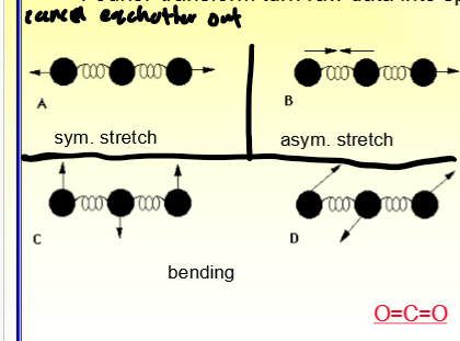
Types of Bond Stretching
symmetrical, asymmetrical, and bending

Chart of Characteristic IR Absorption Bands
where are the bonds typically at: absorption bands assigned to functional groups
wavenumber (cm-1) is more frequently used in IR spectrum = 1/lambda (lambda = wavelength)

What do shifts in the frequency of absorption bands and changes in relative band intensities indicate?
there are changes in the chemical structure or changes in the environment around the sample
Attenuated Total Reflectance (ATR) - FTIR
provides more surface specific info (1-5um)
advantages - inexpensive, quick, no sample prep., can look at kinetics
disadvantages - not highly surface sensitive (bc layers so small), have to make really good contact w/ IRE so need flat surface

IRE
internal reflective element
X-ray Photoelectron Spectroscopy (XPS) aka ESCA (Electron Spectriscopy for Chemical Analysis)
extremely surface selective/sensitive; based on photoelectric effect, interactino of x-rays w/ atoms causes the emission of inner shell e-’s which its kinetic E can be measures and the BE can be calulated (used as the characteristic parameter of the specific element)
Photoelectric Effect
photon comes in and knocks an electron out
BE = hv - KE
BE - binding energy of electron to atom
KE - kinetic energy of emitted electron
hv - energy of X-ray

ESCA/XPS Picture

Why is ESCA/XPS very surface sensitive?
even though x-rays can penetrate deep, elctrons emitted deep lose their energy in elastic collisions and never emerge from the surface → only electrons emitted near surface lose no energy and will contribute to signal; ones that lose some energy but still emerge contribute to background signl (noise)
ESCA/XPS Surface Sensitivity Picture
A/B - lose no energy and emerges from the surface so contributes to signal
C - loses some energy but still emergs from the surface so contributes to background signal (noise)
D/E/F/G - lose their energy in elastic collisions and never emerge from the surface

ESCA/XPS Example Spectra Picture
ESCA/XPS Advantages
surface sensitivty (~8 nm)
high info content (element id, semi-quantitative, molecular environment, finger print of materials)
depth profile
spatial resolution (5 um) → doesn’t damage specimen
low damage potential
little sample prep. (just needs to be completely dry)
ESCA/XPS - High Info Content Advantage
element ID
semi-quantiative
molecular environment (oxidation state, bonding atoms, etc.)
fingerprint of materials (some kind of pattern)
ESCA/XPS - Depth Profile Advantages
use a strong X-ray to oblitate and then keep going
ESCA/XPS Disadvantages
expensive
need vacuum compatibility
not good for complex surface
Secondary Ion Mass Spectrometry (SIMS)
based on the generation of secondary ions by bombardment of a solid surface by incident beam of accelerated ions (primary)
mass/charge of secondary ions measures using time-of-flight mass analyzer

Dynamic SIMS
used for obtaining compositional info as a function of depth below surface
high ion doses
surface erodes fast
only atomic fragments detected
more artifacts as beam erodes faster
Static SIMS
used for sub-monolayer elemental analysis
low ion does (adjusted so less than 1 monolayer of surface atoms is sputtered)
large fragments can be ejected and analyzed (don’t break down)
Example of SIMS Spectra Picture
SIMS Advantages
most surface sensitive (15A or smaller)
usesful w/ isotopes
can achieve high spatial resolution (40 nm or less) by focusing primary ion beam magnetically or electrostatically
imaging SIMS
SIMS Disadvantages
expensive
complex interpretation (need an expert)
What is the key advantage of ATR-FTIR compared to standard transmission FIT, and what is its main limitation?
it provides more surface-specific info and requires no sample prep., but is not highly surface sensitive and needs good contact w/ the IRE
Why is ESCA/XPS very surface sensitive?
electrons emitted deep in the sample lose energy to inelastic collisions and don’t escape. so only electrons from near the surface contribute to the signal w/o energy loss
Scanning Elctron Miscroscopy (SEM)
focusing/rastering a high energy e- beam on a specimen and detecting the emitted low energy secondary e'-’s then reconstructing intensitity of secondary e-’s on a phosphor screen; good for qualitative surface roughness/texture eval.; differs for conductive vs nonconductive materials; e’ beam instead of x-ray
also under vacuum
SEM for Conductive Samples
energy dispersive X-ray (EDX) analysis can be used for bulk elemental analysis (sampling from a few micron deep)
SEM for Non-conductive Materials
coated w/ thin electrcallt grounded layers (metal or carbon) to minimize the negative charge accumulation; surface chem. info will be contaminated; when this, SEM will heat up and destroy the sample so needs coating
Low-voltage or Environment SEM (ESEM
permits the use of uncoated specimen and ESEM allows for wet sample measurement but magnification and resolution drops
SEM of Polypyrrole Surfaces Picture
conductive material; can use chemical or electrochemical approach; regular SEM

ESEM Picture
environment, low vacuum, and low voltage

Atomic Force Microscope
measures topography w/ a force prope
laser beam reflection offers a convenient and sensitive method of measuring cantilever deflection
AFM cantilevers have ranges of spring constants
micromachning techniques produce inexpensive, reasonably sharp tips
piezotube positions the sample w/ high resolution (indvidual atoms)
essenitally put cantilever on surface and move it so the laser detects the changes in the cantilever; can be use din air (no vacuum needed)
Atomic Forcce Microscopy
can be used w/both conductive and non-conductive surfaces, in water, in air, and in vacuum
has atomic level resolution but usually can’t be obtained for organic/bio samples
tip shape limitation
contact vs. tapping mode
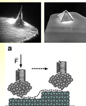
Tip Shape Limitation - Atomic Force Microscopy
determined by shape of the tip, if surface is dynamic/static, and how soft the surface is
Contact vs Tapping Mode - Atomic Foce Microscopy
contact - touches the sample
tapping - no direct contact w/ sample so it doesn’t damage the surface (stays very smal distance away and vibrates)
What distinguishes ESEM from conventional SEM, and AFM from both?
ESEM permits imaging of uncoated and wet samples, while AFM measures surface topography directly w/ a physical probe rather than an electron beam
Both EDX and XPS can be used for elemental analysis, but they differ in how they sample a surface how?
EDX (paired w/ SEM) proves several microns deep for bulk elemental analysis, while XPS is highly surface sensitive (~8 nm) and based on the photoelectric effect
Protein Adsoption
the first phenomena on a biomaterial; can be measures immediately after implantation (< 1 second) → monoplayer of proteins adsrobs to the surface in seconds → mins
When does protein adsorption occur?
immediately after implantation (< 1 sec) and well before cells arrive at the surface
What does the nature of the absorbed protein layer determine?
the cellular response to the surfaces → adhere, release active compounds, recruit other cells, or grow; essentially by the time the cells get there, the surface is covered in proteins that tell them what to do
Proeznyme Activation and Localization
this happens through surface adsoprtion which initiates coagulation, fibrinolysis, etc.
Good vs Bad Protein Adsorption
good: artifical organs
bad: too much protein can clog artifical organs, clotting, ELISA, biosensors (need a clean surface to get a good signal)
Factors that Influence Protein Asorption
protein and surface
Protein Properties that Influence Protein Adsorption
protein structure, size, and surface charge
folding and stability
bulk conc.
Surface Properties that Influence Protein Adsorption
wettability, charge, roughness, surface mobility, chemical composition, crystallinity, heterogeneity to biological rxns
Structure of Proteins
primary, secondary, tertiary, and quaternary
Primary Structure of Proteins
amino acid sequence with peptide bonds

Secondary Structure of Proteins
use non-covalent forces; ex. alpha helix and beta sheet; FTIR can tell you how many alpha-helices vs beta-sheets are in a protein

Non-covalent Forces of Secondary Proteins
H-bonding, hydrophobic interactions, electrostatic interactions, and Van der Waals forces
Hydrophobic Interactions
the hydrophobicity of certain amin acid R-groups tend to drive them away from the exterior of proteins and into the interior; hydrophobic on inside and hydrophilic on outside
Electrostatic Interactions
charge-charge (K, H, and R (+) and then D and E (-))
charge-dipole (charged R-group and water dipole)
Tertiary Structure of Proteins
complete 3d structure of the polypeptide

Quaternary Structure of Proteins
2 or more diff. polypeptide chains held together in association by non-covalent factors or disulfide bonds

Complex Proteins
glycoproteins and apolipoprotein A-I
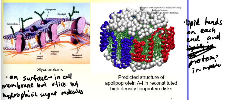
Protein Adsorption Irreversibility
essentially it can’t be undone but you cna remove some proteins from the sirface using a buffer wash but need to use a surfactant wash to get them all off

Why do surfactants remove proteins from the surface?
because they have a higher affinity for the surface so it kicks the protein off
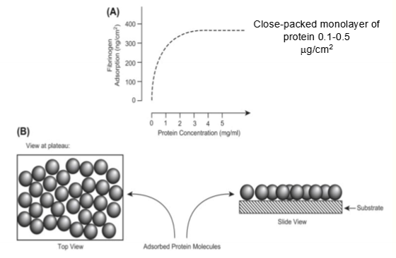
Protein Adsorption Isotherms
relates the amount of adsorbed protein to the soln. of protein at equilibrium; can be difficult to reach true equilibrium though (might start stacking on top of each other instead so multiple layers instead of a monolayer where it wkuld just be a plateau)
Thermodynamic Driving Force on Hydrophobic Surfaces
hydrophobic effect happens; these surfaces have a tsrong tendency to absorb protein irreversibly; unfolding of the protein on the surface increases entropy (delta S) and therefore releases many structured water molecules from the interface (increases delta S) so large entropy gain for the system; overtime some adsorbed protein can reaarange to expose more hydrophobic residue to the surface; delta G = delta H - T (delta S)

Polyelectrolytes
what proteins can be considered because of the large number of charged amino acid residues
What happens when a protein is minimal (near pI)?
protein adsorption exhibits a max on the neutral or slightly charged surfaces
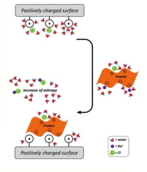
Major Thermodynamic Driving Force on Charged Surfaces
a combo of ion-ion coulombic interactions (-deltaH) and entropy gain (+deltaS) due to release of counterions along with their bound water
Protein Adsoprtion on Charged Surface Example Picture

Summary of Thermodynamics Picture

Selecitvity of Protein Asoprtion Process
protein compete for sites on surface
diff. ones have diff. surface activity
when in contact woth blood (plasma) surface protein comp. can be diff. than bu.k so some of low conc. surface active proteins can dominate surface
Selecitvity of Protein Asoprtion Process Picture
higher number = more protein adhered to surface

Enrichment
surface fraction / bulk fraction; higher = more protein on surface
Vroman Effect
protein asorption from blood plasma involves complex series of adsorption and displacement steps; initial deposition depends on bulk conc. and trasnport rate to surface → later, proteins that binds to surface strongly can replace weaker binding ones
Vroman Effect Example/Picture
2 proteins on the same surface
B has a higher a conc. in soln. so it is more readily available to absorb on the surface
A has a higher affinity for surface and can kick B out

Conformational Differences on Different Surfaces
protein goes through conformational changes in order to best adhere to the surface

Conformation of Adsorbence Protein
when adsorped onto diff. surfaces, proteins expose diff. domains = diff. bioactivity
Enhanced Bioactivity - Conformation of Adsorbed Proteins
adsorption renders clotting factor XII active as a protease and a conformational change induced by the absorption activates the protein; only happens in some cases do you get the enhancement
Conservation of Bioactivity - Conformation of Adsorbed Proteins
in many cases; ex. immunoasassy
Reduction of Bioactivity - Conformation of Adsorbed Proteins
in some cases; due to denaturization at the surface
What is the degree of denaturization dependent on?
the protein, surface, length of time protein has spent on surface, solvent, etc.
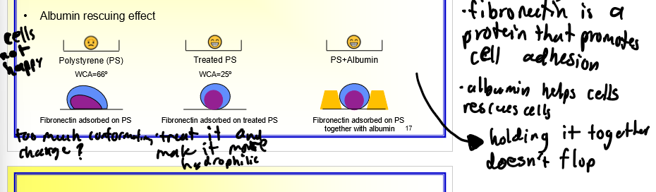
Albumin Rescuing Effect
????????????????????
Principles of Protein Adsorption to Biomaterials
not all proteins in plasma phase can be equally repped on surface
intrinisic surface activity and bulk conc. of proteins drive porcess
surfaces vary in selectivity of adsorption
bioactivity varies
Why can’t all proteins in plasma phase be equally represented on the surface?
due to monolayer adsorption and consequent competition for binding sites
Means to Study Protein Adsorption
radioactive labeling, AFM, XPS and SIMS, ATR/FTIR, etc.

Nonfouling Surface
a specialized material interface engineered to resist the unwanted adhesion of proteins, cells, bacteria, and other biomolecules

What are nonfoudling surfaces important for?
biofilm(bacteria) prevention
blood compatible biomaterials (prevent blood clots)
electrochemical sensors
What did we learn from nature to help woth designing nonfouling surfaces?
make the surface hydrophilic and mimic the cell membrane
Nonfouling Surfaces are Biomolecule-based
passivating proteins (albumin and casein)
polysaccharides (hyaluronic acid) → hydrophilicity comes from these
liposaccharides
phospholipid bilayer (mimic cell membrane)
glycoproteins (mucin)
Synthetic Nonfouling Surfaces
hydrophilic polymer and Zwitterionic polymers
Hydrophilic Polymer - Synthetic Nonfouling Surfaces
Poly(ethylene glycol) aka PEG
has resistance of polymer coil to compression
has resistance to release bound and free water from hydrated coil

Zwitterionic Polymers - Synthetic Nonfouling Surfaces
better option; has ionic solvation → strong hydration (binds water and repels protein); has a neutral charge bc each repeat unit has a positive and negative charge (prevents charged domains so minimizes charge-charge interactions)
