APC 100 Lecture Midterm 2
 Call Kai
 Learn
 Practice Test
 Spaced Repetition
 Match
 Flashcards
 Knowt Play
1/56

There's no tags or description
Looks like no tags are added yet.
57 Terms
4 Basic Steps of Fertilization
contact and recognition
regulation of sperm entry
fusion of pronuclei
activation of egg metabolism
Step 1 of Fertilization: Contact and Recognition
egg releases attractants to guide sperm
sperm develops acrosomal filament in marine invertebrates
acrosome in mammals is activated by contact with zona pellucida (outer layer of egg)

Step 2 of Fertilization: Regulation of Sperm Entry
contact with the vitelline envelope (nonmammals) or zona pellucida (mammals)
sperm fuses to egg
egg changes so no additional sperm can enter

Step 3 of Fertilization: Fusion of Pronuclei
fertilization envelope is created
prevents poly sperm
sperm head becomes trapped in fertilization envelope
two pronuclei (male and female) fuse inside the egg
sperm centriole acts as a microtubule organizing center
fusion forms diploid zygote
Step 4 of Fertilization: Activation of Egg Metabolism
happens concurrently with sperm fusion
sperm entry alters Ca2+ levels in the egg, which activates the egg
Fast Block to Polyspermy
membrane potential change of the egg cell
calcium released inside the egg
sodium channels closed preventing Na+ entry/exit
resting membrane potential changes from -70mV to +20mV after fertilization
sperm cannot fuse with these eggs
Slow Block to Polyspermy
cortical granule reaction
triggered by Ca2+ release
granules release enzymes and particles that make the membrane physically impossible for sperm to access
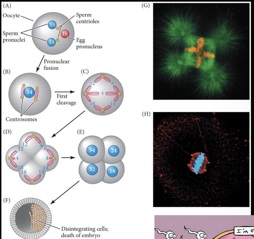
Result of Polyspermy
creates a polyploid nucleus
sperm’s centriole creates multiple planes of division
cells either die or develop abnormally
Amniote
animal whose embryo develops in an amnion and chorion and has an allantois
a mammal, bird, or reptile
Anamniote
any vertebrate animal, such as a fish or amphibian, that lacks an amnion, chorion, and allantois during embryonic development
Gastrula
an embryo undergoing gastrulation
Neurula
an embryo whose neural tube is closing to form the CNS
Potency
a cell’s ability to differentiate into (become) other cell types
decreases as embryo development progresses
Totipotent
an embryonic stem cell that can differentiate into all the cell types required for the formation of tissues and organs in developing embryo AND EXTRAEMBRYONIC TISSUES
high potency
cleavage stages
Pluripotent
an embryonic stem cell that can differentiate into all the cell types required for the formation of tissues and organs in the developing embryo
blastocyst
less potency
Ectoderm
the superficial tissue that forms things like the CNS (brain and spinal cord), skin, and sensory system
Mesoderm
the middle tissue that forms bone, muscle, blood, kidneys, and urogenital tract
Endoderm
the internal tissue that forms the digestive tract, thyroid, and lungs
Mullerian/Wolffian Ducts
embryonic tissues that make male and female sex organs
Cell Fate
the future identity of a cell determined by environment (signals they receive) and differential gene expression

Gastrulation
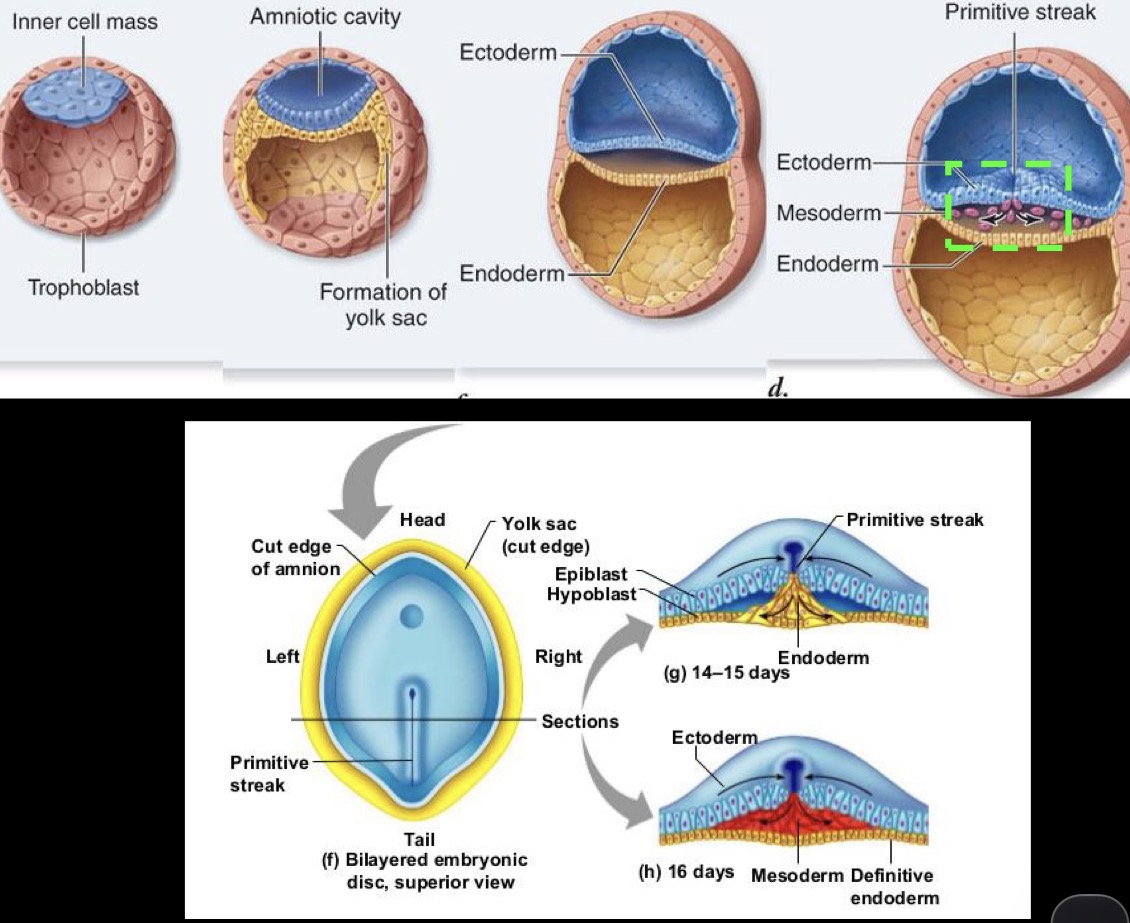
formation of the three germ layers (endoderm, mesoderm, ectoderm)
occurs during 2nd week of embryogenesis and after implantation into uterine wall
in externally developing animals (fish, frogs, amphibians, etc), occurs within days of fertilization
Endoderm Development
in amniotes, endodermal cells replace hypoblasts as they migrate during gastrulation

Amniote Primitive Gut
both anus/cloaca and mouth openings must be created
tube is blind at each end and openings are formed via interactions between ectoderm and endoderm
mid-gut remains continuous with the embryonic yolk sac
allantois (becomes umbilical cord in mammals)

Allantois
combination of midgut and hindgut
becomes the umbilical cord in mammals
collects liquid waste and exchanges gas for the embryo
Amphibian Primitive Gut
posterior gut is already “open” to outside environment (anus/cloaca), while anterior opening (mouth) forms later
gut becomes more complex as tadpoles near metamorphosis
Gut Regionalization and Differentiation
endoderm is surrounded by mesoderm
mesoderm and endoderm signal to each other to induce the formation of different structures
Primary Sex Determination
based on chromosomes received during fertilization
bipotential gonad becomes
XX → ovaries and Müllerian Ducts persist
XY → testis and Wolffian Ducts persist
Bipotential Gonad
gonad that has not yet differentiated that contains the different bits for both the male and female reproductive system. It loses or retains certain ducts depending on chromosomes received during fertilization
Müllerian Ducts
retained by XX chromosomes
differentiate into
oviduct
uterus
cervis
upper portion of vagina
Wolffian Ducts
retained by XY chromosomes
differentiate into o
vas deferents
epididymis
seminal vesicle
Secondary Sex Determination
based on hormones that are secreted from gonads
androgens and estrogen
identify the sex-based phenotype of an individual
Differentiation of Male Gonads
gonad rudiments
appear at week 4 (sexually indifferent until week 7)
wolfian duct becomes vas deferents
XY- mesodermal cells surrounding germ cells proliferate and become sertoli cells
Week 8- sertoli cells surround germ cells and become testis cords → become the seminiferous tubules
SRY
activates the Sox9 gene that leads to testis development
Sox9 is both necessary and sufficient for the development of male genitalia
activation of this gene in an XX individual still leads to the development of male genitalia, however sperm development will be impossible since XX prevents sperm formation
Differentiation of Female Gonads
sexually indifferent until week 7
XX- premature Wolffian duct degenerates
Müllerian duct becomes oviducts, uterus, cervix, and vagina
germ cells become eggs
surrounding tissues are supporting cells
follicles that surround the germ cells and secrete estrogen
Nondisjunction of Sex Chromosomes
usually no severe developmental abnormalities
individuals can have abnormal features, but often reach maturity and in some cases may be infertile
XXX - triple X females
XXY - males (kleinfelter)
XO - females (turner syndrome)
OY - nonviable zygotes
XYY - males (jacob syndrome)
Analogous Structures
traits that serve a similar function but arose from separate evolutionary trajectories
Homologous Structures
similar physical features in organisms that share a common ancestor
may serve completely different functions due to divergent evolution
Phylotypic Stage
the time point in embryonic development when different species of embryos from the same phylum (e.g., vertebrates) most closely resemble each other
morphological similarity is highest at this stage
Neurulation
when the neural plate folds and fuses to make the neural tube
neural crest cells specified at the dorsal side of the neural tube
3 Main Ectoderm Lineages
Epidermal Ectoderm
epidermis / mouth / cloaca
Neural Plate Ectoderm
brain, eyes, and spinal chord (neural tube)
Neural Crest Ectoderm
cranial muscles, teeth, visceral skeleton
Neural Crest Cells
transient, embryonic pluripotent stem cells that differentiate into many tissues (bones, cartilage, neurons, melanocytes, etc)
vertebrate specific and contribute key elements of vertebrate anatomy like craniofacial bone
inverts lack neural crest cells
develop cranial to caudal
Epithelial to Mesenchymal Transition (EMT)
transition of cells from tightly adherent and epithelial to mesenchymal, migratory, and invasive
done by neural crest cells to migrate through the embryo
Cranial Neural Crest Cells
colonize the brachial arches, which fuse and form cranial bones and cartilage
craniofacial skeleton
teeth
thyroid cells
Cardiac Neural Crest Cells
outflow tract of the heart
smooth muscles cells
cardiac septa
Vagal Neural Crest Cells
parasympathetic nervous system of digestive tract
Trunk Neural Crest Cells
sympathetic neurons, melanocytes, etc (two migratory pathways
Sacral Neural Crest Cells
parasympathetic nervous system of digestive tract
Neural Crest Cells → Pigment Cells
become melanocytes in mammals and birds
become chromatophores in fish, amphibians, and reptiles
Neurocristopathies
congenital disorders caused by abnormal neural crest development
deafness
albinism
cleft palate
heart defects
cancer
Function of the Larynx
air passageway
aids vocalization
prevents inspiration of foreign material
swallowing
Structure and Function of Conducting Upper Airways
oral and nasal cavities, pharynx, larynx, trachea, and bronchi
function: filter, warm, humidify, sense
Structure and Function of Pulmonary/Alveolar Part of Lungs
alveoli/gills
function:
gas exchange
blood pH
reduce work of breathing
immune cell recruitment
Epithelial Cells in the Trachea
ciliated pseudostratified columnar with goblet (mucous) cells
Epithelial Cells in the Bronchi
ciliated simple columnar with goblet cells
Epithelial Cells in Secondary Bronchus
cliliated simple columnar epithelium
Bronchioles
small airways
lack cartilage or glands
have smooth muscle
secretory cells are club cells
Epithelial Cells of Bronchioles
ciliated simple columnar epithelium