14. Synaptic Integration Part 1-Postsynaptic potentials

0.0(0)
Studied by 0 people
call kaiCall Kai
learnLearn
examPractice Test
spaced repetitionSpaced Repetition
heart puzzleMatch
flashcardsFlashcards
GameKnowt Play
Card Sorting

1/15

encourage image

There's no tags or description

Looks like no tags are added yet.

16 Terms

1
New cards

How does synaptic integration occur in the CNS and how does it differ from the neuromuscular junction?

  • CNS neurons communicate via chemical synapses (like neuromuscular junctions).

  • Each CNS neuron receives thousands of excitatory & inhibitory inputs.

  • Muscle cells: innervated by only one motor neuron → no integration needed.

  • CNS neurons must integrate all inputs to determine output (firing).

  • Knowledge of synaptic integration largely from alpha motor neurons in vertebrate spinal cord.


<ul><li><p>CNS neurons communicate via <strong>chemical synapses</strong> (like neuromuscular junctions).</p></li></ul><ul><li><p>Each <strong>CNS neuron receives thousands of excitatory &amp; inhibitory inputs</strong>.</p></li><li><p>Muscle cells: innervated by <strong>only one motor neuron</strong> → no integration needed.</p></li><li><p>CNS neurons must <strong>integrate all inputs</strong> to determine output (firing).</p></li><li><p>Knowledge of synaptic integration largely from <strong>alpha motor neurons</strong> in vertebrate spinal cord.</p></li></ul><p></p>
2
New cards

What are the main synapse types in the CNS?

  • Axosomatic: axon → soma.

  • Axodendritic: axon → dendrite.

  • Axo-axonic: axon → another axon.


<ul><li><p><strong>Axosomatic:</strong> axon → soma.</p></li><li><p><strong>Axodendritic:</strong> axon → dendrite.</p></li><li><p><strong>Axo-axonic:</strong> axon → another axon.</p></li></ul><p></p>
3
New cards

How might axo-axonic synapses influence action potential generation?

  • Presynaptic axon synapses directly onto another axon terminal.

  • This direct input depolarizes the terminal enough to open voltage-gated Ca²⁺ channels.

  • Ca²⁺ influx → neurotransmitter release even without a full action potential in the postsynaptic neuron’s axon.

  • Eliminates reliance on graded potentials (no decay) and bypasses need for an AP to reach the terminal.

  • Result: any signal from the presynaptic neuron reliably triggers neurotransmitter release.


4
New cards

How does the patellar (knee-jerk) reflex work?

  • Tapping the patellar tendon stretches the quadriceps, activating stretch receptors.

  • These receptors send signals through an afferent neuron to the spinal cord.

  • That afferent neuron does two things:

    • Excites an efferent motor neuron → contracts the quadriceps → leg kicks forward.

    • Excites an inhibitory interneuron, which hyperpolarizes the efferent motor neuron to the hamstring → hamstring relaxes.

  • This spinal reflex happens without involving the brain, enabling fast, automatic movement.
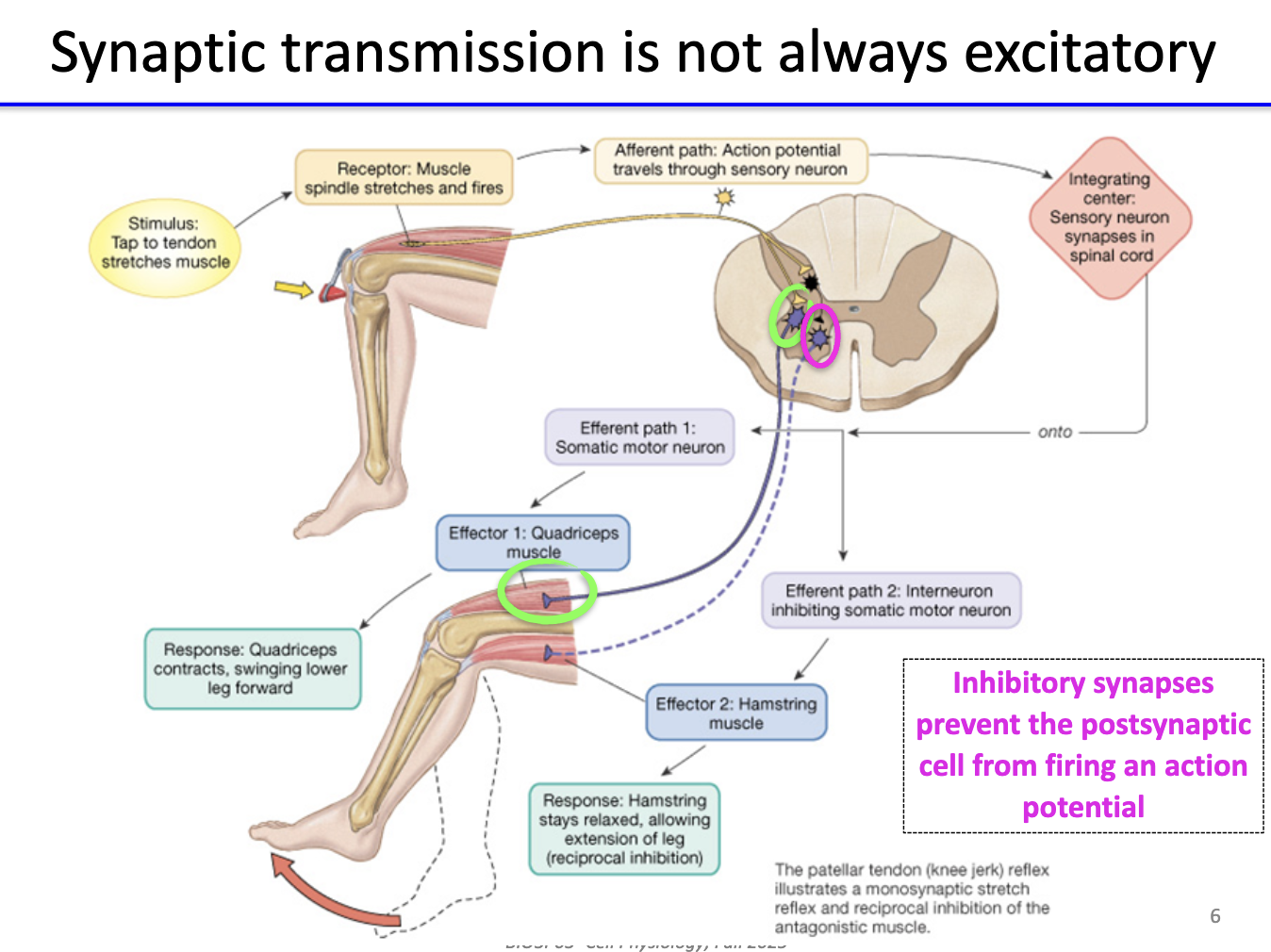

<ul><li><p>Tapping the patellar tendon stretches the quadriceps, activating stretch receptors.</p></li><li><p>These receptors send signals through an afferent neuron to the spinal cord.</p></li><li><p>That afferent neuron does two things:</p><ul><li><p>Excites an efferent motor neuron → contracts the quadriceps → leg kicks forward.</p></li><li><p>Excites an inhibitory interneuron, which hyperpolarizes the efferent motor neuron to the hamstring → hamstring relaxes.</p></li></ul></li><li><p>This spinal reflex happens without involving the brain, enabling fast, automatic movement.</p></li></ul><p></p>
5
New cards

What is the function of inhibitory synapses?

  • Inhibitory synapses prevent the postsynaptic neuron from firing an action potential.

  • They hyperpolarize or stabilize the membrane potential below threshold.


<ul><li><p><strong>Inhibitory synapses</strong> prevent the postsynaptic neuron from firing an <strong>action potential</strong>.</p></li><li><p>They <strong>hyperpolarize</strong> or <strong>stabilize</strong> the membrane potential below threshold.</p></li></ul><p></p>
6
New cards

How can excitatory transmission be studied in the knee-jerk reflex?

  • Each stretch receptor connects to multiple afferent neurons.

  • Stimulating one afferent neuron produces a small EPSP in the postsynaptic motor neuron—not enough for an action potential.

  • Stimulating all afferent neurons together causes their EPSPs to summate, reaching threshold.

  • This summation triggers a full action potential in the motor neuron → muscle contraction.

  • Therefore, the reflex relies on the combined input of many afferent neurons, not just a single one.


<ul><li><p>Each stretch receptor connects to <strong>multiple afferent neurons</strong>.</p></li></ul><ul><li><p>Stimulating <strong>one</strong> afferent neuron produces a small <strong>EPSP</strong> in the postsynaptic motor neuron—not enough for an action potential.</p></li><li><p>Stimulating <strong>all</strong> afferent neurons together causes their EPSPs to <strong>summate</strong>, reaching threshold.</p></li><li><p>This summation triggers a <strong>full action potential</strong> in the motor neuron → <strong>muscle contraction</strong>.</p></li><li><p>Therefore, the reflex relies on the <strong>combined input</strong> of many afferent neurons, not just a single one.</p></li></ul><p></p>
7
New cards

How do inhibitory interneurons ensure hamstring relaxation during the patellar reflex?

  • One inhibitory interneuron firing → small IPSP in the hamstring motor neuron → slight hyperpolarization (not enough to prevent contraction).

  • Stimulating all sensory afferents → strong excitation of the inhibitory interneurons → large IPSP in the hamstring motor neuron.

  • This strong hyperpolarization prevents any action potential, even if excitatory signals arrive.

  • Key point: Inhibitory interneurons themselves receive EPSPs (they must be excited), but they produce IPSPs in the hamstring motor neuron.


<ul><li><p>One inhibitory interneuron firing → small <strong>IPSP</strong> in the hamstring motor neuron → slight hyperpolarization (not enough to prevent contraction).</p></li></ul><ul><li><p>Stimulating <strong>all</strong> sensory afferents → strong excitation of the inhibitory interneurons → large <strong>IPSP</strong> in the hamstring motor neuron.</p></li><li><p>This strong hyperpolarization <strong>prevents any action potential</strong>, even if excitatory signals arrive.</p></li><li><p>Key point: <strong>Inhibitory interneurons themselves receive EPSPs</strong> (they must be excited), but they produce <strong>IPSPs</strong> in the hamstring motor neuron.</p></li></ul><p></p>
8
New cards

What are EPSPs and what causes them?

  • EPSPs: depolarizations that increase likelihood of postsynaptic AP.

  • Caused by neurotransmitter release → binds to postsynaptic receptors → membrane depolarization.

  • Single EPSPs: small (<1 mV), below threshold.

  • Thousands of EPSPs can sum in space & time to reach threshold.

  • Ion flux: likely Na⁺ influx (sometimes Ca²⁺).


<ul><li><p><strong>EPSPs:</strong> depolarizations that <strong>increase likelihood</strong> of postsynaptic AP.</p></li><li><p>Caused by <strong>neurotransmitter release</strong> → binds to postsynaptic <strong>receptors</strong> → <strong>membrane depolarization</strong>.</p></li><li><p>Single EPSPs: <strong>small (&lt;1 mV)</strong>, below threshold.</p></li><li><p><strong>Thousands of EPSPs</strong> can <strong>sum in space &amp; time</strong> to reach threshold.</p></li><li><p><strong>Ion flux:</strong> likely <strong>Na⁺ influx</strong> (sometimes Ca²⁺).</p></li></ul><p></p>
9
New cards

What are IPSPs and how do they affect postsynaptic neurons?

  • IPSPs: hyperpolarizations that decrease likelihood of AP.

  • Can sum with EPSPs → reduce EPSP amplitude.

  • Can reduce probability of reaching threshold.

  • Ion flux: likely Cl⁻ influx or K⁺ efflux.


<ul><li><p><strong>IPSPs:</strong> hyperpolarizations that <strong>decrease likelihood</strong> of AP.</p></li><li><p>Can <strong>sum with EPSPs</strong> → reduce EPSP amplitude.</p></li><li><p>Can <strong>reduce probability of reaching threshold</strong>.</p></li><li><p><strong>Ion flux:</strong> likely <strong>Cl⁻ influx</strong> or <strong>K⁺ efflux</strong>.</p></li></ul><p></p>
10
New cards

How do EPSPs and IPSPs interact to determine if an action potential occurs?

  • Balance between excitatory (EPSP) and inhibitory (IPSP) inputs changes over time.

  • Neurotransmitters bind receptors → open/close ion channels → change conductance → alter current flow.

  • Summation of all inputs determines membrane potential → whether threshold is reached → AP generation.


<ul><li><p>Balance between <strong>excitatory (EPSP)</strong> and <strong>inhibitory (IPSP)</strong> inputs <strong>changes over time</strong>.</p></li><li><p><strong>Neurotransmitters bind receptors</strong> → open/close ion channels → change <strong>conductance</strong> → alter <strong>current flow</strong>.</p></li><li><p><strong>Summation</strong> of all inputs determines <strong>membrane potential</strong> → whether threshold is reached → AP generation.</p></li></ul><p></p>
11
New cards

How do spatial and temporal summation influence action potential generation?

  • Spatial summation: EPSPs from different synapses on dendrite combine → may reach threshold.

  • Temporal summation: repeated EPSPs from same synapse in quick succession overlap → may reach threshold.

  • Real cases: Spatial + temporal summation through same sets of axons


<ul><li><p><strong>Spatial summation:</strong> EPSPs from <strong>different synapses</strong> on dendrite combine → may reach threshold.</p></li><li><p><strong>Temporal summation:</strong> <strong>repeated EPSPs</strong> from same synapse in quick succession overlap → may reach threshold.</p></li><li><p><strong>Real cases: </strong>Spatial + temporal summation through same sets of axons</p></li></ul><p></p>
12
New cards

How do postsynaptic currents spread, and where is integration most effective?

  • Currents spread electrotonically toward axon hillock (spike initiation zone).

  • Potentials decay with distance from synapse.

  • Axon hillock: lower threshold due to high density of voltage-gated Na⁺ channels → key site for AP initiation.

  • If current starts at point A (further) vs point B (closer), A decays more.


<ul><li><p><strong>Currents spread electrotonically</strong> toward <strong>axon hillock</strong> (spike initiation zone).</p></li><li><p>Potentials <strong>decay with distance</strong> from synapse.</p></li><li><p><strong>Axon hillock:</strong> lower threshold due  to <strong>high density of voltage-gated Na⁺ channels</strong> → key site for AP initiation.</p></li><li><p>If current starts at <strong>point A (further)</strong> vs <strong>point B (closer)</strong>, <strong>A decays more</strong>.</p></li></ul><p></p>
13
New cards

How does synaptic integration use action potential frequency to encode signal strength?

  • Action potentials are all-or-nothing, so their amplitude never changes.

  • What does change is frequency: stronger depolarization at the soma/axon hillock → more frequent APs.

  • Weak depolarization just above threshold → low-frequency firing (longer gaps due to relative refractory period).

  • Strong depolarization → high-frequency firing, APs occur as soon as channels recover.

  • Postsynaptic cells interpret frequency, not AP size, as the signal strength (e.g., intense pain = very high firing rate).


<ul><li><p>Action potentials are <strong>all-or-nothing</strong>, so their <strong>amplitude never changes</strong>.</p></li><li><p>What <em>does</em> change is <strong>frequency</strong>: stronger depolarization at the soma/axon hillock → <strong>more frequent APs</strong>.</p></li><li><p>Weak depolarization just above threshold → <strong>low-frequency firing</strong> (longer gaps due to relative refractory period).</p></li><li><p>Strong depolarization → <strong>high-frequency firing</strong>, APs occur as soon as channels recover.</p></li><li><p>Postsynaptic cells interpret <strong>frequency</strong>, not AP size, as the signal strength (e.g., intense pain = very high firing rate).</p></li></ul><p></p>
14
New cards

What happens to firing frequency as depolarization increases?

  • Increased depolarization → more action potentials.

  • Beyond a certain depolarization, firing rate plateaus (absolute refractory period).


<ul><li><p>Increased depolarization → <strong>more action potentials</strong>.</p></li><li><p>Beyond a certain depolarization, <strong>firing rate plateaus</strong> (absolute refractory period).</p></li></ul><p></p>
15
New cards

What happens in a muscle fiber when a single motor neuron fires one action potential?

  • One motor neuron AP → one AP in the skeletal muscle fiber.

  • This produces a single muscle twitch, shown on a myogram.

  • A twitch has two phases:

    • Contraction phase: muscle tension rises.

    • Relaxation phase: tension falls back to baseline.

  • The exact timing varies by muscle type, but the key idea is: one AP → one brief contraction (twitch).


<ul><li><p>One motor neuron AP → one AP in the skeletal muscle fiber.</p></li></ul><ul><li><p>This produces a <strong>single muscle twitch</strong>, shown on a <strong>myogram</strong>.</p></li><li><p>A twitch has two phases:</p><ul><li><p><strong>Contraction phase:</strong> muscle tension rises.</p></li><li><p><strong>Relaxation phase:</strong> tension falls back to baseline.</p></li></ul></li><li><p>The exact timing varies by muscle type, but the key idea is: <strong>one AP → one brief contraction (twitch)</strong>.</p></li></ul><p></p>
16
New cards

What is wave summation and how does it lead to tetanus?

  • Wave summation: repeated stimulation → less relaxation time → stronger contraction.

  • Caused by high AP frequency in motor neuron.

  • Tetanus: sustained contraction (no relaxation) due to rapid stimuli.

  • Useful for sustained tension (e.g., lifting), but can cause cramps if uncontrolled.


<ul><li><p><strong>Wave summation:</strong> repeated stimulation → less relaxation time → stronger contraction.</p></li><li><p>Caused by <strong>high AP frequency</strong> in motor neuron.</p></li><li><p><strong>Tetanus:</strong> sustained contraction (no relaxation) due to <strong>rapid stimuli</strong>.</p></li><li><p>Useful for <strong>sustained tension</strong> (e.g., lifting), but can cause <strong>cramps</strong> if uncontrolled.</p></li></ul><p></p>



