KAAP310 - Exam 3 - Set #2 (urinary system)
 Call Kai
 Learn
 Practice Test
 Spaced Repetition
 Match
 Flashcards
 Knowt Play
1/192

There's no tags or description
Looks like no tags are added yet.
193 Terms
organs of the urinary system
2 kidneys, 2 ureters, urinary bladder, urethra
function of ureters
pass urine from the kidney to the bladder
function of the urinary bladder
store urine
function of urethra
connects bladder to exterior and expels urine
primary function of the kidneys
filter blood and excretion
other functions of the kidney
regulate water output, electrolyte balance, secrete erythropoietin, vitamin D synthesis, clear hormones and drugs, detoxify free radicals, gluconeogenesis
what do the kidneys filter
nitrogenous wastes
what is waste
any substance that is useless to the body or in excess to the body's needs
what is metabolic waste
waste made in the body
what form are metabolic wastes mostly in
nitrogenous wastes
nitrogenous wastes are
highly toxic
1/2 of the waste forms urea and is measured by
blood urea nitrogen (BUN)
azotemia
high BUN and is related to a decease in renal function where urea is not being filtered out
byproduct of urea cycle in protein breakdown
urea
what happens with ammonia
highly toxic and gets converted immediately to less toxic urea
uric acid
breakdown of nucleic acids
creatinine
breakdown of creatine phosphate
excretion definiton
process of separating waste from the body fluids and eliminating them from the body
4 organ systems involved in excretion
respiratory, integumentary, digestive, urinary
what is excreted in the respiratory system
CO2 and H2O
what is excreted in the integumentary system
sweat (H2O, salts, urea, lactate)
what is excreted in the digestive system
H2O, salts, CO2, lipids, cholesterol, bile pigments, metabolic wastes
what is excreted in the urinary system
metabolic wastes, toxins, drugs, hormones, salts, H20, H+
3 layers that protect the kidney (outermost to inner)
fibrous renal fascia, perirenal fat capsule, fibrous capsule
fibrous renal fascia
attaches to other organs, helps hold kidneys in place
perirenal fat capsule
provides cushioning (adipose tissue)
fibrous capsule
enclose kidney to protect from trauma and infection
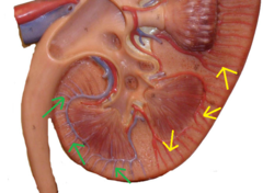
look at slide 53 for where parts of the kidney are
renal cortex
contains parenchyma where nephrons are found

renal medulla
separated into pyramids and contains part of the nephron where urine goes to be collected

minor calyx
collect urine from associated pyramid

major calyx
where 2-3 minors converge to dump into sinus that goes to ureter

renal column
between pyramids, separates medulla into 6-10 pyramids

cortical radiate artery
90 degree angle to arcuate artery, permeate through cortex and branch
interlobar artery
segmental arteries separate, go through renal columns

segmental artery
move blood from renal artery to interlobar artery

arcuate artery and vein
wrap around wide portion of pyramids

renal circulation steps

nephron
functional unit of kidney
step 1 of renal circulation
afferent arteriole into glomerulus
step 2 of renal circulation
whole blood is filtered at the glomerulus
step 3 of renal circulation
filtered blood comes out of the efferent arteriole
step 4 of renal circulation
blood moves towards the renal tubule and forms capillary bed
step 5 of renal circulation
peritubular capillaries around renal tubule moves filtrate back into blood to return reabsorbed filtrate items to blood
efferent arteriole becomes
vasa recta
where most blood flow goes
into the cortex where the parenchyma are
percentage of blood that goes to the medulla
1-2% because the vasa recta are very small
what makes up the corticomedullary junction
arcuate artery and vein
type of nephron that is 85% of nephrons
cortical nephron
juxtamedullary nephron
15%, next to medulla and is responsible for maintaining osmotic gradient of medulla to conserve water
nephron make up
renal corpuscle and renal tubule
renal corpuscle
glomerulus surrounded by capsule

why the afferent arteriole is larger
more blood enters then leaving
parietal layer of glomerular capsule
outside layer, simple squamous
visceral layer of glomerular capsule
inner layer
Podocytes of visceral layer
wrap around arteries and filter the blood that runs through them and retain large molecules (ex. proteins)
urinary pole
filtrate foes to renal tubule to eventually become urine

mesangial cells
pack space around glomerular capillaries and support capillaries to help maintain blood flow and clean
mesangial cells are similar to
macrophages
4 regions of the renal tubule
PCT, Nephron loop, DCT, collecting duct

each nephron will have their own
PCT, nephron loop, and DCT
multiple nephrons share
collecting ducts
Proximal Convoluted Tubule (PCT)
Aries from renal corpuscle, most coiled, simple cuboidal with brush border

what is the PCT good at
reabsorption due to increased surface area
where the nephron loop is mostly found
medulla

thick segments of nephron loop location
1st part of descending limb and most of ascending limb,
thick segments nephron loop purpose
used for active transport of salts and has a high metabolic activity due to lots of mitochondria
thin segments of nephron loop location
ladder portion of descending limb made of simple squamous
thin segments of nephron loop purpose
have low metabolic activity which is highly permeable to water and good for water reabsorption
distal convoluted tubule (DCT)
less coiled, re enters cortex, no microvilli, passageway that moves filtrate to collecting duct

collecting duct make up
from multiple nephrons, numerous ducts converge at tip of papilla, drain into minor calyx, simple cuboidal
renal plexus defined
receives sympathies innervation from superior mesenteric and celiac ganglions
what the renal plexus does
decrease urine production and glomerular blood flow and stimulus release of renin to increase blood pressure
parasympathetic input from vagus nerve
no known function
4 stages from blood to urine
glomerular filtration, tubular reabsorption, tubular secretion, water conservation
what happens in glomerular filtration
create a plasma like filtrate in the blood that goes into the nephron
purpose of barriers in glomerular filtration membrane
prevents all protein from leaving the blood and small molecules get filtered out
fenestrated endothelia of capillary (barrier 1)
has large filtration pores that are highly permeable to small molecules
basement membrane (barrier 2)
made of proteoglycan gel that excludes molecules larger than 8nm
charge of basement membrane and what that does
negatively charged, repels anions that have a negative charge even if they are under 8nm
filtration slits (barrier 3)
within pedicles of podocytes and are negatively charged
size of molecules that can pass freely in glomerular filtration
less than 3nm
protein percentage in plasma
7%
protein percentage in filtrate
0.03%
proteinuria/albuminuria/hematuria
protein (typically albumin) in the urine, sign of kidney dysfunction
blood hydrostatic pressure in glomerulus
extremely high - 60mmHg out
colloid osmotic pressure in glomerulus
-32 mmHg in
capsular pressure in glomerulus
-18 mmHg in
net flitration pressyre in glomerulus
10 mmHg out
percentage of filtrate reabsorbed
99%
urine produced daily
1-2 L/day
why can't you rid of all filtrate
you would be in the bathroom all day
glomerular filtration rate equation
GFR = NFP x Kf
how to adjust glomerular filtration rate
change the glomerular blood pressure
3 mechanisms for changing glomerular filtration rate
renal auto regulation, sympathetic control, hormonal control
renal auto regulation
nephrons adjust own blood flow without external input
myogenic mechanism
glomerulus receives feedback on status of downstream fluid and rely on juxtaglomerular apparatus
what is the glomerular apparatus
next to the glomerulus at the very end of nephron loop as it re enters the cortex affecting the afferent arteriole
how renal auto regulation starts
cells of macula dense
tubuloglomerular feedback
glomerulus receives feedback on the status of downstream tubular fluid and adjusts filtration rate accordingly