Anatomy Exam 3 - Brain and Cranial Nerves
 Call Kai
 Learn
 Practice Test
 Spaced Repetition
 Match
 Flashcards
 Knowt Play
1/70

There's no tags or description
Looks like no tags are added yet.
71 Terms
neural tube
develops into the central nervous system (CNS) which consists of the brain and spinal cord
rostral vs. caudal neural tube
rostral neural tube: anterior end that develops into the brain (toward the head)
caudal neural tube: posterior end that develops into the spinal cord (toward the tail)
4 major parts of the brain
1) cerebrum
2) cerebellum
3) diencephalon
4) brainstem
cerebrum
largest part of the brain
responsible for conscious thought, memory, sensory perception, language, and voluntary movement
divided into left and right cerebral hemispheres

cerebellum
located posteriorly and inferiorly, behind the brainstem
coordinates balance, posture, and smooth, accurate movements
helps with motor learning

diencephalon
located between the cerebrum and brainstem
includes the thalamus and hypothalamus
processes sensory information and regulates homeostasis

brainstem
connects the brain with the spinal cord
includes the midbrain, pons, and medulla oblongata
controls the essential function such as breathing, heart rate, and alertness

meninges
connected tissue membranes that protect the skull
3 meninges of the skull
1) dura mater: outermost, tough protective membrane
2) arachnoid mater: middle, web-like membrane
3) pia mater: innermost, delicate membrane that closely follows the brain’s surface
meningeal spaces
1) epidural space
2) subdural space
3) subarachnoid space
epidural space
between the skull and dura mater
can form when blood separates the dura from the skull
subdural space
between the dura and arachnoid
can form when blood separates these membranes
subarachnoid space
between arachnoid and pia mater
contains cerebrospinal fluid (CSF) and blood vessels
choroid plexus
produces cerebrospinal fluid (CSF)
consists of blood capillaries and ependymal glial cells that form a specialized barrier + secrete CSF
cerebrospinal fluid (CSF)
cushions the brain and spinal cord, provides buoyancy, helps maintain a stable chemical environment, and transports waste products
brain ventricles
interconnected cavities within the brain that contain CSF
CSF flow pathway
1) choroid plexus produces CSF
2) lateral ventricles
3) interventricular foramina
4) third ventricle
5) cerebral aqueduct
6) fourth ventricle
7) openings into the subarachnoid space
8) subarachnoid space around the brain and spinal cord
9) arachnoid villi → dural venous sinuses → blood
the choroid plexus is found in the lateral, third and fourth ventricles; amount of CSF increases as it travels down

arachnoid villi (granulations)
structures that allow CSF to be reabsorbed into the venous blood

dural venous sinuses
channels b/w layers of dura mater that collect venous blood and receive reabsorbed CSF
cerebral hemispheres
left and right halves of the cerebrum

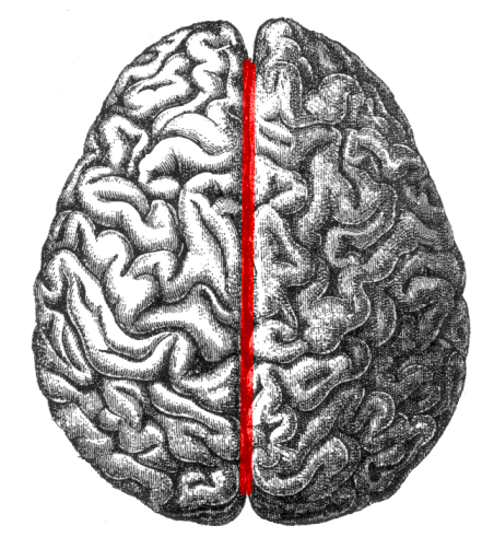
longitudinal fissure
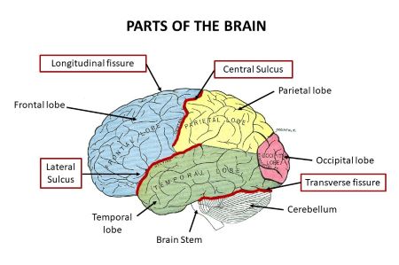
deep groove separating the right and left hemispheres
transverse fissure
separates the cerebrum from the cerebellum
sulcus (plural sulci)
groove in the brain’s surface
gyrus (plural gyri)
raised ridge b/w sulci
cerebral cortex
outer layer of gray matter of the cerebrum
folds of the cerebrum increase the surface area available for neurons
central sulcus
separates the frontal lobe from the parietal lobe

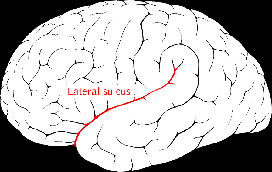
lateral sulcus
separates the temporal lobe from the frontal + parietal lobes
precentral gyrus
located immediately anterior to the central sulcus; contains the primary motor cortex

postcentral gyrus
located immediately posterior to the central sulcus; contains the primary somatosensory cortex

gray vs white matter
gray matter:
contains neuron cell bodies, dendrites, and synapses
processes/integrates information
forms the cerebral cortex + deeper nuclei
white matter:
contains primarily myelinated axons
carries information b/w regions
forms tracts connecting different CNS regions
4 cerebral lobes
1) frontal lobe
2) parietal lobe
3) temporal lobe
4) occipital lobe
frontal lobe
voluntary motor control
planning, decision-making, judgment, and personality
speech production in the dominant hemisphere (Broca’s area)
contains the primary motor cortex

parietal lobe
processes touch, pressure, pain, temperature, and body position
contains the primary somatosensory cortex
helps with spatial awareness

temporal lobe
hearing and auditory processing
memory and aspects of emotion
language comprehension in the dominant hemisphere (often Wernicke’s area)

occipital lobe
primarily responsible for visual processing
contains the primary visual cortex

motor homunculus vs. sensory homunculus
homunculus: maps of the body represented across the cerebral cortex
motor - represents control of voluntary movement
sensory - represents bodily sensations
body parts requiring detailed sensation + precise movements (hands, lips, face) receive more cortical representation

3 types of white matter fibers
1) commissural fibers
2) association fibers
3) projection fibers
commissural fibers
connect the left and right cerebral hemispheres
ex: corpus callosum

association fibers
connect different areas within the same hemisphere
ex: fibers connecting the frontal and parietal cortical areas

projection fibers
connect the cerebral cortex w/ lower CNS structures, including the brainstem and spinal cord
ex: internal capsule fibers

major limbic structures
1) cingulate gyrus
2) hippocampus
3) amygdala
cingulate gyrus
curves along the medial surface of the cerebral hemisphere
involved in emotion, motivation, and attention

hippocampus
located in the medial temporal lobe
important for forming new memories + spacial memory

amygdala
located in the medial temporal lobe, near the hippocampus
helps process emotional significance (fear and threat)

primary functions of the limbic system
involved in emotion and emotional responses, learning and memory, motivation and reward, and linking emotions w/ memories and behavior
functions of the cerebellum
coordinating voluntary movement
maintaining balance and posture
regulating muscle tone
improving the timing and accuracy of movements
supporting motor learning

thalamus vs. hypothalamus
thalamus: major relay and processing station for most sensory information traveling to the cerebral cortex; also participates in motor and attention circuits
hypothalamus: regulates homeostasis, autonomic activity, endocrine function, body temperature, hunger, thirst, and circadian rhythms

brainstem components (rostral to caudal)
1) midbrain
2) pons
3) medulla oblongata

midbrain
involved in visual and auditory reflexes
helps control eye movements
participates in motor control
pons
relays information between the cerebrum and cerebellum
participates in breathing regulation
contains nuclei associated with several cranial nerves
medulla oblongata
regulates vital functions (heart rate, blood pressure, breathing)
contains reflex centers for swallowing, coughing, and sneezing
connects w/ spinal cord
reticular formation
network of neurons extending through the brainstem
maintains alertness and wakefulness, regulates the sleep-wake cycle, helps control attention and consciousness, filters incoming sensory information, contributes to muscle tone and autonomic regulation
RAS (reticular activating system) is especially important for arousal + maintaining consciousness
cranial nerves
12 pairs
originates in the brain or brainstem
distributes to the head and neck, with CN X extending to thoracic/abdominal organs
can function as sensory, motor, autonomic, or mixed
spinal nerves
31 pairs
originates in the spinal cord
distributes to trunk and limbs + some internal structures
all 31 spinal nerves are mixed nerves (carry sensory + motor fibers)
motor nerve
carries commands to skeletal muscle
autonomic nerve
carries parasympathetic signals to glands, smooth muscle, or cardiac muscle
general sensory nerve
carries touch, pain, temperature, and other general sensations
special sensory nerve
carries specialized senses such as smell, vision, hearing, balance, or taste
CN I
olfactory nerve - sensory
function: smell
modality: special sensory
CN II
optic nerve - sensory
function: vision
modality: special sensory
CN III
oculomotor nerve - motor + autonomic
function: moves most eye muscles, elevates the upper eyelid, constricts the pupil, and helps the lens accommodate for near vision
modality: motor and parasympathetic
CN IV
trochlear nerve - motor
function: controls the superior oblique eye muscle, which helps direct the eye downward when it is turned inward
modality: motor
CN V
trigeminal nerve - mixed
function: facial sensation and chewing
modality: general sensory and motor
3 branches: ophthalmic, maxillary, and mandibular
CN VI
abducens nerve - motor
function: controls the lateral rectus muscle, moving the eye laterally (abduction)
modality: motor
CN VII
facial nerve - mixed
function: facial expressions, taste, parasympathetic control of the lacrimal and several salivary glands
modality: motor, special sensory (taste), and parasympathetic
CN VIII
vestibulocochlear nerve - sensory
function: hearing and balance
modality: special sensory
CN IX
glossopharyngeal nerve - mixed
function: taste/sensation of tongue, swallowing, parasympathetic stimulation of the parotid salivary gland
monitors blood pressure through sensory receptors
modality: sensory, motor, parasympathetic
CN X
vagus nerve - mixed
function: parasympathetic regulation of thoracic + abdominal organs; contributes to swallowing + voice production
modality: sensory, motor, parasympathetic
CN XI
accessory nerve - motor
function: controls the sternocleidomastoid and trapezius muscles
actions: head rotation and shoulder elevation
modality: motor
CN XII
hypoglossal nerve - motor
function: controls most tongue muscles involves in tongue movement
modality: motor
cranial nerve mnemonic
Old - olfactory
Opie - optic
ocassionally - oculomotor
tries - trochlear
trigonometry - trigeminal
and - abducens
feels - facial
very - vestibulocochlear
gloomy, - glossopharyngeal
vague, - vagus
and - accessory
hypoactive - hypoglossal