hypothalamic - pituitary axis
 Call Kai
 Learn
 Practice Test
 Spaced Repetition
 Match
 Flashcards
 Knowt Play
1/51

There's no tags or description
Looks like no tags are added yet.
52 Terms
describe the endocrine system
key regulatory system - made up of glands and other tissues to produce and release hormones
regulates longer term, ongoing processes throughout the body including metabolism, growth and reproduction
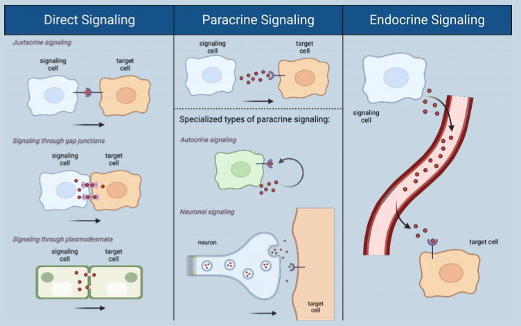
two ways of hormone secretion
hormone secretion by endocrine cells
hormone secretion by neurosecretory neurones
pure endocrine glands
pituitary
pineal
thyroid
adrenal
organs containing endocrine cells
pancreas
thymus
gonads
hypothalamus
what are endocrine cells
glandular secretory cells that release hormones directly into the blood, lymphoid system or interstitial fluids
production of hormones and synthesis/secretion
each hormone produced by specific gland and acts through specific receptors on target organs
synthesis, secretion and storage of hormones is dependent on feedback mechanisms
4 classes of hormones (based on chemical structure)
amino acid derivatives
peptide and protein hormones
steroid hormones
fatty acid compounds
describe amino acid derivatives
derivatives of tyrosine
thyroid hormones and catecholamines (dopamine, norepinephrine, epinephrine)

describe steroid hormones
derived from cholesterol
not stored - released as synthesised (receptors inside cell)
reproductive organs, adrenal cortex, placenta

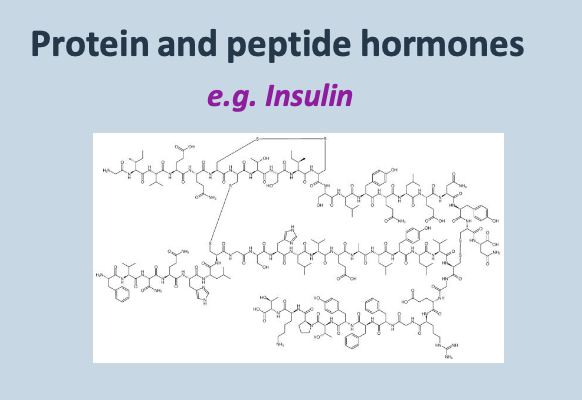
describe protein and peptide hormones
chains of amino acids - the largest group
formed as preprohormones, cleaved to prohormones and then active hormones
proteins (insulin) - all pituitary hormones
describe fatty acid compounds
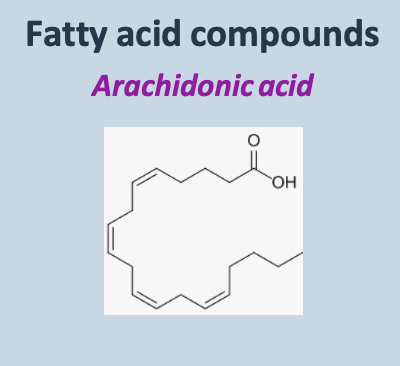
derived from arachidonic acid
eicosanoids (prostaglandins and leukotrienes)
coordinate cellular activities and affect enzymatic activity
what is endocrinopathy
disease of endocrine glands/tissues associated with imbalances in hormone concentrations
treatment - using replacement hormones or drugs which prevent production or action of hormones
examples of endocrinopathies in animals
pituitary dwarfism (secrete too little growth hormones in young animals)
hypo/hyperthyroidism (secrete too little/ much thyroid hormone)
hypo/hyperparathyroidism (secrete too little/much parathyroid hormone)
diabetes insipidus (too little antidiuretic hormone)
diabetes mellitus (too little insulin/ insulin resistance)
insulinoma (tumour secreting too much insulin)
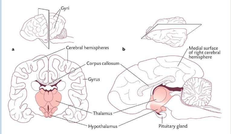
describe the hypothalamus
located at the base of the brain central to the thalamus
plays a role in maintaining body’s internal balance by linking nervous and endocrine system
produces peptides and amines that influence the pituitary gland
describe the pituitary (hypophysis)
located in a small pocket in the sphenoid bone of the skull in base of braincase
linked to hypothalamus by tissue called infundibulum
consists of anterior lobe (adenohypophysis), intermediate lobe (melanocyte stimulating factor), posterior lobe (neurohypophysis - oxytocin and ADH)
describe the lobes of the pituitary
posterior lobe - extension of the hypothalamus and part of the CNS - ADH and oxytocin are released from axon terminals
anterior and intermediate lobes are epithelial tissue and contain endocrine cells
neural and vascular communications between hypothalamus and pituitary gland
Neural communication with the posterior pituitary - ADH and oxytocin are produced in hypothalamic neuroendocrine cells - travel down the axons to the posterior pituitary, where they are stored in nerve endings and released directly into its capillary bed
Vascular communication with the anterior pituitary - Hypothalamic releasing and inhibiting hormones enter capillaries and travel through the hypothalamic-pituitary portal system to a second capillary bed in the anterior pituitary. They regulate the endocrine cells, which then release anterior pituitary hormones into the systemic circulation

describe the hypothalamic pituitary portal system
posterior pituitary - ADH and oxytocin are produced in the neuroendocrine cells in hypothalamus - transported down axons which extend into posterior pituitary - stored in nerve endings and released directly into capillary bed of posterior pituitary
anterior/intermediate pituitary - releasing/inhibiting hormones are produced in neuroendocrine cells of hypothalamus and released into portal vessels surrounding endocrine cells of anterior and intermediate lobes - hormones bind to receptors on pituitary endocrine cells and release hormones onto 2nd capillary bed

describe how the hypothalamic pituitary portal system works
release of hormones controlled by neural inputs from CNS
direct sensing of blood composition
local neurotransmitter and peptide signalling within the hypothalamus
negative feedback by target gland hormones

describe feedback control of hormones
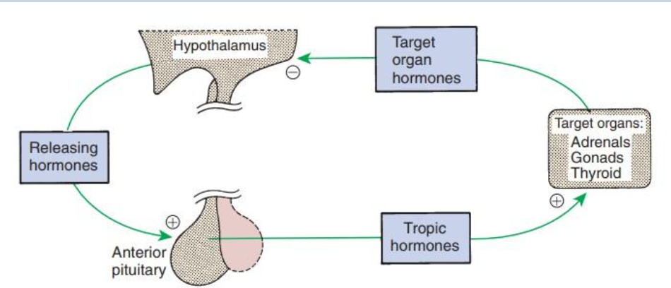
function of posterior pituitary function
releases ADH (water balance) and oxytocin (contraction of smooth muscle cells in uterus and mammary glands)
ADH and oxytocin are produced in neurosecretory neurons with the cell bodies residing within the hypothalamus in two defined nuclei (supraoptic nucleus and paraventricular nucleus)

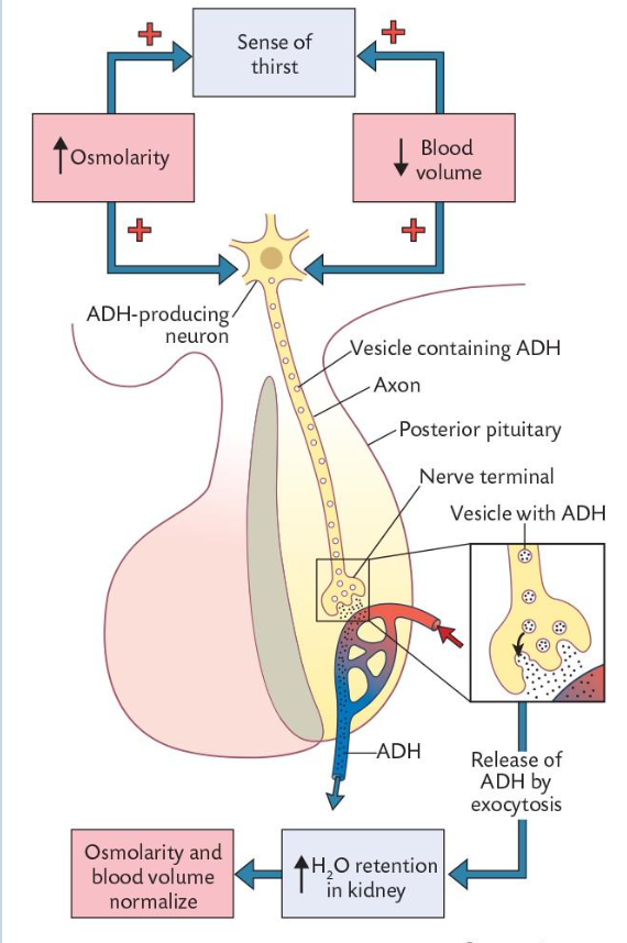
describe feedback mechanism of anti diuretic hormone (vasopressin)
negative feedback mechanism
hypothalamus detects decrease in blood volume and increase in osmolarity
stimulates ADH producing neurones in the hypothalamus
ADH released into blood and acts on tubular cells of collecting ducts in kidney
when ADH concentrations are high, more aquaporins are incorporated into luminal membranes of the tubular cells so more water absorbed in collecting ducts
blood volume increases and osmolarity decreases
ADH production decreases
lower ADH concentration in blood = fewer aquaporins and less water reabsorption (diuresis)
function of anti diuretic hormone
regulation of water permeability of renal distal tubules and collecting ducts (via insertion of aquaporin channels)
causes contraction of arteriolar smooth muscles
extracellular osmolarity and extracellular fluid volume are primarily regulators of ADH secretion
water deprivation = increases osmolarity of plasma
hyperosmolarity - induces loss of intracellular water from osmoreceptors in hypothalamus > release of ADH
water ingestion supresses firing of osmoreceptors
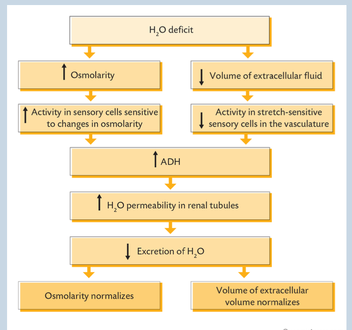
anti diuretic hormone linked to blood pressure/ volume - hypovolaemia
ADH is also released when there is a decrease in blood volume/pressure
hypovolaemia detected by pressure sensors
carotid and aortic baroreceptors and stretch receptors in the walls of the left atrium and pulmonary veins
impulse carried to supraoptic and paraventricular nuclei oh hypothalamus - normally inhibits ADH release (drop in 15-25% in blood volume inhibits impulse and ADH is released)
describe alterations in ADH
deficiencies - central diabetes insipidus (decreases ADH from posterior pituitary caused by head trauma, infections, tumour) & nephrogenic diabetes insipidus - normal ADH but kidneys unresponsive
excess - syndrome of inappropriate ADH secretion - continued secretion - caused by CNS damage, certain drugs or infections (hyponatraemia)
what does the supraoptic and paraventricular nucleus secrete
supraoptic - secrete oxytocin
paraventricular - secrete ADH
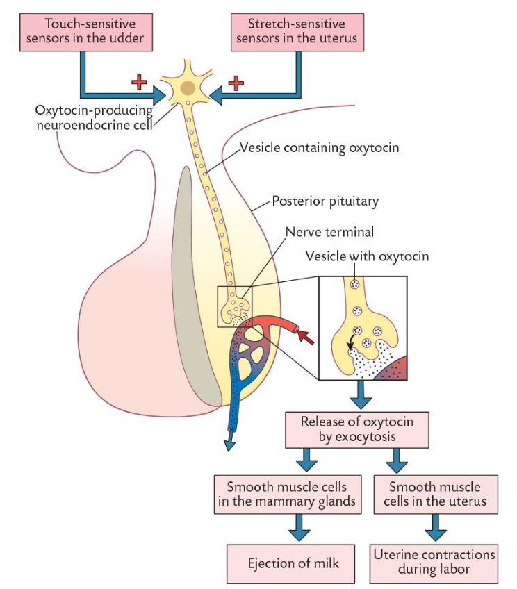
describe oxytocin feedback mechanism
positive feedback mechanism
essential for uterine contractions during parturition
contraction of myoepithelial cells (epithelial cells which have contraction ability) in mammary alveoli for milk let down (release milk from glands)
hypothalamus detects stimulation of sensors in mammary gland or uterus - suckling
neuroendocrine cells within paraventricular and supraoptic nuclei produce and release oxytocin by exocytosis
act to contract smooth muscle cells
describe anterior pituitary function
controlled by releasing and inhibiting hormones from hypothalamus
neurohormones are produced in the hypothalamus and secreted within it
hormones are transported to anterior pituitary via tiny blood vessels and hypothalamic pituitary portal system
in anterior pituitary the neurohormones bind to receptors on endocrine cells, controlling secretion of hormones from anterior lobe

regulation of anterior pituitary hormone secretion
releasing/inhibiting hormones produced by neurosecretory neurones in the hypothalamus enter the hypothalamic capillaries
hypothalamic capillaries rejoin to form the hypothalamic-hypophyseal portal system (vascular link to anterior pituitary)
portal system branches into capillaries of anterior pituitary
releasing/inhibiting hormones leave blood across anterior pituitary capillaries, control the release of anterior pituitary hormones
when stimulated, anterior pituitary secretes a given hormone into capillaries
anterior pituitary capillaries rejoin to form a vein, anterior pituitary hormones leave for ultimate distribution throughout the body by systemic circulation

describe hypophysiotropic hormones (inhibiting/releasing hormones)
secretion regulated by one or more hypothalamic hypophysiotrophic hormones
small peptide hormones
either releasing (increase secretion) or inhibiting (decrease secretion)
named based on pituitary hormones they regulate
what are the anterior pituitary hormones
gonadotrophs (FSH and LH)
corticotrophs (ACTH)
thyrotrophs (TSH)
somatotrophs (GH)
lactotrophs (PRL)

function of ACTH/ CRH
stimulates secretion of ACTH
function of TRH releasing hormone
stimulates secretion of TSH and prolactin
function of GHRH
GH releasing hormone - stimulates secretion of growth hormone
function of GHIH (somatostatin)
inhibits secretion of growth hormone and TSH
describe GnRH
gonadotropin releasing hormone - stimulates secretion of LH and FSH
PRL releasing hormone function (PRLRH)
stimulates secretion of prolactin
function of dopamine/ PRL inhibiting hormone (PRLIH)
inhibit secretion of prolactin
describe different feedback pathways
long loop feedback - hormones released from peripheral endocrine gland provides feedback to pituitary or hypothalamus
short loop feedback - hormone from pituitary provides feedback to hypothalamus
ultra-short feedback - hormone released by endocrine directly provides feedback to endocrine organ of origin
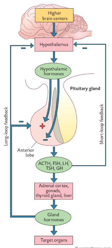
describe function of growth hormone
peptide hormone (191 amino acids)
most abundant hormone
age dependent secretion
lipid and glucose metabolism
stimulates chondrocyte differentiation and elongation of bones
reduced concentration of amino acids in plasma

describe the mediation of growth potential of GH
indirectly stimulated - requires another hormone (thyroid stimulating hormone)
Gh stimulates release of peptide hormone that is structurally similar to insulin called IGF-1 (aka somatomedins) - released into blood on stimulation by GH
liver is major producer of IGF-1
describe the control of growth hormone secretion
regulated by two hypothalamic - hypophysiotrophic hormones
GHRH - releasing - stimulates synthesis and secretion of GH
GHIH - inhibiting - somatostation - inhibits Gh secretion
factors that stimulate GH secretion
GHRH
decreased blood glucose
decreased free fatty acids
increased amino acids
starvation or fasting
trauma/stress
exercise
sex hormones
deep sleep
ghrelin (hunger hormone)
factors that inhibit GH secretion
GHIH
increased blood glucose
increased free fatty acids
decreased amino acids
obesity
IGF
exogenous GH
alterations in GH secretion
deficiencies - hypothalamic dysfunction - lack of GHRH
pituitary defect - defect in production of GH
failure of targets cells to respond normally - receptor mutation
excess - tumour of GH, defect in mechanism regulating GH secretion
over production - after growth plates have fused, thicker bones, skin and connective tissue proliferation
describe function of prolactin (PRL)
peptide hormone (198 amino acids)
originates from anterior pituitary
stimulates development of mammary gland and milk production
involved in reproductive function
regulation of secretion in controlled by PRH and PIH (dopamine)
stimulated by oestradiol, stretch receptors in vagina and cervix
effects of prolactin
before and after sexual maturity - stimulates proliferation and branching of ducts in mammary epithelial tissue
during pregnancy - cause development of lobules of alveoli where milk is produced
after parturition - stimulates milk synthesis
reproduction - excess PRL blocks synthesis and release of GnRH - role of gonadal hormones reduced
alterations in prolactin secretion
deficiencies - caused by destruction of anterior pituitary
excess - caused by hypothalamic dysfunction or pituitary tumours
signs - infertility, decreased testosterone - decreased sperm production, decreased libido
describe intermediate lobe function
regulates camouflague in fish, amphibians and reptiles
melanotropes produce MSH
MSG scatters melanin granules and concentrates light reflecting particles called iridophores around nucleus of melanocytes
increased MSG can result in darker pigmentation
structurally similar in ACTH - excessive production of ACTH can also caused hyperpigmentation (addinsons disease)
describe pituitary pars intermedia dysfunction (cushings disease)
melanotrope precursor protein can be cleaved to form corticotropin-like intermediate lobe protein (cushingoid type disease)
signs of equine cushings - laminitis, weightloss, ulcers in mouth, excessive thirst, excessive urination, impaired wound healing and susceptibility to infections
posterior pituitary hormones
ADH and OT

anterior pituitary hormones
GnRH (LH/FSH)
TRH
PRH
GHRH
CRH
