3. Assessment of nonstrabismic binocular vision
 Call Kai
 Learn
Learn Practice Test
 Spaced Repetition
 Match
Match Flashcards
 Knowt Play
1/78

There's no tags or description
Looks like no tags are added yet.
Name | Mastery | Learn | Test | Matching | Spaced | Call with Kai | Chat |
|---|
No analytics yet
Send a link to your students to track their progress
79 Terms
What major functions are evaluated during an assessment of binocular vision?
Ocular alignment
Versions
Phoria testing, which is required for the sensorimotor examination
Fixation disparity and associated phoria
AC/A relationship
Fusional ranges and convergence amplitude
Vergence facility
Sensory integration
Stereoacuity
Suppression
How are versions tested and recorded?
Hold a penlight or transilluminator at approximately 30 cm
Move the target through each extraocular muscle’s diagnostic action field
Observe:
Binocular eye movements and corneal reflexes
Overaction (OA) or underaction (UA)
Eyelid changes or globe retraction
A- or V-patterns
Lift the eyelids in downgaze if necessary to see the eyes fully
Record:
OA as positive
UA as negative
Grade from 1 to 4, with larger numbers indicating a greater deviation from normal
If an OA or UA is detected, follow up by assessing monocular ductions
What additional abnormalities should be observed during versions, and how is an A- or V-pattern identified?
Look for:
Globe retraction
Narrowing or widening of the palpebral aperture
Eyelid retraction or other lid changes
An A- or V-pattern, indicated by an approximately 10 to 15 prism-diopter change in horizontal alignment between upgaze and downgaze
What is the difference between a muscle’s diagnostic action field and its muscle action?
Diagnostic action field, or DAF:
The position of gaze in which a particular extraocular muscle is the primary mover of the eye. Versions use these gaze positions to isolate and compare yoked muscles.Muscle action:
The rotational effect produced when the muscle contracts while the eye starts in primary position.
Key distinction: The DAF tells you where to test the muscle; the muscle actions describe what the muscle does to the eye.
Which muscles are primarily evaluated in each diagnostic position during versions?
Right gaze: right LR and left MR
Left gaze: right MR and left LR
From right gaze:
Up and right: right SR + left IO
Down and right: right IR + left SO
From left gaze:
Up and left: right IO + left SR
Down and left: right SO + left IR
What are the primary, secondary, and tertiary actions of the six extraocular muscles from primary position?
Medial rectus: adduction
Lateral rectus: abduction
Superior rectus: elevation, intorsion, adduction
Inferior rectus: depression, extorsion, adduction
Superior oblique: intorsion, depression, abduction
Inferior oblique: extorsion, elevation, abduction
What information should be obtained when measuring a phoria?
Direction: exophoria, esophoria, or vertical phoria
Magnitude: measured in prism diopters
Viewing distance: distance and near
Comitancy: whether the deviation remains similar across different gaze positions and with either eye fixating
How is phoria comitancy evaluated, and what finding indicates an incomitant deviation?
Measure ocular alignment in more than one gaze position and compare measurements with a change in:
Direction of gaze
Fixating eye
A deviation is considered incomitant if the magnitude changes by 5 prism diopters or more with a change in gaze or fixating eye.
Why must accommodation be controlled during cover testing, and how does underaccommodation affect the measured phoria?
Accommodation stimulates accommodative convergence, so inaccurate or inconsistent accommodation changes ocular alignment.
Underaccommodation causes:
Increased exophoria, or
Decreased esophoria
Therefore, perform the cover test using the patient’s best optical correction and an accommodative target that keeps the patient actively engaged.
How should an accommodative cover-test target be selected for children versus adults and school-age children?
For younger children: Use an interesting, detailed target and keep the child engaged by:
Asking the child to identify or describe details
Having the child read when possible
Changing or moving the target
Using a sticker or other attention-maintaining target
For adults: Use approximately a 20/30 letter target to stimulate and maintain accommodation.
Key principle: Simply looking toward the target is insufficient. The patient must remain visually engaged to prevent underaccommodation and an artificially exo-shifted phoria result.
How is the unilateral cover test performed, and what does it determine?
First assess fixation stability
Cover one eye for 2 to 3 seconds
Observe the uncovered eye for a refixation movement
Repeat on the opposite eye
The unilateral cover test, or cover-uncover test, determines whether a manifest deviation, or strabismus, is present.
If strabismus is present, document:
Frequency: constant or intermittent
Laterality: unilateral or alternating
Direction
Magnitude
How is the magnitude of a manifest deviation measured without dissociating an underlying phoria?
Use the simultaneous prism cover test (SPCT):
Place prism before the deviating eye while simultaneously covering the fellow eye
Increase prism until the refixation movement is neutralized
Because the prism and cover are introduced simultaneously, the test measures the manifest tropia while minimizing dissociation
How is the alternate cover test performed, and what does it measure?
Alternate the cover between the two eyes, using 1 to 2 second cover strokes
Do not allow binocular fusion between cover movements
Estimate the deviation, then add prism until movement is neutralized
Continue alternating for approximately 20 seconds, allowing the phoria to reach a stable dissociated position
The endpoint is a consistent reversal of movement around neutral
What is “high neutral” during prism-neutralized alternate cover testing?
High neutral is the highest prism value that produces no observable movement before the direction of movement reverses.
Why should the unilateral cover test be repeated after alternate cover testing in a patient with a large phoria?
Alternate cover testing strongly dissociates fusion. A large phoria may temporarily break down into a manifest deviation after prolonged dissociation.
Repeating the unilateral cover test determines whether the patient can:
Re-establish binocular alignment, or
Remain manifestly deviated after dissociation
This provides information about the patient’s control of the deviation.
How is the Modified Thorington test performed, and what are its major advantages and limitation?
Test with the patient’s best correction
Place a Maddox rod over the right eye
Orient the rod with horizontal striations, producing a vertical streak
Center a penlight through the hole in the card
Ask where the streak crosses the numbered scale
Use the appropriate card for:
Distance: 10 ft
Near: 16 in
Use an accommodative card or target to stimulate accommodation
Advantages:
Direct measurement of horizontal phoria
Repeatable
Can measure both distance and near phoria
Limitation:
The examiner cannot observe the eyes as well as during cover testing, so the result depends more heavily on the patient’s subjective response.
What testing considerations and limitations are important during von Graefe phoria measurement?
Keep the target clear at all times to maintain accommodation.
The examiner cannot observe the eyes as well as during cover testing.
The technique can have greater variability, reduced repeatability, and increased testing time.
Increasing target stability may improve the reliability of the response.

How is the double Maddox rod test performed to measure cyclodeviation?
Place a vertically oriented Maddox rod before each eye in a straight trial frame.
The rods may be the same color or different colors, such as red, white, or green, to help distinguish each eye’s line.
Use a distant light as the target.
A small vertical prism may be used to separate the two horizontal lines.
Ask whether the two lines are horizontal and parallel.
Have the patient rotate one or both rods until both lines are:
Parallel to each other
Parallel to the horizon, like railroad tracks
Record the amount and direction of rotation in degrees.
How is incyclodeviation versus excyclodeviation identified when recording the double Maddox rod test?
Observe the direction in which the top of the Maddox rod must be rotated to make the perceived line horizontal:
Top rotated inward toward the nose → incyclodeviation
Top rotated outward toward the temple → excyclodeviation
What does the double Maddox rod test measure, and how does subjective torsion differ from objective torsion?
The double Maddox rod test measures subjective torsion, meaning the patient’s perceived orientation of the images.
The test is particularly useful for:
Torsion occurring after visual maturity
Evaluating cyclodeviation based on the patient’s visual perception
Objective torsion is assessed by directly observing or documenting ocular rotation using:
Binocular indirect ophthalmoscopy
Fundus photography
What important artifacts can affect double Maddox rod findings?
Paradoxical torsion: The reported subjective torsion is opposite the expected direction. The slide notes that this may occur when the paretic eye has better visual acuity than the nonparetic eye and becomes the preferred eye under dissociation.
Artifactual localization to the red line: The patient may incorrectly attribute the perceived torsion to the red line because color makes that line especially noticeable.
What are the expected distance and near phoria values for children and young adults?
Distance: 1Δ exophoria, SD ±1Δ
Near: 3Δ exophoria, SD ±1Δ
How do expected phoria values in presbyopes differ from those in children and young adults?
Presbyopes are expected to have a greater distance-to-near exo shift, with substantially more exophoria at near.
What is fixation disparity, and how can binocular vision be maintained despite it?
Fixation disparity is a small binocular misalignment in which the images of a fixated object do not fall precisely on corresponding retinal points but remain within Panum’s fusional area, so the patient maintains single binocular vision.
Usually <6 minutes of arc, approximately <0.25Δ
Almost always <0.75Δ
1 prism diopter = 34.2 minutes of arc
What visual features must a fixation disparity target contain?
A fixation disparity target requires:
A binocular fusion lock visible to both eyes
One monocular alignment target visible only to OD
A second monocular alignment target visible only to OS
The perceived position of the monocular targets relative to the binocular scale measures the fixation disparity in minutes of arc.
On a Wesson or Borish card, how do the perceived target positions distinguish exo from eso fixation disparity?
The monocular lines are physically centered, but fixation disparity makes them appear displaced.
Exo fixation disparity
OD line, usually the upper line, appears to the left
OS line appears to the right
Eso fixation disparity
OD line appears to the right
OS line appears to the left
Memory aid:
Exo = lines spread outward
Eso = lines cross inward
How is horizontal fixation disparity measured at near with the Borish card?
Mount the Borish card on the phoropter
Have the patient view through polarized filters
The numbers, letters, horizontal scale, and fusion targets are visible to both eyes
The upper triangle is visible only to the right eye
The lower triangle is visible only to the left eye
Ask where each triangle appears relative to the binocular scale
Recording
The horizontal scale consists of vertical reference lines
Major intervals represent 10 minutes of arc
Subdivisions represent 5 minutes of arc
The perceived triangle positions indicate the magnitude and direction of horizontal fixation disparity
How is near fixation disparity measured with the Wesson card?
Use the patient’s appropriate correction and Polaroid glasses
The card is handheld or placed on a stand, not mounted on the phoropter
Illuminate the card with a stand lamp
Testing distance:
25 cm for pre-presbyopes
40 cm for presbyopes
The black arrow is seen by OS
The colored reference lines are seen by OD
Ask where the arrow appears relative to the colored lines
If the arrow falls between lines, interpolate the fixation disparity value
How is exo versus eso fixation disparity identified on the Wesson card?
Based on the perceived position of the black arrow:
Arrow displaced to the right → exo fixation disparity
Arrow displaced to the left → eso fixation disparity
The card converts the arrow’s position into fixation disparity in minutes of arc, with separate scales for:
40 cm
25 cm
How is the Saladin Near Point Balance Card used, and what information can it provide?
Test at near using polarized glasses
Illuminate the card with a penlight or backlight
Have the patient report the positions of the monocular targets relative to the binocular scales
The horizontal scale measures fixation disparity from approximately:
18 minutes of arc exo
Through zero
To 18 minutes of arc eso
The card also contains targets for evaluating:
Horizontal fixation disparity
Vertical fixation disparity, including right hyper and right hypo
Near visual acuity and binocular balance features
What is a disparometer used to measure?
A disparometer measures fixation disparity while binocular fusion is maintained.
Monocular targets are presented separately to each eye, typically through polarized filters.
A binocular background or fusion lock maintains fusion.
The patient aligns or compares the monocular targets.
The resulting displacement indicates the direction and magnitude of fixation disparity.
What is associated phoria, and how is associated phoria measured?
Associated phoria is the amount of prism required to reduce fixation disparity to zero while binocular fusion is maintained.
Procedure:
Present a fixation-disparity target with a binocular fusion lock.
Add base-in or base-out prism.
Adjust the prism until the monocular targets appear aligned.
Record the prism amount and base direction in prism diopters.
Key distinction:
Fixation disparity is recorded in minutes of arc.
Associated phoria is recorded in prism diopters.
Why is associated phoria measured clinically?
The measurement is especially useful for:
Evaluating vertical deviations
Helping determine an appropriate prism prescription
Assessing binocular alignment without fully dissociating fusion
High-yield comparison:
Dissociated phoria: Deviation measured when fusion is disrupted
Fixation disparity: Small residual misalignment while fusion remains
Associated phoria: Prism required to eliminate that residual fixation disparity
How is associated phoria measured with the Borish card at near?
The circles and central X are visible to both eyes and provide a binocular fusion lock.
Through the polarized system:
OS sees: lower and left bars
OD sees: upper and right bars
Add prism until the appropriate monocular bars show vernier alignment:
Horizontal associated phoria: Add horizontal prism until the vertical lines align.
Vertical associated phoria: Add vertical prism until the horizontal lines align.
What is the clinical rule for choosing prism orientation when measuring associated phoria?
Use prism in the same plane as the measured deviation, but align lines oriented perpendicular to that deviation:
Horizontal associated phoria
Add horizontal prism, BI or BO
Align the vertical lines
Vertical associated phoria
Add vertical prism, BU or BD
Align the horizontal lines
Which targets can be used to measure associated phoria at near and distance?
Near
Borish card
Wesson card
Saladin Near Point Balance Card
Distance or near
Vectograph slide
Distance
Canela visual acuity system
What is forced-vergence fixation disparity testing, and how do base-out and base-in prism affect fixation disparity?
Forced-vergence fixation disparity testing measures fixation disparity while systematically changing the patient’s vergence demand with prism.
Increasing base-out (BO) prism increases exo fixation disparity
Increasing base-in (BI) prism increases eso fixation disparity
Expose the patient to each prism demand for approximately 15 seconds, alternating between conditions
Primarily evaluates horizontal fixation disparity
When is forced-vergence fixation disparity testing especially clinically useful?
Forced-vergence testing is particularly useful when:
The cause of the patient’s symptoms is unclear
The clinician wants to determine whether binocular stress is related to the symptoms
The fixation disparity curve has a steep slope
The patient has a large associated phoria
Significant fixation disparity corresponds with the patient’s symptoms
What is the AC/A ratio, and why is accommodation control essential when measuring it?
The AC/A ratio is the change in accommodative convergence, in prism diopters, produced by a given change in accommodation, in diopters.
Expected value: 4:1 ± 2
Approximately 4Δ of accommodative convergence per 1.00 D of accommodation
Used in binocular vision diagnosis and management, such as evaluating near esophoria
How is the calculated AC/A ratio determined from distance and near phoria measurements?
Calculated AC/A=IPD (cm)+NFD (m)(Hn−Hf)
Where:
IPD = interpupillary distance in centimeters
NFD = near fixation distance in meters
Hn = near heterophoria
Hf = distance heterophoria
Sign convention
Esophoria = positive
Exophoria = negative
How do calculated and gradient AC/A differ, and which is more useful when considering a lens addition?
Calculated AC/A
Derived from IPD, fixation distance, and distance versus near phoria:
AC/A = IPD (cm) + NFD (m) × (Hn − Hf)
Includes effects of changing viewing distance, including proximal vergence
Uses the accommodative stimulus, so a lag of accommodation can affect accuracy
Gradient AC/A
Measures the change in phoria after changing lens power while keeping the same viewing distance
Better isolates the effect of accommodation because proximal demand remains constant
More clinically useful for predicting how a lens addition will change near alignment
What do positive and negative fusional vergence ranges measure, and which prism direction tests each system?
Positive fusional vergence, or convergence: tested with base-out prism
Negative fusional vergence, or divergence: tested with base-in prism
These ranges assess the patient’s ability to use fusional vergence to compensate for a phoria and maintain single binocular vision.
Compensating vergence:
Exophoria is compensated by positive fusional vergence
Esophoria is compensated by negative fusional vergence
How are step fusional vergence ranges measured with a prism bar?
Use a 20/30 column of letters.
Place the prism bar with the flat side toward the patient.
Make sure the patient looks through the center of the prism.
Increase prism at approximately 2Δ per second:
BO for positive fusional vergence
BI for negative fusional vergence
Instruct the patient to report when the target:
Becomes blurred
Becomes double
Returns to single and clear
Watch the eyes for an objective loss or recovery of fusion.
Record the blur, break, and recovery, when present.
What is the difference between step and smooth fusional vergence testing, and why must recovery always be recorded?
Step vergence: measured with a BI/BO prism bar, increasing demand in discrete prism steps
Smooth vergence: commonly measured with von Graefe vergences, changing prism continuously
Recovery is the prism value at which the patient regains single binocular vision after the break.
Recovery must always be recorded because recovery reflects the patient’s ability to re-establish fusion, not merely the maximum vergence demand the patient can briefly tolerate.
What are the expected von Graefe smooth fusional vergence values at distance and near?
Values are recorded as blur / break / recovery in prism diopters.
Base-in, negative fusional vergence
Distance: X / 7 / 4
No expected blur, 7Δ break, 4Δ recovery
Near: 13 / 21 / 13
Base-out, positive fusional vergence
Distance: 9 / 19 / 10
Near: 17 / 21 / 11
What targets can be used to measure NPC, and how can the target affect the result?
NPC may be measured with:
An accommodative target, such as a 20/30 letter
A nonaccommodative target, such as a line
A penlight with red-green glasses
A penlight with a red lens
An accommodative target stimulates both:
Accommodative convergence
Fusional convergence
A penlight or other nonaccommodative target provides less accommodative stimulation and may produce a somewhat more remote NPC.
What are the expected NPC values?
Accommodative target
Break: 2.5 cm ± 2.5 cm
Recovery: 4.5 cm ± 3 cm
Penlight with red-green glasses or a red lens
Break: 3 cm ± 4 cm
Recovery: 5 cm ± 5 cm
What finding suggests a clinically abnormal or symptomatic NPC?
Using an accommodative target, an NPC of 5 cm or less is expected by the Maples criterion.
School-aged children with an NPC break greater than 6 cm are approximately 2.5 times more likely to be symptomatic than children with a break of 6 cm or less.
A remote break, remote recovery, or progressively worsening NPC on repeated trials suggests reduced convergence ability and may support convergence insufficiency when consistent with the patient’s other findings and symptoms.
What does vergence facility measure, and when is the test especially useful?
Vergence facility, also called fusional facility or vergence dynamics, measures how rapidly and repeatedly a patient can alternate between convergence and divergence while maintaining clear, single binocular vision.
It provides an indirect measure of:
Vergence speed and flexibility
Ability to sustain repeated vergence responses
Dynamic binocular function that may not be apparent from static fusional ranges
The test is especially useful when a patient has symptoms of unclear etiology despite relatively normal static binocular vision findings.
How is near vergence facility tested and recorded?
Use a vertical row of 20/30 letters at near.
Place a 12Δ BO/3Δ BI prism flipper or split prism before both eyes.
Alternate between BO and BI after the patient reports the target is single and clear.
Continue for 1 minute.
Record the number of completed cycles per minute, or cpm.
How do difficulty with BO versus BI during vergence facility testing help localize the binocular vision problem?
Difficulty clearing BO indicates reduced or slow positive fusional vergence, or convergence.
Difficulty clearing BI indicates reduced or slow negative fusional vergence, or divergence.
Difficulty clearing both directions suggests a more generalized vergence facility dysfunction.
What are the expected near vergence facility values for commonly used prism flippers?
Using 12Δ BO/3Δ BI
Traditionally expected: approximately 15 cpm
An alternative value highlighted in the slides: approximately 10 cpm
Using 8Δ BO/8Δ BI
Expected: approximately 7 cpm
What do NRA and PRA measure, and what are their expected values?
Negative relative accommodation (NRA)
Measures the ability to relax accommodation while maintaining clear, single binocular vision
Tested by adding plus lenses
Expected: +2.00 D ± 0.50 D
Indirectly assesses positive fusional vergence (PFV)
Positive relative accommodation (PRA)
Measures the ability to increase accommodation while maintaining clear, single binocular vision
Tested by adding minus lenses
Expected: −2.37 D ± 1.00 D
Indirectly assesses negative fusional vergence (NFV)
Why is NRA an indirect measure of positive fusional vergence, and what can cause a low NRA?
During NRA:
Plus lenses relax accommodation
Relaxing accommodation reduces accommodative convergence, creating a divergence tendency
The patient must use positive fusional vergence, or convergence, to keep the target single
A low NRA may indicate:
Poor ability to relax accommodation
Poor positive fusional vergence
A combination of both
Why is PRA an indirect measure of negative fusional vergence, and what can cause a low PRA?
During PRA:
Minus lenses stimulate accommodation
Increased accommodation produces accommodative convergence, creating a convergence tendency
The patient must use negative fusional vergence, or divergence, to keep the target single
A low PRA may indicate:
Reduced accommodative amplitude or poor accommodative stimulation
Poor negative fusional vergence
A combination of both
How does target size affect NRA and PRA measurements?
The standard target size is approximately 20/20.
Using a larger target increases the measured ranges:
20/25 target
NRA increases by approximately +0.375 D
PRA increases in magnitude by approximately −0.75 D
20/30 target
NRA increases by approximately +0.375 D
PRA increases in magnitude by approximately −0.625 D
Clinical implication: A larger target is easier to resolve through blur, so the patient may accept more plus or minus before reporting sustained blur. Always interpret NRA/PRA relative to the target size used.
How can binocular accommodative facility with ±2.00 D flippers indirectly assess fusional vergence?
During binocular accommodative facility:
+2.00 D lenses
Relax accommodation
Reduce accommodative convergence
Require positive fusional vergence (PFV) to maintain single vision
−2.00 D lenses
Stimulate accommodation
Increase accommodative convergence
Require negative fusional vergence (NFV) to maintain single vision
Interpretation:
Difficulty with plus may reflect poor accommodative relaxation and/or poor PFV.
Difficulty with minus may reflect poor accommodative stimulation and/or poor NFV.
How can monocular versus binocular accommodative response help identify a binocular vision problem?
An accommodative response test, such as MEM retinoscopy, can determine whether there is:
Accommodative spasm or excess
An excessive accommodative lag
A significant difference between monocular and binocular accommodative lag
A larger monocular-binocular difference is more likely to be associated with symptoms.
What is suppression, and how should a suppression finding be characterized?
Suppression is cortically mediated inhibition of all or part of one eye’s normally visible field that occurs specifically during simultaneous binocular stimulation.
Characterize suppression by:
Size or location
Foveal: ≤1°
Central: >1° to ≤5°
Peripheral: >5°
Laterality: which eye suppresses
Frequency: constant or intermittent
Depth: strength of the suppression
Effect on other binocular tests
How is suppression evaluated with the orthoscope, and what findings suggest suppression?
Test using the appropriate distance or near orthoscope card:
Set the patient’s interpupillary distance
Ensure any prism dials and occluders are positioned correctly
Ask about the central dots:
Do any dots disappear?
Do any dots flash on and off?
Ask the patient to identify the surrounding numbers
Test at both distance and near
Interpretation:
A missing target indicates suppression of the eye to which that target is presented.
A target that flashes on and off indicates intermittent suppression or binocular rivalry.
Central dots help evaluate central suppression, while the surrounding numbers assess a wider retinal area.
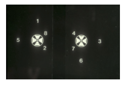
What is the main limitation of a standard Worth 4 Dot flashlight when assessing suppression at different distances?
The standard Worth 4 Dot flashlight is not standardized for visual angle:
At distance, the dots subtend a smaller visual angle and primarily test a more central retinal area
At near, the dots subtend a larger visual angle and test a more peripheral retinal area
Therefore, different findings at distance and near may reflect the size or location of the suppression scotoma, not simply a change in binocular status.
How are standard Worth 4 Dot responses interpreted through red-green glasses?
Four dots: binocular fusion
Two red dots: suppression of the eye viewing through the green filter
Three green dots: suppression of the eye viewing through the red filter
Five dots: diplopia
Dots that alternate between red and green responses suggest alternating suppression or binocular rivalry
What advantages does an adjustable Worth 4 Dot app provide over a standard flashlight?
The app can adjust:
Dot diameter
Dot separation
Red-green color contrast
Viewing distance
The app calculates the subtended visual angle using the viewing distance and separation between the outside edges of opposing dots.
This allows the examiner to:
More systematically estimate the size of the suppression scotoma
Evaluate the depth of suppression by changing color contrast
Compare findings under more controlled conditions than with a standard flashlight
Testing caution: Check that the red-green glasses adequately filter the app’s displayed colors before interpreting the result.
How does the Worth 4 Dot app calculate the target’s subtended visual angle?
The app calculates the subtended visual angle using:
The viewing distance
The distance between the outside edges of two opposing dots

What is the difference between contour stereopsis and random-dot stereopsis, and what are common examples of each?
Contour or local stereopsis
Uses recognizable, separated contours that may provide monocular cues.
Examples:
Stereo Fly
Randot circles
BVAT circles
Borish card circles
Random-dot or global stereopsis
Requires the visual system to detect a form embedded within random dots, providing stronger evidence of global binocular integration.
Examples:
Random Dot E
Random Dot E on the BVAT or Borish card
Randot Preschool
Randot Stereosmile
Lang test
How can a patient with strabismus appear to pass a contour stereoacuity test, and why should random-dot stereopsis also be tested?
A patient with strabismus may identify contour targets using monocular cues rather than true stereopsis.
Some patients with deviations greater than 20Δ can still appear to demonstrate stereopsis on Titmus fly, animal, or circle targets.
True stereopsis is uncommon when strabismus exceeds approximately 4Δ.
Random-dot targets minimize monocular contour cues and better assess genuine global stereopsis.
How should near stereoacuity testing be performed to prevent invalid results from monocular cues?
Put the polarized glasses on before the patient sees the test book.
Place the polarized glasses over the patient’s habitual correction.
Maintain the proper testing distance.
Do not allow the patient to:
Move or tilt the head
Move or tilt the test book
Use a cotton swab for pointing so the test is not marked or touched.
Ask the patient to identify or describe the stereoscopic target, not merely select a box.
What stereoacuity targets are available on the Borish card, and what range do they test?
The Borish card includes:
Contour circles, which may contain local or monocular cues
A random-dot E, which assesses global stereopsis with fewer monocular cues
The card tests disparities from 1280 to 10 seconds of arc:
1280, 640, 320, 160, 80, 40, 20, and 10 seconds of arc
How should Randot Preschool stereoacuity results be interpreted according to the patient’s age?
Randot Preschool assesses global stereopsis from 800 to 40 seconds of arc and has high interobserver and test-retest reliability.
Approximate age norms:
Age 3: 100 arcsec
Subnormal: worse than 400 arcsec
Ages 4 to 5: 60 to 100 arcsec
Subnormal: worse than 200 arcsec
Age 6: 60 arcsec
Subnormal: worse than 100 arcsec
Ages 7 to 17: 40 arcsec
Subnormal: worse than 60 arcsec
How is the Random Dot E test performed, and what constitutes a passing response?
Place the appropriate polarized glasses over the patient’s correction.
Present the random-dot E target together with a comparison target.
Randomize the target’s position between trials so the patient cannot guess based on location.
Ask the patient to identify or point to the target containing the E.
Require 4 out of 4 or 4 out of 5 correct responses to pass.
The disparity varies with testing distance:
504 arcsec at 50 cm
165 arcsec at 5 ft
138 arcsec at 6 ft
A patient performs well on contour circles but fails the Random Dot E. What does this pattern suggest?
The result suggests that the patient may:
Use monocular or local contour cues to pass the circles
Lack reliable global stereopsis
Have suppression, strabismus, or impaired binocular sensory integration
Before diagnosing abnormal stereopsis, confirm:
Proper polarized-filter placement
Appropriate testing distance
Correct target orientation and randomization
No movement or tilting of the head or test
Adequate visual acuity to see the target
How is the Pediatric Assessment of Stereopsis with a Smile (PASS) administered and interpreted?
Designed for pediatric stereoacuity screening
Test at 40 cm using the attached measuring string
Present the target in randomized positions
Passing criterion: 4/4 or 4/5 correct
Screening target: 720 arcsec
Preschool screening set: 480 and 240 arcsec
Full examination set: 480, 240, 120, and 60 arcsec, with an optional 40 arcsec target
Expected finding: Children ages 3 to 5 years should achieve approximately 120 arcsec or better.
What is the major advantage of the Lang stereotest?
The Lang test evaluates global stereopsis without requiring polarized or red-green glasses.
Uses a built-in lenticular surface to present separate images to the two eyes
Useful for young children or patients who will not tolerate special glasses
Approximate disparity range: 1200 to 550 arcsec
What stereoacuity ranges are assessed by the Stereobutterfly test, and what is a major testing concern?
Butterfly target
Tests coarse stereopsis from approximately 2000 to 700 arcsec:
Top of wings: 2000 arcsec
Bottom of lower wings: 1150 arcsec
Tip of abdomen: 700 arcsec
Circle targets
Test approximately 800 to 40 arcsec.
Important limitation: The large butterfly is a contour stereotest, so monocular cues may allow a patient to appear to pass without reliable global stereopsis.
How does the Frisby stereotest differ from polarized contour and random-dot stereotests?
The Frisby test is a real-depth stereotest:
Uses the physical thickness of transparent plates to create disparity
Does not require polarized or red-green glasses
Stereoacuity is changed by altering:
Plate thickness
Testing distance
The patient identifies the region that appears to have a different depth
Advantage: Because the test uses real physical depth and requires no glasses, the Frisby test can be useful for young children and may provide stronger evidence of stereopsis than contour tests containing monocular cues.
Testing caution: Prevent head movement and control the viewing distance because motion parallax and changes in distance can provide nonstereoscopic clues.
Why can an apparently positive Stereo Fly result be misleading in a patient with large-angle strabismus?
The Stereo Fly, animals, and circles are contour stereotests, so visible contours may provide monocular cues.
Approximately 6% to 35% of patients with strabismus greater than 20Δ may appear to demonstrate stereo on the Titmus test.
A positive result may therefore reflect recognition of monocular cues, not true binocular stereopsis.
Verify unexpected Stereo Fly performance with a global random-dot stereotest.
What does the Distance Randot test assess, and what stereoacuity range does the test measure?
The Distance Randot evaluates global stereopsis at distance using random-dot targets.
Range: approximately 400 to 60 seconds of arc
Useful for detecting binocular sensory deficits that may be less evident during near stereoacuity testing
How is dynamic visual acuity measured and compared with static visual acuity?
Measure static best-corrected visual acuity using a full Snellen chart.
Keep the patient’s best-corrected line near the bottom of the displayed chart, typically with 20/20 to 20/50 visible.
Measure visual acuity during head movement:
Approximately 2 Hz
Approximately 20° in each direction
Compare dynamic visual acuity with static visual acuity.
Other testing approaches may use:
A treadmill
A moving visual-acuity target traveling approximately 30° to 90° per second
Sports-vision equipment
A patient’s dynamic visual acuity is more than two lines worse than static visual acuity. What does this finding suggest, and what follow-up should be considered?
A decline of more than two lines during head movement is considered abnormal and suggests difficulty maintaining stable retinal images during motion, potentially involving the vestibulo-ocular reflex, or VOR.
Consider:
Referral for vestibular physical therapy
If vestibular physical therapy has already been completed, incorporating more challenging head movement during vision therapy to further train the VOR
Patient finding:
Static VA of 20/20 with dynamic VA of 20/50 represents a decline greater than two lines and warrants further vestibular or VOR evaluation.