Practical #2 Ortho
 Call Kai
 Learn
Learn Practice Test
 Spaced Repetition
 Match
Match Flashcards
 Knowt Play
1/78

There's no tags or description
Looks like no tags are added yet.
Name | Mastery | Learn | Test | Matching | Spaced | Call with Kai | Chat |
|---|
No analytics yet
Send a link to your students to track their progress
79 Terms
genu valgum
proximal tibia is angle toward midline more than 5° relative to femur

genu varum
proximal tibia is angle away midline more than 5° relative to femur

genu recurvatum
tibiofemoral extension greater than 0°

vastus intermedius
o: anterior surface of shaft of femur
ins: tibial tuberosity, via patella, and patellar ligament
a: knee extension
inn: femoral (L2, L3, L4)

vastus medialis
o: medial lip of lines aspera of femur
ins: tibial tuberosity, via patella, and patellar ligament
a: knee extension
inn: femoral (L2, L3, L4)

rectus femoris
o: anterior inferior iliac spine and just above acetabulum of os coxae
ins: tibial tuberosity, via patella, and patellar ligament
a: knee extension
inn: femoral (L2, L3, L4)

vastus lateralis
o: greater trochanter and lateral lip of linea aspera of femur
ins: tibial tuberosity, via patella, and patellar ligament
a: knee extension
inn: femoral (L2, L3, L4)

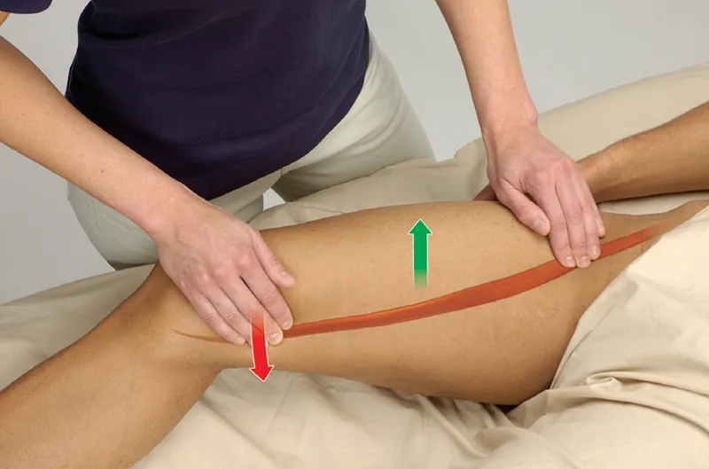
sartorius
o: anterior superior iliac spine
ins: proximal medial surface of tibia, below tuberosity
a: knee flexion and internal rotation
inn: femoral (L2, L3, L4)
gracilis
o: inferior ramus of pubis
ins: medial surface of tibia
a: knee flexion and internal rotation
inn: obturator (L3, L4)

semitendinosus
o: ischial tubersoity
ins: medial surface of proximal end of tibia
a: knee flexion and internal rotation
inn: tibial (S1, S2)

popliteus
o: lateral condyle of femur
ins: posterior surface of tibia below tibial plateau
a: knee flexion and internal rotation
inn: tibial (L4, L5, S1)
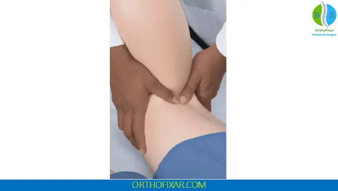
biceps femoris
o: long head - ischial tuberosity, short head - lateral lip of linea aspera
ins: lateral surface of head of fibula and lateral condyle of tibia
a: knee flexion and external rotation
inn: sciatic (L5, S1, S2)

semimembranosus
o: ischial tuberosity
ins: medial surface of proximal end of tibia
a: knee flexion and internal rotation
inn: tibial (S1, S2)

gastrocnemius
o: lateral head - posterior lateral condyle of femur, medial head - popliteal surface of femur above medial condyle
ins: posterior surface of calcaneus
a: knee flexion
inn: tibial (S1, S2)

patella

tibial tuberosity

medial joint line

medial tibial plateau

medial femoral condyle

medial femoral epicondyle

lateral joint line

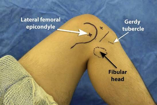
fibular head

gerdy’s tubercle

lateral femoral condyle

lateral femoral epicondyle
ischial tuberosity

patellar tendon

quadriceps tendon

pes anserine tendon

medial meniscus

pes anserine bursa

medial collateral ligament (MCL)

lateral collateral ligament (LCL)

iliotibial band

popliteal fossa

popliteal artery
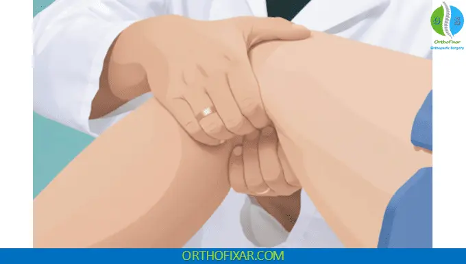
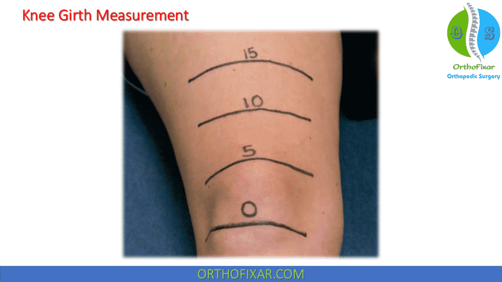
girth measurements
positive test: difference of ±1 cm compared bilaterally
implications: increased girth on injured side across joint line - edema, decreased muscular girth on injured side - atrophy
sweep test
positive test: reformation of edema on medial side of knee when pressure is applied to lateral aspect
implications: swelling within joint capsule indicating possible ACL trauma, osteochondral fracture, synovitis, meniscal lesion or patellar dislocation

ballotable patella
positive test: patella depresses and strikes patellar groove (femoral trochlea) and returns back to its former position
implications: effusion within joint capsule

anterior drawer test
positive test: increased amount of anterior tibial translation compared w/ opposite (uninvolved) limb or lack of firm endpoint
implications: ACL sprain

lachman test
positive test: increased amount of anterior tibial translation
implications: ACL sprain

prone lachman
positive test: excessive anterior translation relative to uninvolved knee indicates ACL sprain
implications: positive test results for anterior drawer and/ or lachman test and in alternate lachman test indicate ACL sprain, positive anterior drawer test and/ or lachman test result and negative alternate lachman test result implicate PCL sprain

posterior drawer test
positive test: increased amount of posterior tibial translation compared w/ opposite (uninvolved) limb or lack of firm endpoint
implications: PCL sprain

godfrey’s test
positive test: unilateral posterior (downward) displacement of tibial tuberosity
implications: PCL sprain

valgus stress test
positive test: increased laxity, decreased quality of endpoint, and/ or pain compared w/ uninvolved limb
implications: in complete extension - MCL sprain, medial joint capsule, and cruciate ligaments; distal femoral epiphyseal fracture, in 25° flexion - MCL sprain
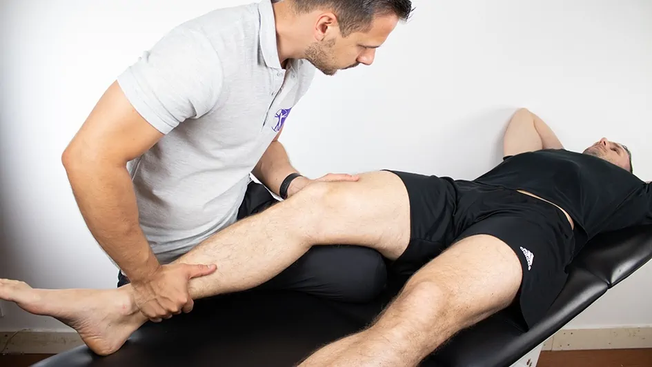
varus stress test
positive test: increased laxity, decreased quality of endpoint, and/ or pain compared w/ uninvolved limb
implications: in complete extension - LCL sprain, lateral joint capsule, and cruciate ligaments, and related structures indicating possible rotational instability of joint; distal femoral epiphyseal fracture, in 25° flexion - LCL sprain
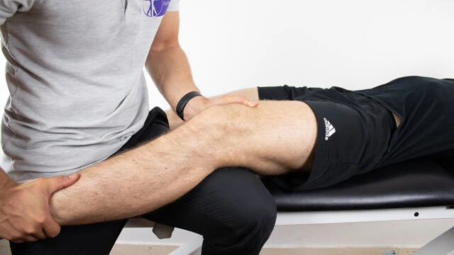
lever sign
positive test: heel remains flat on table
implications: ACL rupture
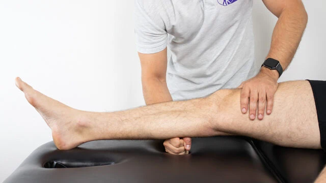
quadriceps active test
positive test: anterior translation of tibia on femur
implications: grade II or III PCL sprain

slocum drawer test
positive test: increased amount of anterior tibial translation compared w/ opposite (uninvolved) limb or lack of firm endpoint
implications: test for anterolateral instability - damage to ACL, anterolateral capsule, LCL, IT band, popliteus tendon, posterolateral complex, test for anteromedial instability - damage to MCL, anteromedial capsule, ACL, posteromedial capsule, pes anserine

crossover test
positive test: patient reports pain, instability, or apprehension
implications: ALRI - instability of lateral capsular restraints, AMRI - instability of medial capsular restraints

pivot shift test/jerk test
positive test: tibia’s position on femur reduces as leg is flexed in range of 30°-40°, jerk test - during extension, anterior reduction is felt in same range
implications: ACL anterolateral capsule, LCL biceps femoris, popliteus, posterolateral capsule

slocum ALRI test
positive test: appreciable “clunk” or instability as lateral tibial plateau sublimates or pain or instability is reported
implications: tear of ACL, LCL, anterolateral capsule, arcuate ligament complex, biceps femoris, tendon, and/ or IT band

flexion-rotation drawer test
positive test: femur is reducing itself on tibia by moving anteriorly and internally, rotating on tibia
implications: tears of ACL, LCL, anterolateral capsule, arcuate ligament complex, biceps femoris tendon

external rotation test (dial test)
positive test: increase of external rotation greater than 10° compared w/ opposite side
implications: difference at 30° of knee flexion but not at 90° - injury isolated to posterolateral corner of knee, difference at 30° and 90° of knee flexion - trauma to PCL, posterolateral knee structures, and posterolateral corner, difference at 90° of knee flexion but not at 30° - isolated PCL sprain

external rotation recurvatum test
positive test: marked difference in hyperextension, external femoral rotation, and varus alignment between two knees
implications: posterolateral corner trauma, ACL sprain, and posterolateral rotary instability

posterolateral/posteromedial drawer test
positive test: increased external rotation of lateral (posterolateral) or medial (posteromedial) tibial condyle relative to uninvolved side
implications: posterolateral test - tibia externally rotated 15°, trauma to posterolateral corner and PCL, possible posterolateral rotational instability, posteromedial test - tibia internally rotated 15°, PCL tear, oblique ligament, MCL, posteromedial capsule, semimebranosus

reverse pivot shift test
positive test: appreciate reduction (“clunk”) of tibia on femur
implications: posterolateral rotational instability and/ or trauma to posterolateral corner

dynamic posterior shift test
positive test: “clunk” or “jerk” as knee nears full extension, representing subluxated tibia reducing on femur
implications: posterolateral instability

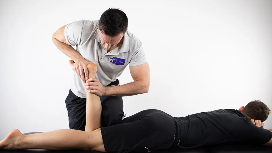
mcmurray test
positive test: popping, clicking, or locking of knee; pain emanating from menisci; or sensation similar to that experienced during ambulation
implications: meniscal tear on side of reported symptoms

apley compression and distraction tests
positive test: pain experienced during compression that is reduced or eliminated during distraction
implications: meniscal tear
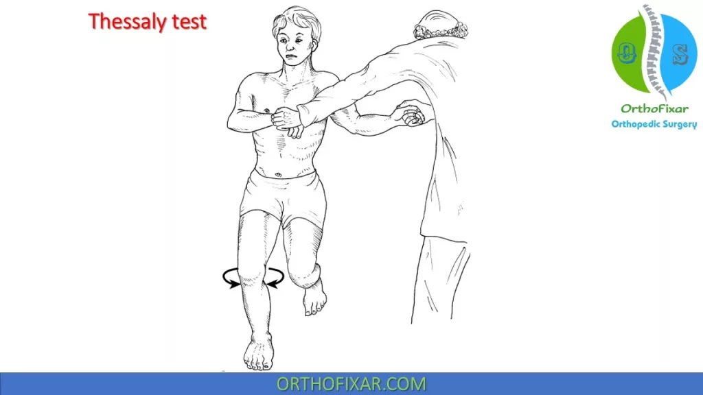
thessaly test
positive test: joint-line discomfort, complaints of “locking” or “catching”
implications: lesion of medial or lateral meniscus, depending on source of pain and/or catching sensation
wilson’s test
positive test: pain experienced during extension w/ internal tibial rotation that is relieved by externally rotating tibia
implications: OCD or osteochondritis dissecans on intercondylar area of medial femoral condyle

noble compression test
positive test: pain under thumb, most commonly as knee approaches 30°
implications: inflammation of IT band or it associated bursa or inflammation of lateral femoral condyle
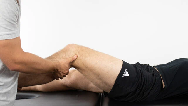
proximal tibiofibular syndesmosis
positive test: hyper or hypomobility of fibula on tibia compared w/ uninvolved side and/ or pain elicited during test
implications: anterior fibular shift indicates damage to proximal posterior tibiofibular ligament; posterior displacement reflects instability of anterior tibiofibular ligament of proximal tibiofibular syndesmosis, hypomobility may alter proximal and distal joint mechanics

step length
distance traveled between initial contacts of right and left foot

step width
distance between points of contact of both feet

stride length
linear distance covered in one stride

foot angle
angle of foot (imaginary line from heel to second toe) relative to line of progression

stance time
amount of time spent in contact w/ surface by single limb
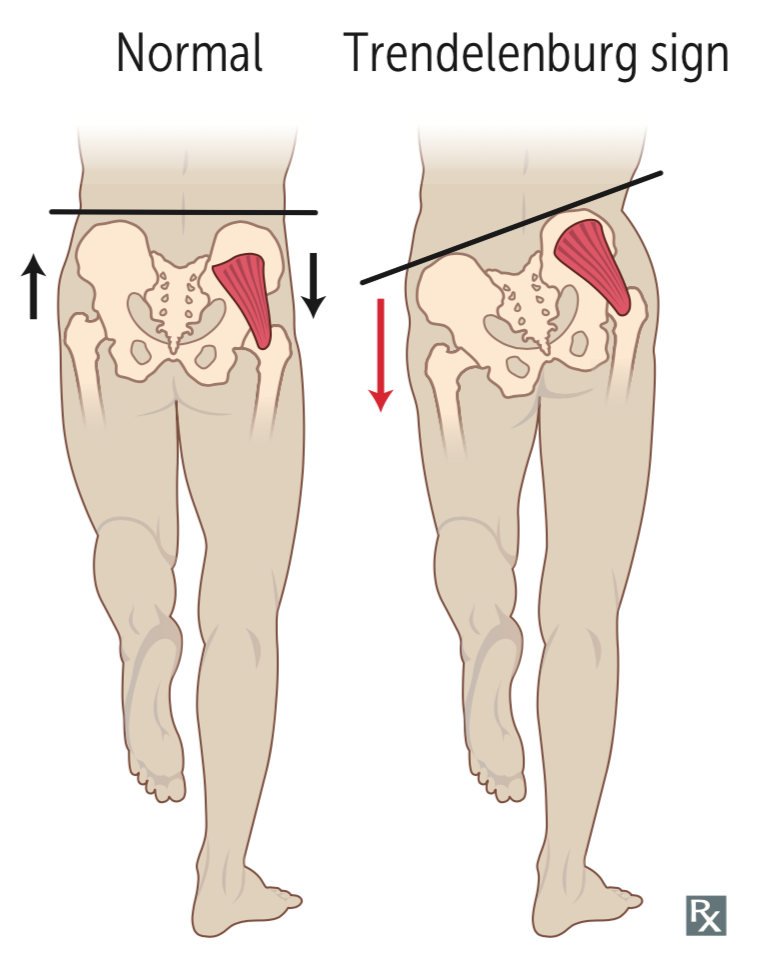
pelvic drop
hip drop that occurs when opposite limb is wb could be result of either leg-length discrepancy (shorter side shows dropped hip) or weak-gluteus medius muscle causing short-leg or tredenelenburg gait
asymmetrical arm swing
arm swing occurs primarily to counterbalance activity of hips and pelvis during walking and running

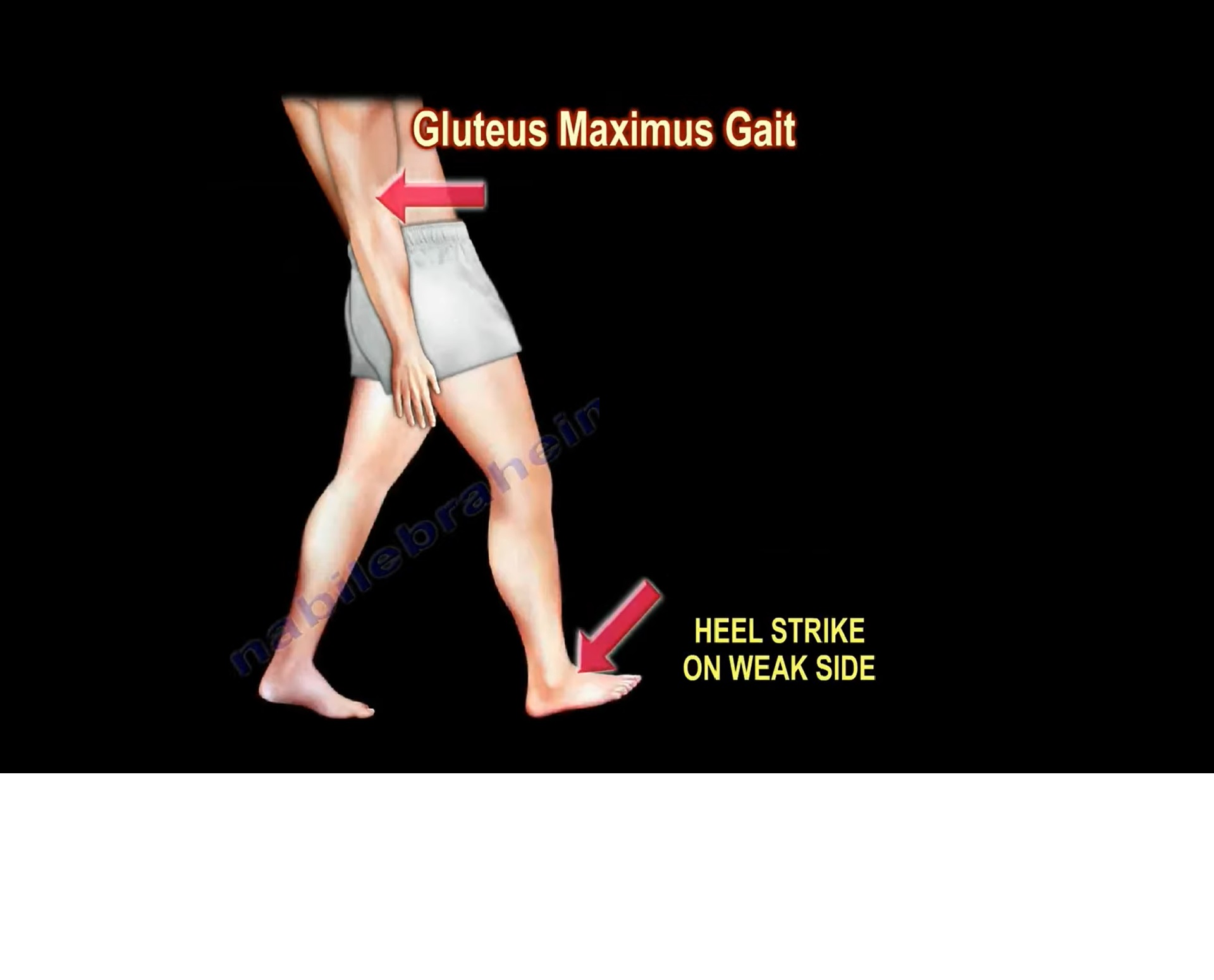
gluteus maximus gait
at initial contact, thorax is posteriorly to maintain hip extension during stance phase often causing lurching of trunk (cause: weakness or paralysis of gluteus maximus muscle)
stiff knee or hip gait
in swing phase, affected extremity is lifted higher than normal to compensate for knee or hip stiffness, to accomplish this uninvolved extremity demonstrates increased plantar flexion (cause: knee pathologies such as meniscal or ligamentous tears or hip pathologies such as bursitis or muscle tears that result in decreased ROM

trendelenburg gait
during stance phase of affected limb, thorax lists toward involved limb, this serves to maintain COM and prevent drop in pelvis on affected side (cause: weakness of gluteus medius muscle)

calcaneal gait
during stance phase, increased dorsiflexion and knee flexion occur on affected side resulting in decreased step length (cause: paralysis or weakness of plantar flexors or pain when wb on forefoot or toes caused by such conditions as blisters, hallux rigid, sesamoiditis, or ankle sprains)

psoatic limp
to compensate during swing phase, lateral rotation and flexion of trunk occurs w/ hip adduction, trunk and pelvic movement are exaggerated (cause: weakness or reflex inhibition of psoas major muscle)

short leg gait
increased pronation occurs in subtalar joint of long leg, accompanied by shift of trunk toward longer extremity (cause: true leg-length discrepancy; right leg is longer)

steppage gait
foot slaps as initial contact owing to drop foot, during swing face unaffected limb demonstrates increased hip and knee flexion to avoid toe dragging, producing “high-step” pattern (cause: weakness or paralysis of dorsiflexors)

abductory twist gait
foot abruptly abducts beginning at heel-off and then rotates into adduction through swing; it’s often seen w/ excessive pronation (cause: weak hip external rotators and abductors; contracture at 1st MTP joint)
