Small animal valvular heart dx
 Call Kai
 Learn
Learn Practice Test
 Spaced Repetition
 Match
Match Flashcards
 Knowt Play
1/43

There's no tags or description
Looks like no tags are added yet.
Name | Mastery | Learn | Test | Matching | Spaced | Call with Kai | Chat |
|---|
No analytics yet
Send a link to your students to track their progress
44 Terms
What is the most common cardiac disease?
Myxomatous degenerative valve disease (MDVD)
List some of the reasons that you can have a valvular leak in the mitral valve?
MDVD
acquired in older animals
small breeds
Mitral dysplasia (congenital young dogs; large breeds)
Mitral regurg in DCM
Endocarditis
Explain what is happening during MVDV
Nodular thickening of valve leaflets
glycosaminoglycan + proteoglycan accum
Chordae lengthen or rupture
What is the aetiology of MVDV? What breed is predisposed
Largely idiopathic but genetic basis
Abnormalities in collagen & extracellular matrix formation
Abnormalities of serotonin signalling
Mechanical stress
Cavalier King Charles Spaniel
What are the pathological consequences of MVDV?
LA dilatation
LV dilatation
Elongation chordae tendinae
Thickened leaflets
Jet lesions —> regurg back up into atrium → damages L atrial wall
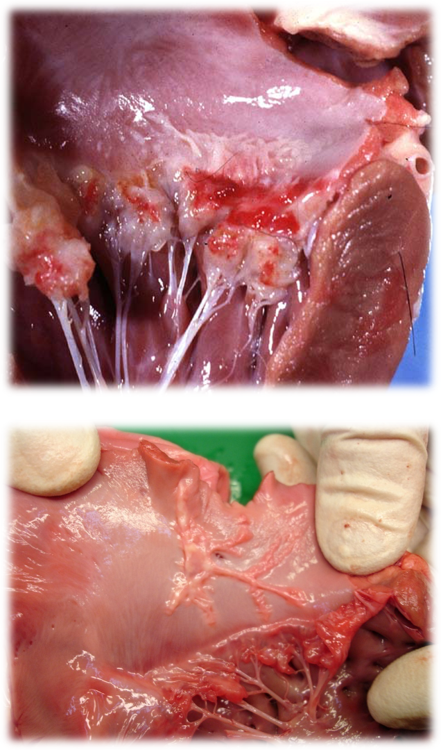
Describe the histopathology of MDVD
Accum of GAGs & arteriosclerosis in myocardium

Explain the pathophysiology of MDVD?
Leakage back into atria
L = mitral
R = tricuspid
Reduction in forward SV and inc in volume entering LV in next diastole due to the leakage from prev cycle
less blood leaving heart = RAAS = retains vol
vol overload
Preload vs afterload
Preload = volume of blood stretching the heart ventricles before they contract
Afterload = pressure / resistance heart must overcome to pump that blood out
Describe hyperdynamic systolic function?
More blood in ventricle @ start
Less blood in ventricle at end
Makes look like contracts well
What are the haemodynamic effects of MDVD?
Reduced afterload (route into LA, easier than into aorta)
Increased preload (due to inc vol in the LV)
SV leaving LV, reduces CO, activating RAAS
Volume overload of LV = eccentric hypertrophy
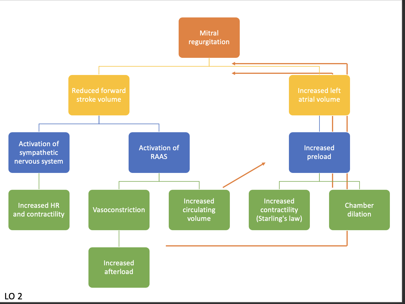
What components of the neurohumoral system activate in response to MDVD?
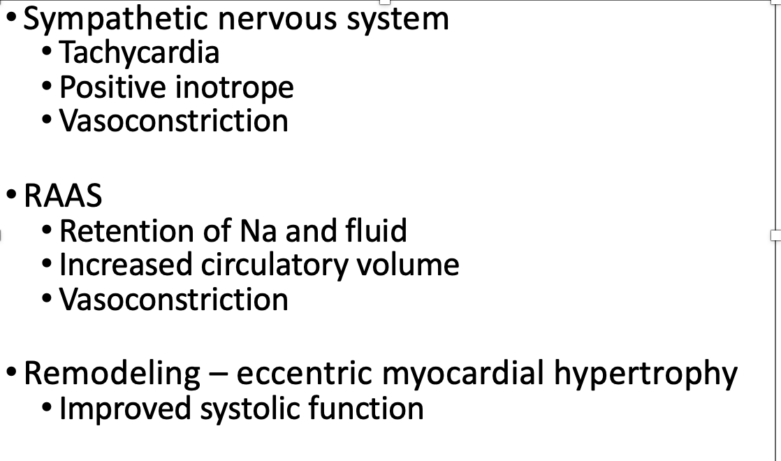
Vasocon = incr. afterload = harder for heart to push against -> more O2 + more ATP needed
Explain the sequelae of MDVD?

Explain the ACVIM staging system for MDVD?
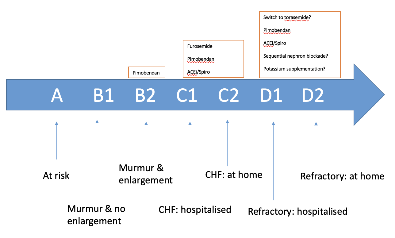
A = e.g. CKCS or any small breed
B1 = heart not remodelled
B2 = heart remodelled —> U/S + BNP bloods (measure of wall stretch)
murmur usually G3 or above
C1 = heart failure —> clinical signs e.g. fluid in lungs = incr. RR, unable to exercise, struggle to breathe
C2 = heart failure but manageable @ home longterm
At what MDVD stage do you introduce medication?
B2
Pimobendan
How would you recognise MDVD in a consult?
More typical in small breeds
Loud harsh heart murmur over LEFT APEX (systolic)
LEFT APICAL SYSTOLIC CONDITION!!!
Adult dog otherwise congenital
incr. likelihood as dog gets older
puppies are NOT degen dx
Resp signs if CHF
dyspnoea, tachpnoea, crackles
Hx —> RR + effort, exercise intol, cough
No clinical signs poss in early stages
How do you diagnose MDVD?
Auscultation
BP (Often normal)
but hypertension incr. afterload → worsens MDVD
but hypotension if forward (systolic) failure —> more likely in DCM
NTProBNP (biomarker in blood)
Rads —> R lateral & DV
ECG / ambulatory ECG if arrhythmia
Echo / TFAST
What blood tests and biomarkers should you do for a MDVD case?
Pre renal azotamia, electrolytes esp if going on diuretics
Biomarker —> NT proBNP
Troponin I —> used less commonly in MMVD
How do you take thoracic rads?
Sedation
DV + right lateral
Look for cardiac size —> tracheal elevation, cd. vena cava direction, bulges?
Pulmonary veins —> enlarged
Lung infiltrate —> interstitial / alveolar
Veins ventral & central

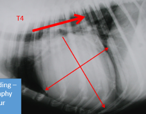
What changes might you see on rads for MVDV?
Cardiomegaly
Auricle bulges
Congested pulmonary veins (veins are ventral and central)
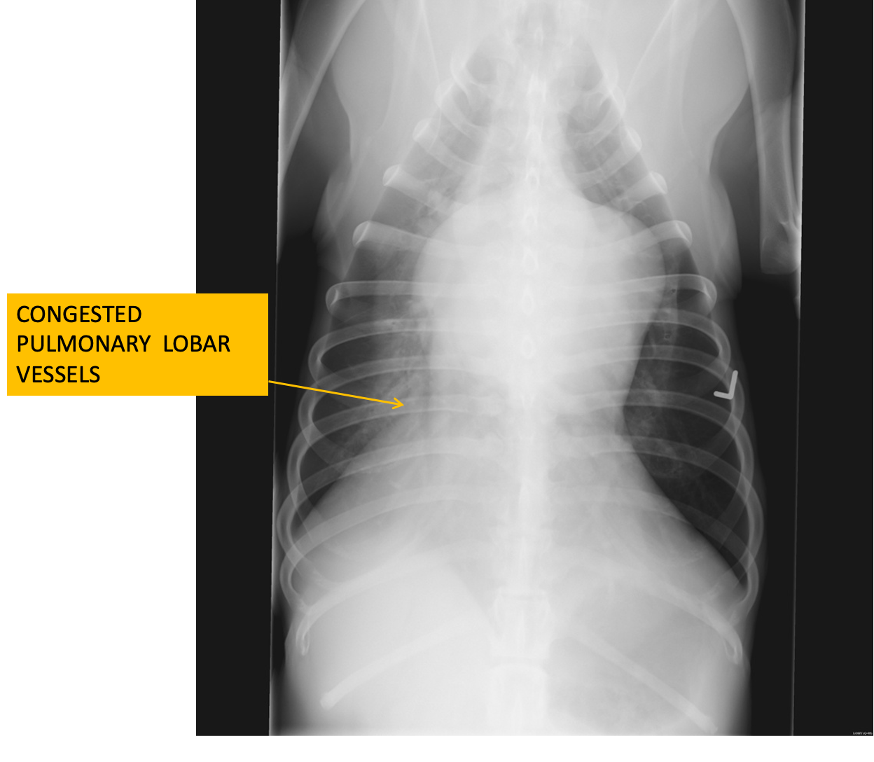
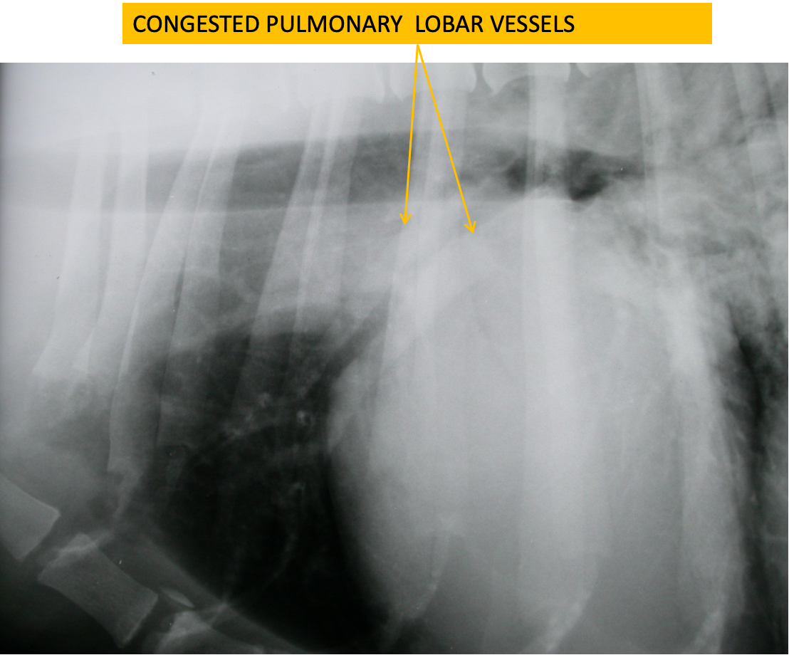
What are the most common arrythmias with MDVD?
Supraventricular premature complexes
Atrial fibrillation (L atrium gets large enough)
Ventricular premature complexes
Which arrythmia indicates that a dog is not in heart failure?
Sinus arrhythmia
What is this showing?

Supraventricular premature complexes
What is this showing?
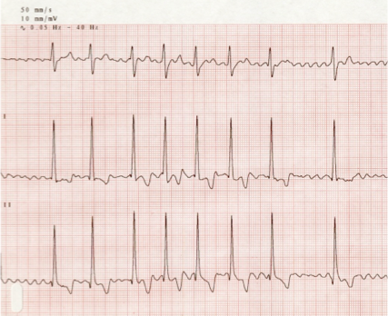
Atrial fibrillation
Tall and narrow, chaotic, no p wave
What is this ecg showing?
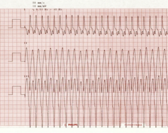
Ventricular tachycardia
What is this ecg showing?

Ventricular premature complexes
Why is echocardiography / TFAST useful in diagnosis of valvular heart dx?
Confirm diagnosis
Exclude other conditions
No sedation needed
Does not tell you about pulmonary oedema accurately
B lines
Progression
Severity
What would you see on echo of MDVD?
Enlarged left atrium LA>LV
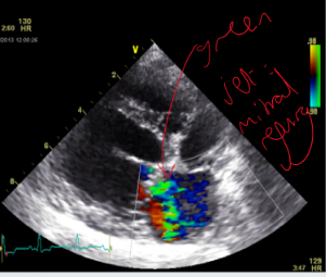
Significant mitral regurgitation
Dilated left ventricle
Rounded left ventricle
Hyperdynamic systolic function (walls coming together)
May also see:
Tricuspid regurgitation
Pulmonary hypertension

What colour is mitral regurg on doppler?
Green
(BART = blue away, red towards)
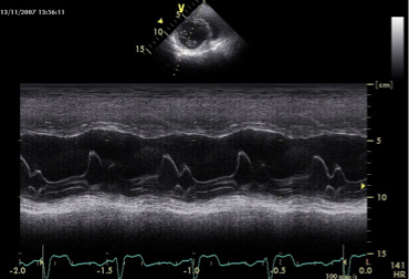
What does MDVD look like on M mode?***
Space can be seen on systole (contraction)
What changes can be seen on this normal vs MDVD echo?
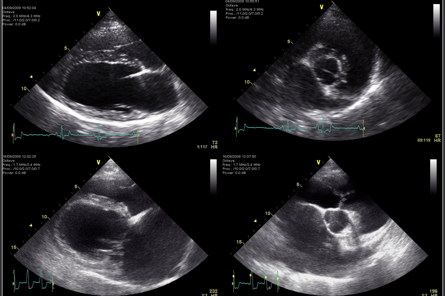
***
La enlargement
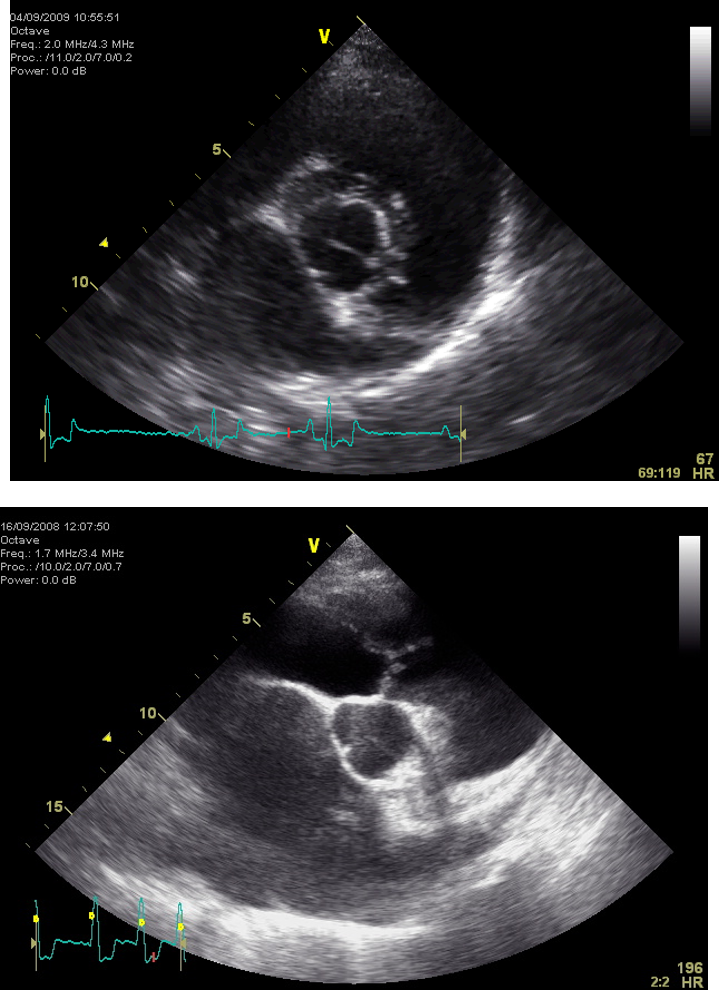
La:Ao (<1.5x)


List the treatments of MDVD based on stage
A —> none, monitor @ routine appts
B1 —> none indicated, murmur present
B2 —> PIMOBENDAN, monitor breathing rates at home
repeat imaging & biomarkers ~6months
C —> Standard CHF therapy (can’t exercise & can’t breathe) ; (1) furosemide (2) pimobendan, (3) ACE inhib, (4) spironolactone (FPAS) + Ongoing home monitoring.
D —> as for C w/ incr. doses & additional therapies e.g. sequential nephron blockade, switch of diuretic to torasemide
Recall CHF therapy (stage C and D MDVD) in order of importance
Diuretic —> Furosemide, torasemide
Pimobendan
ACE inhib
Spirinolactone
FPAS
What additional therapies (other than PFAS) can be added to CHF treatment?
Arrythmias present
Supraventricular
Diltiazem
Digoxin
Ventricular
Sotalol
Pulmonary hypertension
Sildenafil
Nutraceuticals —> omega 3 fish oil, taurine (DCM)?
Cough suppressants (figure out why coughing first)
What is the prognosis of MDVD?
Variable —> some asymptomatic never develop CHF
CKCS better prog if earlier onset
Once in failure = worse
List some of the -ve predictors of outcome of MDVD
Left ventricular dimensions (echo, chest x-ray)
Left atrial enlargement
Rupture of major chorda
NT-proBNP
What is endocarditis?
Bacterial infection (usually 1 or more endocardial surfaces i.e. valves)
Usually affecting left heart (in smallies, right in farm)
mitral > aortic (smallies)
tricuspid > pulmonic (farm)
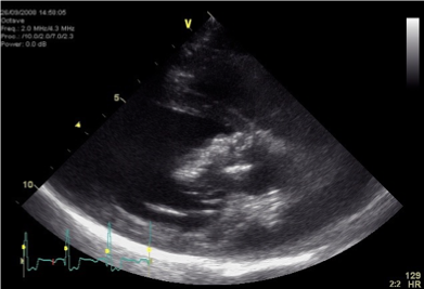
What are some infectious agents that cause endocarditis?
Streptococcus spp
Staphylococcus spp
E. Coli
Pseudomonas
Bartonellaspp
What are the 'requirements' for endocarditis?
Bacteriaemia (infection, IV catheter, surgery)
Damaged endothelium (turbulence, high velocities)
Ability to adhere to endothelial surface
Hypercoagulable states
What is being shown here?
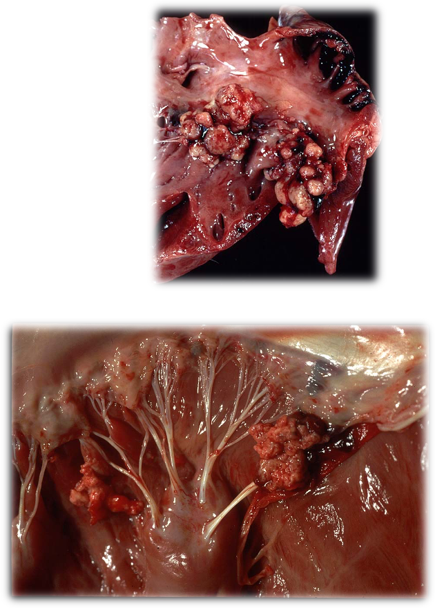
Vegetations endocardial surface of leaflets due to endocarditis
Small nodules
polypoid
Perforated, deformed, calcified
What are the microscopic findings of endocarditis?
Platelets, RBC, WBC, bacteria, fibrin
Fibrous tissue, calcification
Bacteria within vegetation
How is endocarditis diagnosed?
Haem, biochem
Troponin 1
Blood culture (often -ve)
Echo —> valvular vegetations, size, regurg, systolic dysfunction
What does troponin measure in cf to BNP?
Troponin —> damage of myocytes
BNP —> myocardial stretch
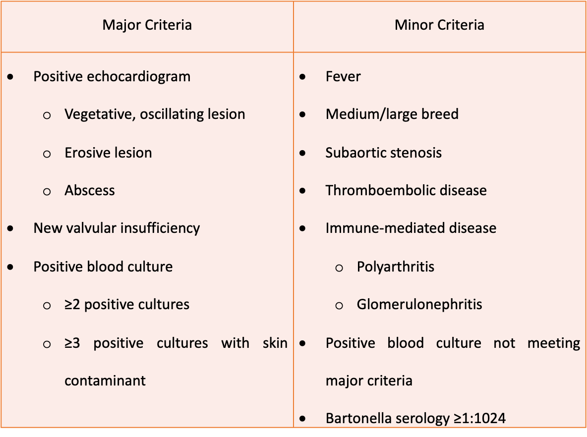
Major and minor criteria of endocarditis
How is endocarditis treated?
IV bacteriocidal AB broad therapy
Long term oral when discharged
+/- Anticoagulant (clopidogrel, aspirin)
+/- CHF tx
Guarded prognosis & recurrence + complications poss, long term valvular damage