Neuropsychology 2
 Call Kai
 Learn
Learn Practice Test
 Spaced Repetition
 Match
Match Flashcards
 Knowt Play
1/97

There's no tags or description
Looks like no tags are added yet.
Name | Mastery | Learn | Test | Matching | Spaced | Call with Kai | Chat |
|---|
No analytics yet
Send a link to your students to track their progress
98 Terms
Association Areas

Three unimodal association areas
As areas get further away from the primary sensory areas, the associative functions become more general
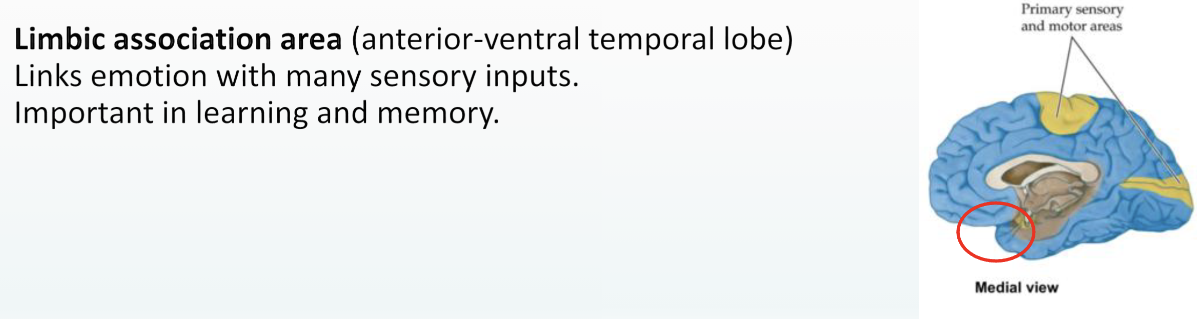
Limbic association area
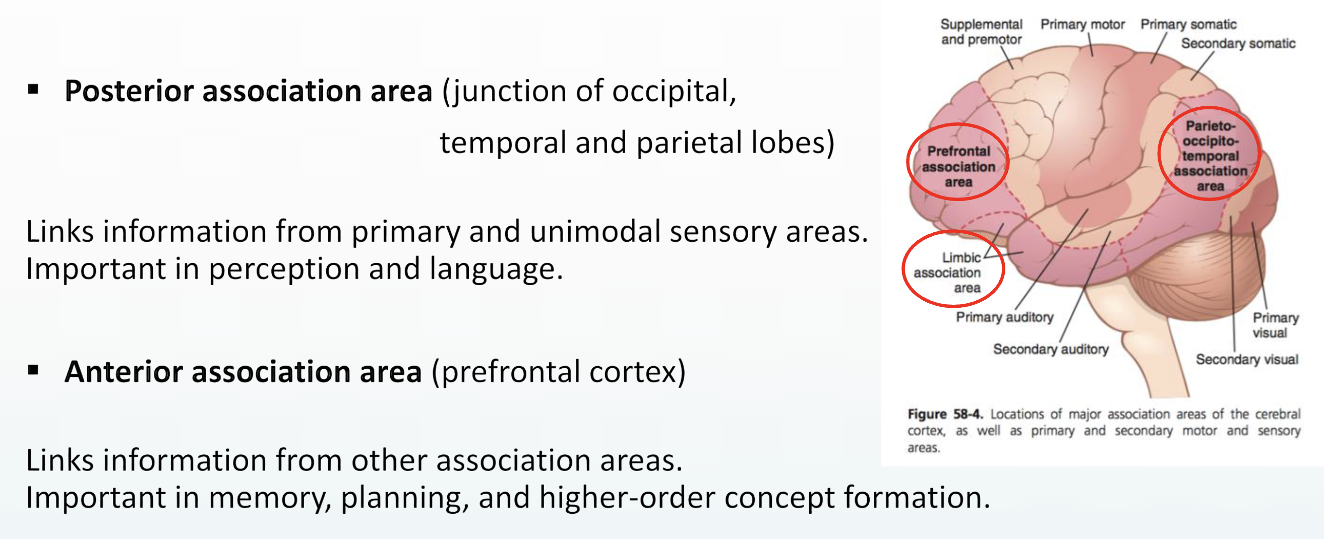
Posterior association area
Anterior association area
Limbic association area
Posterior & Anterior association areas
Parietal lobe functions

Major Functional Regions

Parietal Functions by Zone
Anterior zone = Somatic sensations and perceptions
Somatic = bodily
Posterior zone = Sensory integration and whole-body movements
Networks connect to all other lobes (see later)
Feeds into frontal lobe
Major Functional Regions of the Parietal lobe
1. Superior parietal lobule (SPL)
visuomotor, cognitive, sensory, higher order, working memory and attentional
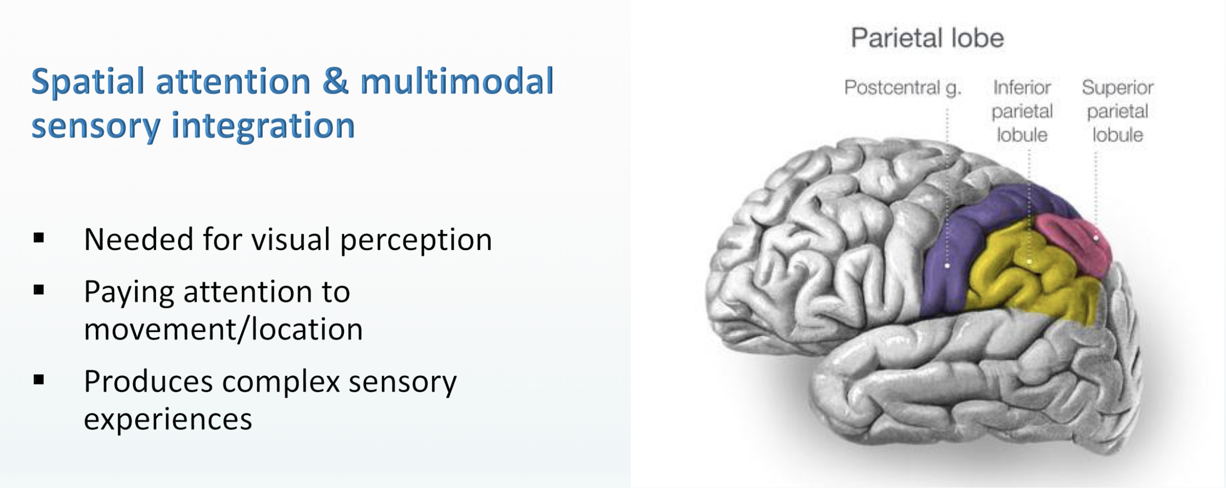
2. Inferior parietal lobule (IPL)
spatial attention, multimodal sensory integration, and oculomotor control
3. Somatosensory cortex
detects sensory information, initiates movement
4. Precuneus
Gestalt integration of information, mental imagery, episodic memory retrieval, self-referential processing

Superior Parietal Lobule
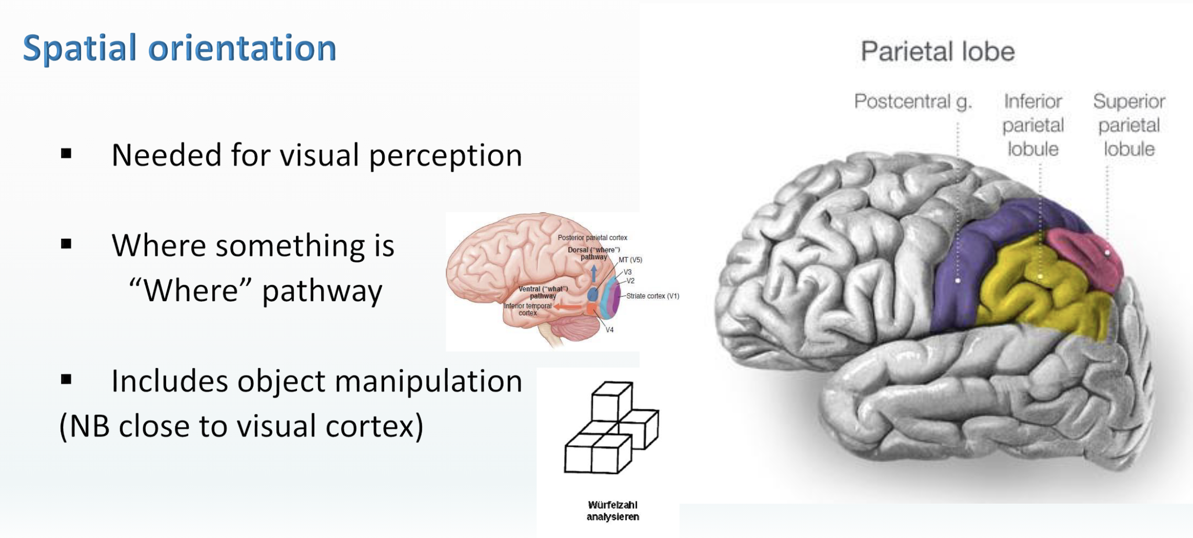
Dorsal Stream Anatomy

Inferior Parietal Lobule
Somatosensory cortex
detects sensory information from the body regarding temperature, proprioception, touch, texture, and pain
receives neuronal projections from the thalamus
adjacent to the primary motor cortex

4 representations of the body

Somatosensory receptors - 3 types

Generating movement
Integrating sensory input
Initiating movement
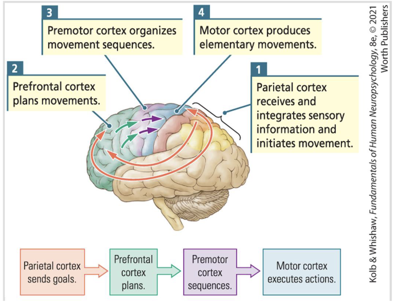
Sequential Production / Initiation of Movement

Sensory-to-motor movement

Hierarchical Control of Movement

Precuneus
Hypothesised that functions can be explained by its central location in the cortical network
May facilitate over 60 processes
Visuo-spatial imagery, episodic memory retrieval, self-processing, first-person perspective taking, experience of agency, self-consciousness (and possibly consciousness)
Default mode network (DMN) = brain processes when brain is at rest; mostly social activations
Precuneus is functionally central to DMN

Default Mode Network

Default Mode Network & MNS

Default Mode Network & Other Networks

Fronto-parietal Network

Parietal Memory Network

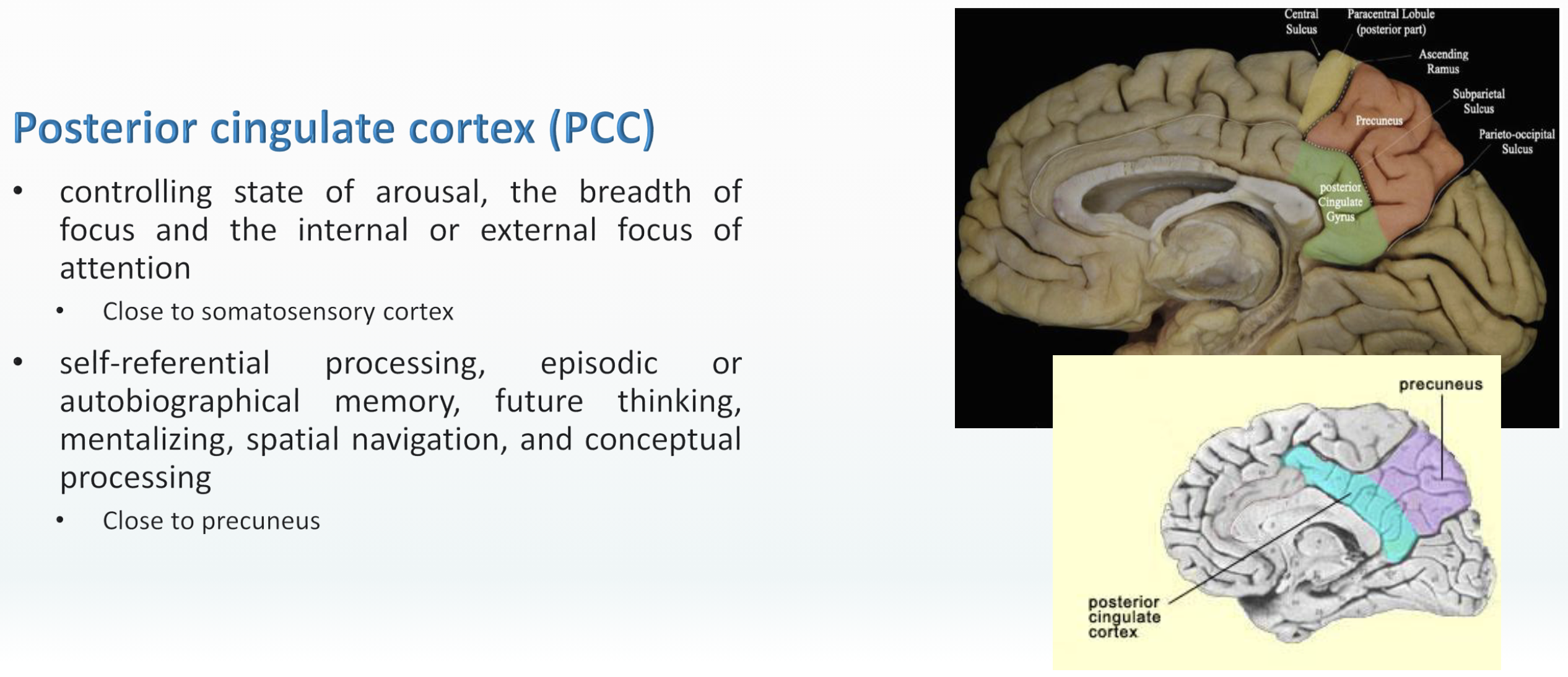
Other Major Functional Regions - Posterior cingulate cortex (PCC)
Controls state of arousal, the breadth of focus and the internal or external focus of attention
Participates in self-referential processing, episodic or autobiographical memory, future thinking, mentalizing, spatial navigation, and conceptual processing
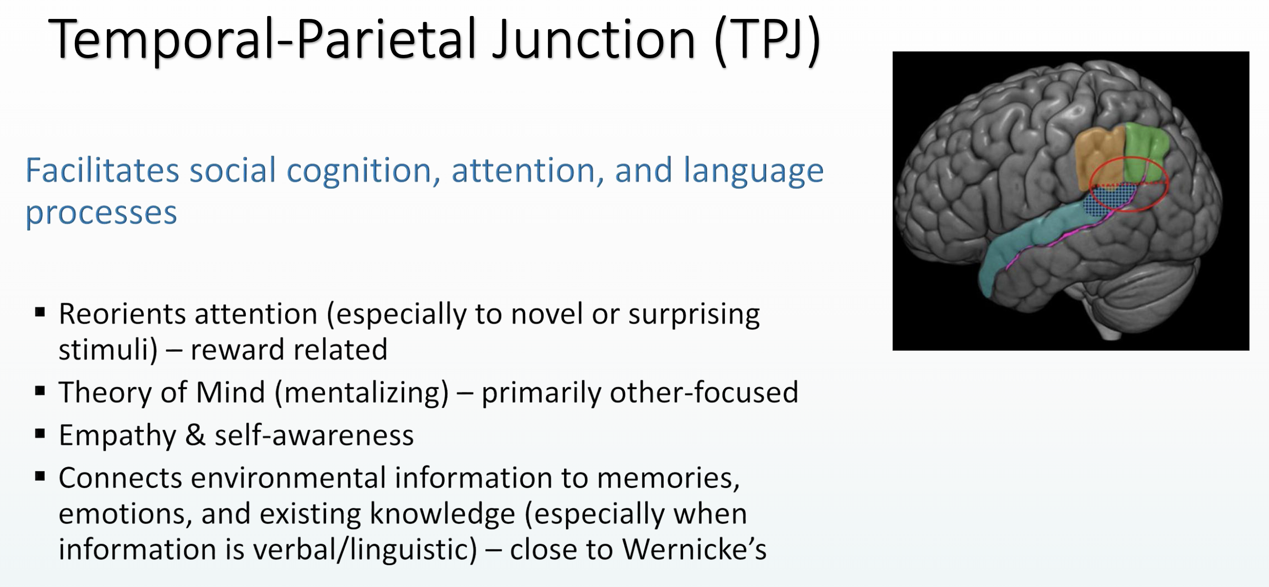
Temporal-Parietal Junction (TPJ)
Also called Temporoparietal Junction
Where the two lobes meet
Forms a bridge at the inferior parietal lobule & posterior superior temporal sulcus
Self and Other - Mentalizing

Common symptoms of parietal lesions and their clinical assessment

Asomatognosis
A loss of knowledge of one’s body, usually considered a type of agnosia caused by parietal lesion
There are several types:
Anosognosia = unawareness/denial of illness
Anosodiaphoria = indifference to illness
Autopagnosia = inability to localize/name body parts
Finger agnosia = the most common type of autopagnosia; unable to identify fingers
Asymbolia for pain = absence of typical pain responses

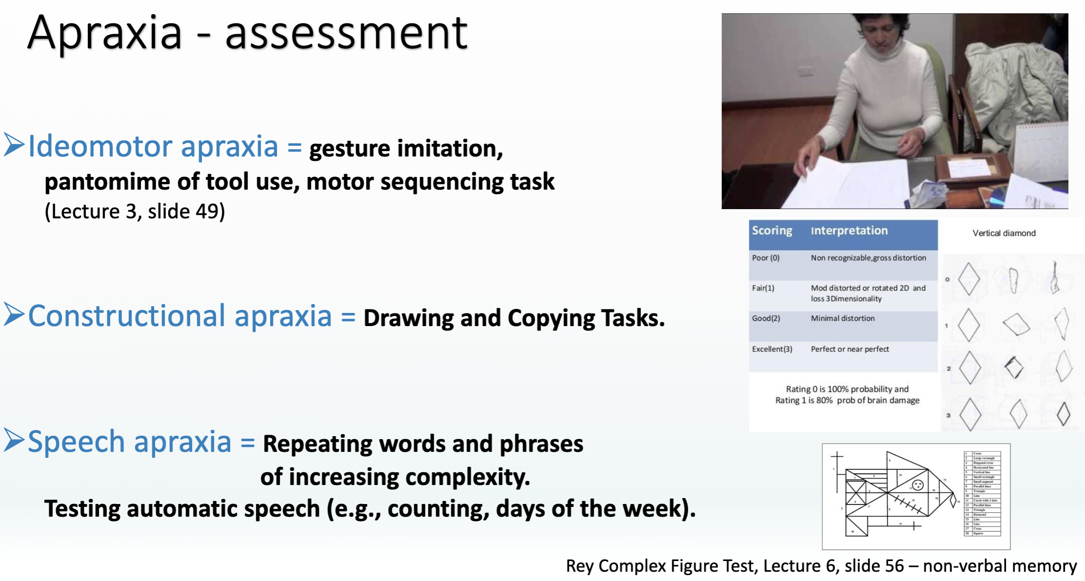
Apraxia
Loss of movement caused by a brain lesion and not by any damage related to the muscles or other body parts/processes
Ideomotor apraxia = Disturbance to physical movements; unable to copy or make gestures (e.g., waving hello)
Constructional apraxia = Disturbance to spatial processing; unable to build a puzzle or draw a picture
Speech apraxia = Disturbance to speech due to brain lesion and not caused by other types of language disorders
Gerstmann's syndrome
Josef Gerstmann (1924) observed a patient with left parietal stroke and unusual symptoms
Finger agnosia
Left-right confusion
Agraphia = inability to write
Acalculia = inability to perform arithmetic tasks
Double dissociation: different regions for subtracting (IPS) and multiplications (AG)

Balint syndrome
Rezsö Bálint (1909) observed a patient with bilateral parietal lesions and unusual symptoms
Could move his eyes but not fixate on visual stimuli
Displayed simultagnosia = could only attend to one stimulus at a time and would not notice other stimuli
Displayed optic ataxia = difficulty in reaching for stimuli even when guided
Parietal Lobe Epilepsy (PLE)
A rare form of epilepsy
Causes bodily sensations during seizures
Somatic illusions = a common symptom that causes misperception of real stimuli (e.g., feeling like body is falsely morphing, moving, or damaged)
Inability to detect space/distance
Dysesthesia = distortion to the sense of touch that causes feeling of pain without nerve stimulation
Vertigo
Disturbs other brain processes because of network connections
E.g., can cause loss of language processes that are in the frontal lobe
Parietal Lobe Seizure
Focal seizures cause bodily distortions
Consider what’s different from a temporal lobe seizure:
Falls to one side, eyes change, loses body control
Because parietal lobe networks across the brain, it is hard to identify the onset location of focal parietal lobe seizures using brain imaging
Electrical disruptions tend to be more spread out
Schizophrenia (not a “Parietal Lobe Disorder”, but...)
Disruptions to frontal and temporal lobe are most pronounced in schizophrenia
Abnormal parietal lobe activations can cause psychotic-like symptoms
I.e., Parietal dysfunction can mimic symptoms of schizophrenia
But parietal lobe disruptions in schizophrenia are usually modest
Parietal lobe may be responsible for several symptoms
Gray matter volumes is reduced across parietal lobe in patients with schizophrenia
During hallucinations, activity in the postcentral gyrus and the inferior parietal lobule increases
Distorted sensory integration / misinterpretation
Illusions of control
Poor distinction between self and other
Poor social cognition
The Acquisition of Culture
Was better social intelligence the cause of the larger brain?
Unclear.
What is clear:
Once the larger brain developed, so did culture
Humans used larger brains for social coordination
Memes
Ideas and behaviors that are passed easily from person to person in a culture
Allow rapid social transmission of ideas
Richard Dawkins (1976) ”Selfish Gene” famously coined the term
Cultural transmission of ideas
3 primary types of cultural transmission:
Vertical = from parents to children
Oblique = from older generation (non-parents) to younger generation
Horizontal = between peers
These transmission structures are commonly seen in social species
Unconcious inference - processes developed through experience but outside of our awareness

Types of Intelligences
Unclear if brain size is tied to intelligence
Species-typical behavior is used to define intelligence
Many types of intelligences and they are hard to measure
Musical
Verbal
Mathematical
Spatial
Logical
Social
IQ tests only measure one or two of these, and are culture-bound
When is brain size relevant among humans?
Normal brain size changes over an individual’s life span
Just as good nutrition early in life can promote larger brain size, a culturally enriched environment is associated with growth of existing brain cells
One way that the brain stores new skills and memories is to add cells and to form new connections among brain cells.
These plastic adaptations in turn contribute to increased brain size.
Size changes from misfortune
Brain injury around birth can cause large shrinkage of brain size
Stress from physical or behavioral deprivation in infancy also leads to decreased brain size
Neurological disorders associated with a mother’s abuse of alcohol or other drugs are associated with conditions such as fetal alcohol spectrum disorder (FASD)
Autism spectrum disorder (ASD), a largely genetic condition affecting development, produces a variety of brain abnormalities, including increases or decreases in brain size in different individuals
Neurological diseases associated with aging accelerate the age-related decrease in brain size
Frontal Cortex

Gross anatomical subdivisions of FC (1)

Gross anatomical subdivisions of FC (2)

Major Frontal Lobe Functions

Prefrontal Cortex Functions

Lateral PFC

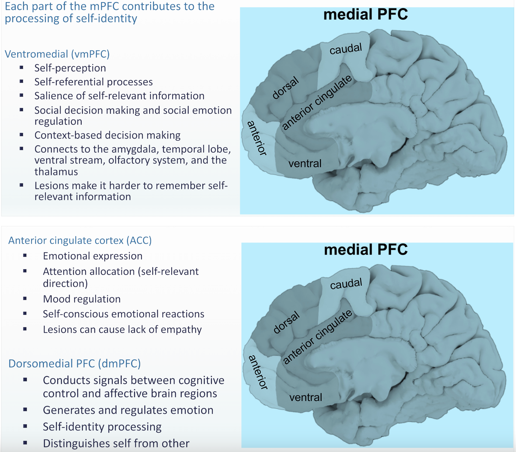
Medial PFC
Premotor cortex
Adjacent to and organised like the somatosensory cortex
Generates signals to initiate body movements
Controls muscle activity
Acquisition and performance of skilled movements
Activated when observing the actions of others (may facilitate skill learning)

Functional zones

Frontal Lobe Networks

Executive functions: planning and generation of novel actions
Prefrontal lobe damage: attentional control, planning, reasoning, working memory, problem solving, abstract thinking, and self-monitoring are intact, same as language and memory; however regulation behavior in a contextually appropriate way is problematiс
Left frontal lobe involved in task setting, the right frontal lobe is more involved in task monitoring;
Hot and Cold executive functions, that rely on different executive subsystems that are anatomically and behaviorally separate.
Behavior Rating Inventory of Executive Function (BRIEF)
Symptoms of Frontal Lobe Lesions
Motor disturbances
Loss of Fine motor movements, speed & strength
Poor Movement programming 🎹
Poor Voluntary eye gaze
Corollary discharge
Motor-related timing (how you know that something has occurred because of your own actions - you have a sense for when you acted and that something occurred after your action)
Speech (Broca’s aphasia) (Agrammatism; mutism – left medial frontal area)
Thinking alterations
Decreases in IQ score
Loss of divergent thinking (i.e., creativity / problem solving abilities)
Loss of behavioral spontaneity (Thurstone Word Fluency Test) + fewer spontaneous facial movements
Loss of strategy formation
Difficulty using environmental cues
Poor response inhibition
“Form is probably the correct solution now so this [sorting to color] will be wrong, and this will be wrong, and wrong again.”
Impaired associative learning (right hand – red light)
Risk taking / rule breaking (MAZE)
Decrease of self-regulation (loss of autonoetic awareness)
Gambling (OFC)
Poor temporal memory
Delayed response
Recency memory 🂦 🃂 🃟
Impaired social & sexual behavior (responses that are dependent on contextual cues)
Pseudodepression / pseudopsychopathy
”Pseudo” = not genuine, false (e.g., depression-like traits but cause is the brain lesion on the left)
Orbitofrontal syndrome = brain lesion (on the right) that causes disinhibition:
Immature behavior, lack of tact and restraint, coarse language, promiscuous sexual behavior , increased motor activity, and a general lack of social graces (dementia), difficulty understanding facial expressions, directing spatially guided behaviors.
Impaired olfactory discrimination
Frontotemporal Dementia
The most common signs of frontotemporal dementia are usually in the form of extreme changes in behavior and personality
Increasingly inappropriate social behavior
Loss of empathy and other interpersonal skills (e.g., insensitivity to another's feelings)
Affects men and women equally
10% - 30% of cases have genetic causes
Runs in families
Clumps of abnormal protein forms inside neurons
Cause of other 70% - 90% of cases is usually unknown

Lateral Brain Tumor
What’s the difference between a tumor and a lesion?
Dementia and lesions have interventions but not treatments
Brain tumors can be benign (non-cancerous) but still disrupt functioning
Tumors can often be removed through surgery
Laterality in the frontal lobe
Left side tumors and lesions affect language and movement
Right side tumors and lesions affect non-verbal processes (like emotion)
Emotions
Emotion is an internal subjective state and involuntary physiological response to an object or a situation.
Emotions have impact on attention, perception, decision-making and memory
Emotionally charged stimuli capture attention (your name, threatening stimuli, a fearful facial expression)
Emotional memories are better remembered and retained (memory)
Emotions guide decision-making by helping to unconsciously make the most advantageous choice (obtaining benefits, avoiding looses)
Cognitions and emotions are inseparable
Emotions inform us about events that matter to us, to our needs, values, and well-being
They determine how we perceive the environment
Emotion and the Social Brain
Emotions = complex reactions
Physiological, experiential, neurobehavioral
Probably had early evolutionary benefits
Social emotions = emotions that depend on other people
People cause emotions
We cause emotions in people
People are often the most salient stimuli
Self-reported cognition → this is a major measure of our internal states (emotions, thoughts, dis/comfort, etc)
Many patient groups have difficulty accessing their internal states
Some healthy people do too
Most healthy people are good at reporting their experiences, but might use a variety of terms to describe them (natural language isn’t as standardized as scientific language is)
Principal behavioral components of emotions
1. Psychophysiology
2. Distinctive motor behavior
3. Self-reported cognition
4. Unconscious behavior
Emotions arise as a result of the influence of subjective and objective factors that interact with the nervous and endocrine systems, trigger the experience of pleasure or avoidance, modulate various cognitive processes (memory, attention), cause physiological changes (heart rate), and lead to behavioral changes that help a person adapt to the situation that caused them
Limbic System & Emotions
Cingulate cortex = links rewards, punishments, and emotional responses
Fornix = episodic memory
Septum = mediates connections with the cortical areas
Olfactory bulb = smell
Amygdala = fear and threat
Hypothalamus = homeostasis; turns emotions physical (e.g., heart rate when scared)
Hippocampus = learning & memory
→ Frontal lobe connects with the limbic system.
Although the entire circuit is important to emotional behavior, the prefrontal cortex (especially the orbitofrontal and ventromedial regions) and amygdala hold the key to understanding the nature of emotional experience
The orbitofrontal cortex (OFC) is especially important in emotion because it represents positive and negative rewards and learns which previously neutral stimuli are associated with positive and negative rewards and when these associations change

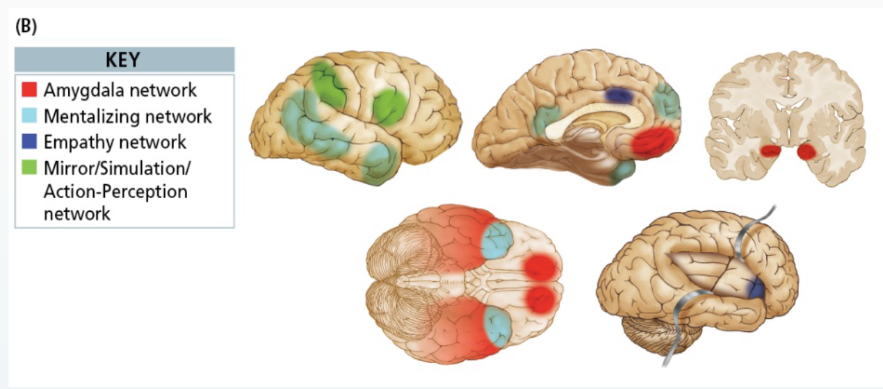
Social Brain Networks - how the brain mediates social interactions
Amygdala Network
Amygdala, orbitofrontal cortex, temporal cortex
Responding emotionally & detecting socially-relevant stimuli
Mentalizing Network
Superior temporal sulcus, anterior temporal cortex
Thinking about the internal states of others
Understanding others’ actions
Empathy Network
Insula, cingulate cortex
Attributing emotion and perspective to others
Mirror / stimulation / action-perception network
Mirror neuron system (MNS) – parietal & premotor regions
Activate when we observe the actions of others
Developing concept of self
N.B.: these networks also recruit subcortical regions such as the nucleus accumbens, globus pallidus, hypothalamus, and ventral tegmentum
Behavioral changes after frontal lesions associated with emotional processes
Reduced social interaction
Loss of social dominance
Inappropriate social interaction
Altered social preference
Reduced affect
Reduced vocalization
Social Brain Lesions
Lesion case studies
Frontal and temporal lesions = deficit in producing facial expression, social speech and processing deficits in facial expression, social context-driven emotions, personality changes
The ability to be humorous and to comprehend humor (social glue): Right-frontal injuries were the most affected in that they reacted less than other patients, with diminished laughter and smiling, and failed to grasp the jokes.
Insula = increase pain threshold, impair pain recognition in others
Anterior insula = emotion awareness
Amygdala = fear recognition in others
Bilateral vmPFC = impaired social conduct, decision making, emotion processing
right sight lesions → pseudo-psychopathy (acquired sociopathy)
left side lesions → not much changes...
WHAT ARE COGNITIVE DISORDERS?

Major Cognitive Processes
Language
Spatial Navigation
Learning & Memory
Attention
And more ...
Perception, Executive functioning, Reasoning, Decision making
Language Disorders
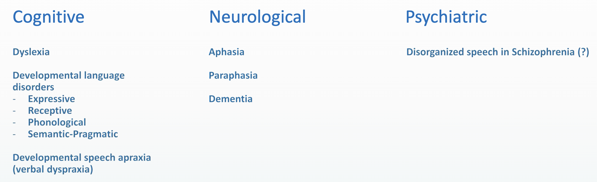
Aphasia's (Broca’s, Wernicke’s)
Paraphasia
Alexia
Agraphia
Dyslexia
Paraphasia

Dyslexia
Characterized by poor fluent / accurate word recognition, poor spelling, and difficulty with word decoding. It is a learning disability and neurobiological in nature.
Attentional dyslexia = multiple letters or words cause difficulty (one letter or one word is fine)
Neglect dyslexia = may misread the first or last part of a word (e.g., whether as smother; strong as stroke)
Letter-by-letter reading = affected persons read words only by spelling them out to themselves (aloud or silently)
Deep dyslexia = semantic errors (e.g., misreading merry as Christmas, because the words are often paired); usually have more trouble with abstract words and have difficulty with short term verbal memory (as we will see later, meaning plays a role in memory)
Phonological dyslexia = inability to read nonwords aloud (most common)
Surface dyslexia = cannot recognize words directly but can understand them by using letter-to-sound relations if they sound out the words. Surface dyslexia does not develop in languages that are totally phonetic and sounded out as they are written (e.g., Italian). Surface dyslexia is a common symptom of children who have difficulty learning to read
Dual route theory of reading
Lexical route
Relies on the activation of picture or sound representations of a whole word
The lexical route can process all familiar words, both regular and irregular, but it fails with unfamiliar words or nonwords because it lacks a means for representing them.
Nonlexical route
Uses a subword procedure based on sound-spelling rules
The nonlexical route can succeed with nonwords (e.g., klanley) and regular words that obey letter-sound rules, but it cannot succeed with irregular words that do not obey these rules (e.g., winding, choir).
Most impaired in developmental dyslexia
Lexical = related to words as whole units, “pictures” (as opposed to grammar or parts of words)
Brain regions for Speech and Sound to Meaning

Key Brain Regions Related to Reading

(Language) Found in the following disorders
Topographic Disorientation

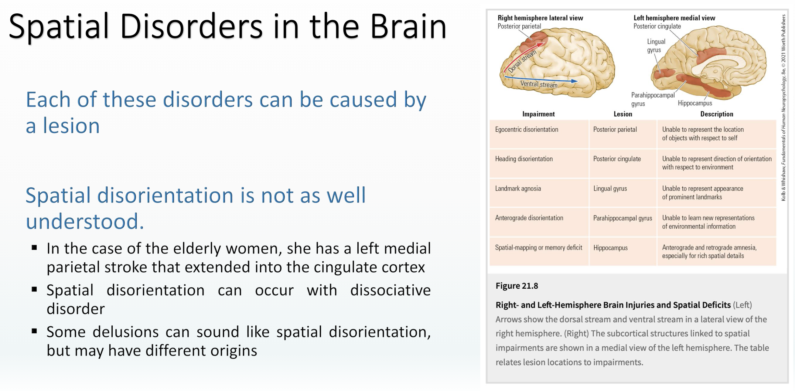
Egocentric Disorientation
Difficulty perceiving the relative location of objects with respect to the self
Able to gesture toward objects with eyes open, but cannot with eyes closed
Performance is impaired on a wide range of other visuospatial tasks
Impaired mental rotation = the ability to visualize the appearance of three-dimensional objects from different perspectives
Impaired ability to judge distances between objects
These patients are uniformly impaired in way-finding tasks both in formerly familiar and in novel environments
Spatial disorientation
Distortions in the perception of the body or environment (PCC)
Alice in Wonderland effect
Out-of-body feeling
Missing places
During a walk down a hospital hallway, an 80-year-old woman who had suffered a stroke stopped and declared that she could go no farther because that is where the hospital ended. When released from the hospital, she refused to return to her home of 20 years because, she declared, it was in a place that was not there. Even after her daughter found a new apartment for her, she became upset because the place did not exist.
Spatial Disorders in the Brain
(Space) Found in the following disorders

Information Processing Model
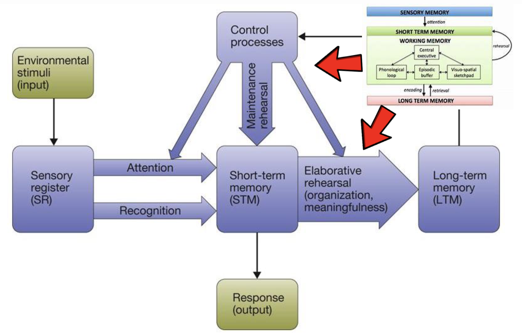
Working and Short-Term Memory

Long-Term Memory

Forgetting - Overload

Types of memory

Learning and the power-law

Learning and long-term potentiation
Neural correlate of the power-law: long-term potentiation (LTP)
Neurons that are activated simultaneously adjust their connection strength over time
They require less and less time to transmit electrical signals between them (myeline)
Connection change follows the same (though inverse) logarithmic relation as the power law of learning
Brain activation changes with skill acquisition
As people become more proficient at a task, they seem to use less of their brains to perform that task (efficient)
Regions activated in the symbol-manipulation task of Qin et al. (2003)
Less is (sometimes) more: The brain seems to have become more efficient
Important to remember that bigger is not always better in brain-terms
Long-term memory - declarative memory

Long-term - non-declarative memory
Procedural memory = memory for skills, motor acts, routines
Summary of memory in the brain
Medial temporal lobe (esp. hippocampus and parahippocampal regions): form, organize, consolidate, and retrieve memories
....and the hippocampus is a brain structure deep in the brain which has a central function for memory:
Storage of new memories
Link together different kinds of information in many regions of brain (perceptual, affective, conceptual etc.)
Prefrontal brain regions: encoding of new memories and retrieval of old memories
Associative regions in the cortex: integrate sensory inputs, enabling to understand the environment and encode memories
Other brain areas (including amygdala, striatum, and cerebellum): other kinds of memories, such as emotional or behavioral memories

Types of amnesia
Anterograde amnesia = an inability to form new memories
Retrograde amnesia = a loss of memory acquired prior to the injury
Time-dependent retrograde amnesia = loss of more recently acquired memory, while memories from the distant past are retained
Childhood (infantile) amnesia = an inability to remember events from the first 4 years and the recall of fewer than expected memories up to 7 to about 11 years of age

Dissociating memory systems
Episodic and procedural memory:
HM learned to mirror draw over a 3-day period
Clive Wearing still plays the piano
Thus, procedural learning can often be intact in amnesia
Episodic and short-term memory
Amnesiacs have normal digit span
HM could remember a number for 15 minutes by continuously repeating, but forgot it within 1 minute of stopping and had no recollection of attempting it (Milner, 1971)
(Memory) Found in following disorders

Types of attentional processes
Selective attention
Paying attention to one thing while ignoring others
Switching attention
From one task to the other
Divided attention
Paying attention to more than one thing at a time
Hard to do sometimes
Similarity, difficulty and practice
Sustained attention
Paying attention over longer time
Focused attention
Paying attention to one task
Attentional capture
Shift of attention to very salient stimuli (e.g., loud noise)
Cocktail party effect: selective attention to one stimulus (conversation) until something catches your attention (hearing your name, spotting a celebrity)
Attention – quantitative limits
There are limits to the amount of information in the world that the mind can attend to and process simultaneously
Unnecessary computational load
Energy inefficient and maladaptive
Serial bottlenecks
A point in the path from perception to action at which people cannot process all the information in parallel
When do they occur? (e.g., parties)
The study of attention and Serial Bottlenecks
Early selection theories
Filter occurs before we perceive the stimulus
Late selection theories
Filter occurs after we perceive the stimulus
Processing capacity & perceptual load

"Blindness"
Caused by selective attention and the attentional bottleneck
Inattentional blindness = not noticing something that is clearly there when paying attention to other stimuli
Change blindness = not noticing changes to stimuli when paying attention to other stimuli
Attentional blink = Difficult to process back-to-back stimuli
Attention and Cognitive control
The ability to control your cognitive functions (sometimes called “executive functions”), which include attention, memory, and other cognitive processes
Frontal lobe
Cognitive load = relative difficulty of the cognitive task
Different from perceptual load
Different from processing capacity because load refers to how many resources you’re using while capacity refers to how much stimuli your can take in
What factors determine to what we attend?

Attention and the Brain
Parietal cortex is activated for attention to location
Occipitotemporal cortex is activated for attention to features such as color and form
Anterior cingulate and prefrontal areas show activation during both visual tasks
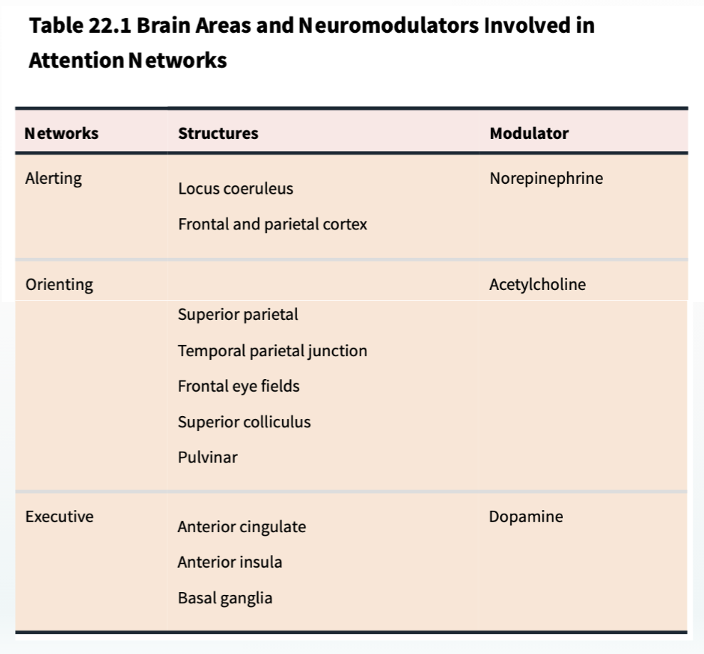
Cognitive control and the Brain

(Attention) Found in the following disorders
Attentional problems are common to many disorders
Often in combination with EF: inhibition, working memory
