622 11/11 GLA pt 4 Skin Infections
 Call Kai
 Learn
 Practice Test
 Spaced Repetition
 Match
 Flashcards
 Knowt Play
1/75

There's no tags or description
Looks like no tags are added yet.
76 Terms
Localized Fungal Infections
fungal infections of the skin
4 Localized Fungal Infections
superficial
cutaneous
subcutaneous
opportunistic
Superficial Fungal Infections (Simple)
involve only the stratum corneum (keratinized outermost layer of epidermis)
cause no inflammation, largely asymptomatic
Cutaneous Fungal Infections (Simple)
deeper layers of the epidermis and dermis but also the hair and nails
can be symptomatic or asymptomatic
Subcutaneous Fungal Infections (Simple)
deeper structures of integument below dermis (blood vessels, nerves), are asociated with trauma
symptomatic, and produce an inflammatory granuloma
Opportunistic Fungal Infections (Simple)
can spread from the site of infection to distant organs to produce inflammatory granulomas and severe systemic disease
When are people most likely to get a fungal infection?
weakened immune systems (immunocompromised)
suffers a direct skin injury
Body Reaction to Fungi Penetrating Stratum Corneum
cell mediated type 4 hypersentitivity response
Main Superficial Fungal Infection
pityriases versicolor (tinea versicolor)
What fungi causes the superficial fungal infection of Pityriases veriscolor?
malassezia furfur
Malassezia furfur Epidemiology
group of very contiagious fungi species, outbreaks are common in otherwise healthy people, transmission is through direct human contact
risk factors are folliculitis, dacrycocytotis, immunocompromisation
Pityriases Versicolor Clinical Presentation
multple oval to round patches or plawues with fine scales
can be hypopigmented, brown, or pink
otherwise asymptomatic with mild itching

Malassezia furfur Pathophysiology
fungi part of normal skin in stratum corneum, depend on lipds for growth and survival
yeast proliferates is presence of oil and converts to hyphal fungal form
How does Malassezia furfur pathophysiology contribute to where it infects?
it occurs in more oil-prone areas of the skin such as the chest, back, neck, and face
mostly in warm, humid climates
Pityriases Versicolor Diagnosis
wood lamp is used to examine growth, and m furfur fungi will emit a yellowish or organe glow
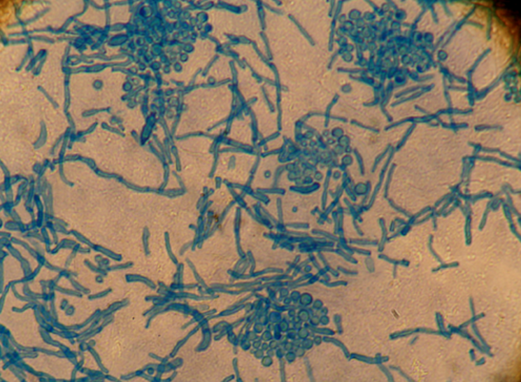
KOH prep of scales would show normal yeast forms and “spaghetti and meatballs” hyphal form
Dermatophytes
filametous fungi that grow in the skin and require keratin for growth
cause cutaneous fungal infections
Tinea
word used to denote dermatophyte infection
presents with annular lesions on the trunk and extremities with a scaly border and central clearing
3 Dermatophytes
microsporum (rubrum and canis)
epidermophyton (floccosum)
trichophyton (rubrum and tonsurans)
Dematophye Infection Epidemiology
common even in healthy
risk factors are persistently damp clothes (athletes), warm and humid climates, sharing person items, poor hygiene
Dematophye Infection Clinical Presentation
itching and scaly rash
Tinea Capitis
dermatophyte infection that presents as dry, scaly patches of alopacia that can sometimes progress to a boggy purulent plaque called keroin

Keroin
progression of tinea capitis

Dermatophytes Infection Pathophysiology
keratinases allow them to break down keratin to invade and replicate in the hair, skin, and nails
Dermatophytes Infection Diagnosis
recognition of rash and symptoms
KOH can reveal septal hypal (cannot differentiate between sepecies)
culture and sporulation can be used to differentiate
wood lamp

Microsporium
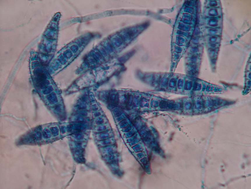
dermatophyte, spindle-shaped
infect skin and hair
Epidermophyton
dematophyte, beaver’s tail
infect skin and nails

Trichophyton
dermatophyte, cigar shaped
infect skin, hair, and nails

Tinea Summary Table

Most Important Subcutaneous Fungal Infection
lymphocutaneous sporotrichosis
aka rose gardener’s disease
What fungi causes lymphocutaneous sporotrichosis?
sporothrix schenckii
Sporothrix schenckii
fungi, dimorphic, found in soil and on plants
Lymphocutaneous sporotrichosis Epidemiology
people who handle plant matter and live in an endemic area (tropical adn subtropical)
minor cuts, punctures, abrasions on skin can serve as entry point for s schenckii fungus
also risk for immunocompromised
Lymphocutaneous sporotrichosis Clinical Presentation
several weeks after injury as ascending, linear, erythematous localized nodulas that travel in lymphatic pattern (sporotrichoid spread)

Lymphocutaneous sporotrichosis Diagnosis
skin biopsy and fungal tissue culture
s scheneckii has cigar-shaped yeast form, asteroid bodies (large eosinophilic spiky structures)

Main Opportunistic Cutaneous Fungal Infection
mucocutaneous cadidiasis
What fungi is responsible for the opportunistic infection mucocutaneous cadidiasis?
cadida albicans
Candida albicans Epidemiology
fungus that can be endogenous or exogenous
risk factors are weakened immune system, cancer, diabetes, chemo, organ transplant, prolonged hostpitalization, catheters, broad-spectrum antibiotics
Candida albicans Clinical Presenation
brightly erythematous patches and plaques, sometimes with satellite papules and pustules
most common where skin rubs together or where it is warm

Other Presentations of Candida albicans
severe rash in diaper area of infants
females with vulvovaginitis
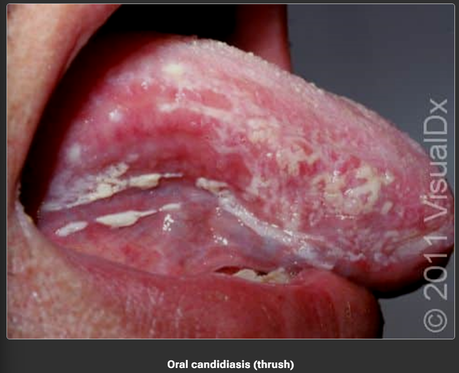
thrush (oral) with white exudate on surface of tongue
Systemic Candida albicans
hypotension, fever, confusion (shock)
may colonize heart valves (endocarditis) leading to heart murmurs or heart failure
Candida albicans Pathophysiology
seen when normal fungis overcomes usual barrier due to compromise of barrier
HIV, AIDS, chronic granulomatous disease, glucocorticoid use, antibiotics
Candida albicans Diagnosis
KOH, dimorphic (yeast in cold, mold in warm)
calcofluor white

4 Fungal Infections and their Fungi
pityriasis versicolor (Malassezia furfur)
dermatophytes
lymphocutaneous sporotrichosis (Sporothrix schenckii)
mucocutaneous candidiasis (Candida albicans)
2 Medically Important Classes of Arthorpods
insecta
arachnida
Class Insecta
flies, fleas, lice, bees
three pairs of walking legs
Class Arachnida
spiders, ticks, mites
four pairs of walking legs
Instars
stages between arthropod molting
Nymph vs Larva vs Pupa
nymph is little adult instar
larva is worm like
pupa is nonfeeding worm (between larva and adult)
Infestation
when an ectoparasite causes disease while feeding or takin up abode on the skin and underlying tissues
Arthropods that Cause Infestations in Humans
itch mite (scabies)
fleas
ticks
bedbugs
louse
maggots
Itch Mite
Sarcoptes scabiei var hominis, ectoparasite that causes scabies
female burrows into skin to lay eggs in upper epidermis, never leaves burrow
females and males die and stay in the skin

Scabies Pathophysiology
arises from close contact, sexual transmission common in young adults
Scabies Clinical Presentation
itching (esp in interdigital folds, sides of fingers, butt, wrist, elbow), short skin lesions, straight or serpingious skin burrow in epidermis
Scabies Hypersensitivity
type 4 reaction to mites or eggs occurs 30 days after infestation (responsible for intense pruritus that is hallmark of disease)
those already sensitized from prior infestation will develop symptoms in 1-4 days
Norwegian Scabies (Crusted)
higly contagious, infested with thousands at a time, scaly areas take on wart-like appearnce
in immunocompromized, old, or physically debilitated
in elbows, knees, palms, scalp, soles of feet
Scabies Mortaility/Morbidity
unlikely to cause mortality or morbidity
spreads in endemic fashion among people in crowded conditions
Scabies Demographic
noone is immune
<15 have highest prevalence, sexually active, debilitated, immunocompromised
When should scabies diagnosis be considered?
severe persistent pruritus
fall and winter months
if multiple family members are involved
Scabies Primary Lesion
short elevated s-shaped tract in superficial epidermis (burrow)
gray-brown wavy channel on skin
occasionally mite is visible to naked eye as small white dot
small vesicle or papule may appear at the end of the burrow or occur independently

Variations in Sacbies Lesions

Scabies Differential
isect bites, atopic dermatitis, contact dermatitis, psoriases
Scabies Diagnosis
mites and its eggs in skin scrapings
elevated IgE titers and eosinophilia
Scabies Treatment
prescribe an appropriate scabicine (5% permethrin cream)
provide relief of symptoms, itching may persist from 1-2 wekks, pruritus may be alleviated partially with oral anihistamine
treat household memberns and personal contacts
provide reassurance that scabies is not a areflection of poor personal hygiene
Scabies Medication
scabicides should be prescribed for patients, household members, and close personal contacts
symptomatic treatment may require antihistamines
more severe symptoms may require a short course of topical or oral steroids
secondary infections may require antibiotics
Bedbugs
hide in household items and attack humans at night through CO2 sensory to get blood meal
Bedbugs Lesions
pruritic erythematous lesions, often in a rough line ro cluster on face, neck, arms and hands perhaps a day or two later
small, red and swollen across the skin with a point or dot in the centera and itching
Inspection for Bedbug Infestation
inspect crevices in walls, mattresses and furniture for the bugs
dark specks – these are bedbug excrement, often found along mattress seams
empty exoskeletons – these are empty skins left behind when the bedbugs molt as they mature
bloody smears – they may find small blood smears on the bed sheets
Bedbugs Itching Treatment
corticosteroid cream or oral antihistamines
Head Lice
Pediculus humanus capitis, live on scalp of humans, hatch from grayish eggs calle nits that female louse attaches to hair shaft with glue like substance
eggs must be warm to hatch, any only hatch near scalp
feed on blood, can life on human 2-3 days, host-specific
Head Lice Transmission
fomites, they can only crawl
Head Lice Itching
caused by irritating saliva injected into bite, mild or intense
pt may also feel ticking feeling of something moving in the hair
occasionally, scratching of the bites can lead to secondary bacterial infection of the site
Head Lice Treatment
medicated shampoos kill lice and eggs
permathrin-based drugs
use fine-tooth nit comb to remove all remaining nits, comb as close to the scalp as possible
repeat if they reappear
all household members and close contacts would be checked
Cleaning Fomites
washable items in hot water and dried in hot drier
unwashable in hot dryer for 10 mins or in a plastic bag for 10 days
dry cleaning
soak combs, brushes, hair clips
Child Prevention of Head Lice
they can go back to school or day care the morning after treatment
don’t borrow anything you put on your head, hang coats so they don’t touch
notify school, parents, and family
Body Lice Epidemiology
people who live in crowded conditins, own one set of clothes do not have access to hot water for regular bathing and laundering of clothes
prevalence has declined in recent years bc people don’t wear the same clothes all the time
greater in higher altitides where cold climate necessitates heavier dressing
Head Lice vs Body Lice Table
