Ocular Anatomy & Physiology
 Call Kai
 Learn
 Practice Test
 Spaced Repetition
 Match
 Flashcards
 Knowt Play
1/58

There's no tags or description
Looks like no tags are added yet.
59 Terms
⭐ Q: How does orbital anatomy differ between dogs, cats, and horses, and what are the functional implications?
Dogs and cats have an incomplete bony orbit that is closed laterally by an orbital ligament, whereas horses have a complete bony orbit. Cats have more extensive frontal and zygomatic processes than dogs, providing greater protection to the globe and requiring a shorter orbital ligament.
🔎 What this means:
Dogs: Incomplete bony orbit with a relatively larger ligamentous portion.
Cats: Incomplete orbit but more extensive bony protection.
Horses: Complete bony orbital rim without an orbital ligament.
An incomplete orbit permits greater jaw mobility but provides less rigid protection to the eye.

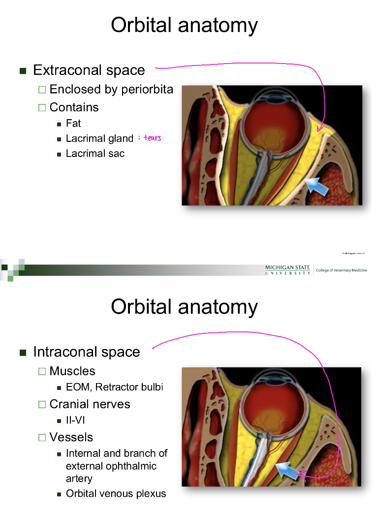
⭐ Q: What are the extraconal and intraconal spaces of the orbit, and what structures do they contain?
The extraconal space is the orbital region outside the extraocular muscle cone, enclosed by the periorbita, and contains fat, the lacrimal gland, and the lacrimal sac.
The intraconal space lies within the muscle cone and contains extraocular muscles, the retractor bulbi, cranial nerves II–VI, and major orbital blood vessels. These spaces are separated by fascia.
🔎 What this means:
Extraconal: Outside the muscle cone; contains fat and lacrimal structures.
Intraconal: Inside the muscle cone; contains major ocular muscles, nerves, and vessels.
This distinction helps clinicians localize an orbital mass based on how it displaces the globe.
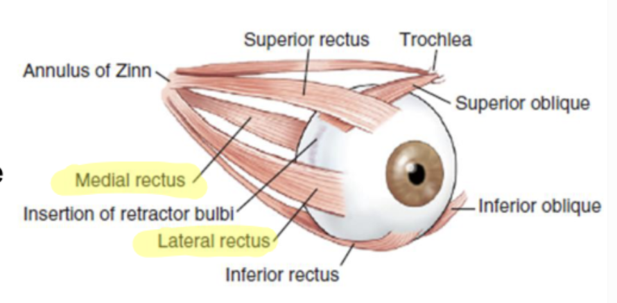
⭐ Q: Which cranial nerves innervate the extraocular muscles, and which muscles does each control?
The oculomotor nerve (CN III, 3) innervates the dorsal, ventral, and medial rectus muscles and the ventral oblique muscle.
The trochlear nerve (CN IV, 4) innervates the dorsal oblique muscle.
The abducens nerve (CN VI, 6) innervates the lateral rectus and retractor bulbi muscles.
🔎 What this means:
CN III: Most extraocular muscles.
CN IV: Dorsal oblique muscle.
CN VI: Lateral rectus and retractor bulbi.
The retractor bulbi retracts the globe backward into the orbit.
💡 Think: CN III does most of the work.
Remember the exceptions: CN IV controls the dorsal oblique, and CN VI controls the lateral rectus and retractor bulbi.
Q: How can dysfunction of cranial nerves III, IV, or VI affect ocular movement?
Dysfunction of CN III can impair movement of the globe by several extraocular muscles,
while CN IV dysfunction affects the dorsal oblique muscle and its rotational action.
Dysfunction of CN VI impairs lateral movement of the globe and its retraction into the orbit.
🔎 What this means:
The pattern of abnormal eye movement can help localize which cranial nerve is affected because each nerve supplies a specific set of muscles.
⭐ Q: What are the major anatomical components of the eyelid, and what are their functions?
The eyelid consists of skin and adnexa, muscular layers, glands, blood vessels, nerves, and palpebral conjunctiva. These structures collectively protect the eye, allow blinking, support tear-film production, and line the inner eyelid with a protective mucous membrane.
🔎 What this means:
Skin and adnexa: External covering and associated structures, including eyelashes.
Muscles: Control eyelid movement.
Glands: Produce components of the tear film.
Palpebral conjunctiva: Mucous membrane lining the inner eyelid.
⭐ Q: Which muscles control eyelid closure and elevation, and how are they innervated?
The orbicularis oculi closes the eyelids and is innervated by the facial nerve (CN VII).
The levator palpebrae superioris elevates the upper eyelid and is innervated by the oculomotor nerve (CN III).
Müller's muscle contributes to upper eyelid elevation and receives sympathetic autonomic innervation.
🔎 What this means:
Orbicularis oculi — CN VII: Closes the eyelids.
Levator palpebrae superioris — CN III: Raises the upper eyelid.
Müller's muscle — sympathetic: Helps maintain upper eyelid elevation.
💡 Think: CN VII closes; CN III opens; sympathetic tone helps keep the eyelid elevated.

⭐ Q: Why can loss of sympathetic innervation cause ptosis in an animal with Horner syndrome?
Loss of sympathetic innervation causes ptosis, or drooping of the upper eyelid, because Müller's muscle no longer receives the stimulation needed to help maintain eyelid elevation. Loss of sympathetic stimulation to the iris dilator muscle also produces miosis, or a constricted pupil.
🔎 What this means:
This explains two findings Dr. Pirie associates with Horner syndrome:
Ptosis: Drooping upper eyelid.
Miosis: Abnormally small pupil.
Both result from loss of sympathetic function.

⭐ Q: What are the meibomian glands, and how do their secretions contribute to tear-film stability?
Meibomian glands are sebaceous glands located in the eyelids that produce a fatty secretion called meibum.
Meibum forms the outer lipid layer of the tear film, reducing evaporation and helping maintain a stable ocular surface.
🔎 What this means:
Dysfunction of these glands can cause a qualitative tear-film problem even when the eye produces adequate aqueous tears.

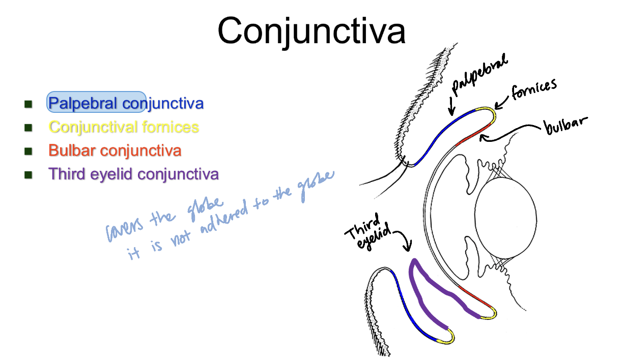
⭐ Q: What are the palpebral conjunctiva, bulbar conjunctiva, and conjunctival fornices, and how do they differ?
The palpebral conjunctiva lines the inner surfaces of the eyelids, while the bulbar conjunctiva covers the anterior surface of the globe outside the cornea. The conjunctival fornices are the folds where the palpebral and bulbar conjunctiva meet.
🔎 What this means:
Palpebral: Inner eyelid lining.
Bulbar: Covering of the globe.
Fornices: Folds connecting the two regions.
The bulbar conjunctiva is generally loosely attached to the globe, except at the limbus, where it becomes firmly attached.
⭐ Q: What are conjunctival goblet cells, and why are they important for the tear film?
Conjunctival goblet cells are specialized cells within the conjunctiva that secrete mucin, which forms the innermost layer of the tear film. Mucin helps the aqueous tear layer spread evenly across the corneal surface and supports tear-film stability.
🔎 What this means:
Chronic conjunctival disease can reduce goblet cell numbers, disrupting tear-film stability even when aqueous tear production is not primarily deficient.
⭐ Q: What is the third eyelid, and what are its major structural and functional components?
The third eyelid, also called the nictitating membrane, is a conjunctiva-covered structure located in the ventromedial orbit. It contains T-shaped cartilage for support, lymphoid tissue for local immune defense, and a third eyelid gland that contributes to aqueous tear production.
🔎 What this means:
T-shaped cartilage: Supports the third eyelid.
Third eyelid gland: Produces part of the aqueous tear film.
Lymphoid tissue: Contributes to ocular surface immune defense.
The professor also describes scrolling or eversion of the T-shaped cartilage as a clinically relevant abnormality.

⭐ Q: What are the three layers of the normal tear film, what produces each, and what are their primary functions?
The lipid layer is the outermost tear-film layer, produced by the meibomian glands to reduce evaporation.
The aqueous layer is the middle layer, produced by the lacrimal gland and third eyelid gland to provide moisture, nutrients, and ocular protection.
The mucin layer is the innermost layer, produced by conjunctival goblet cells to help tears spread and adhere to the corneal surface.
🔎 What this means:
Lipid — meibomian glands: Prevents excessive evaporation.
Aqueous — lacrimal and third eyelid glands: Moisturizes and protects the eye.
Mucin — conjunctival goblet cells: Promotes even tear distribution.
💡 Think: Lipid seals, aqueous hydrates, mucin spreads.

Q: How do quantitative and qualitative tear-film abnormalities differ?
A quantitative tear-film abnormality involves insufficient tear production, particularly the aqueous component, whereas a qualitative abnormality involves altered tear-film composition or stability despite potentially adequate tear volume.
🔎 What this means:
Quantitative: Not enough aqueous tears.
Qualitative: Tears are present but their composition or stability is abnormal.
For example, meibomian gland dysfunction can affect the lipid layer, while chronic conjunctivitis may reduce mucin-producing goblet cells.
⭐ Q: What is the Schirmer tear test (STT), and which component of the tear film does it primarily evaluate?
The Schirmer tear test (STT) is a clinical test used to measure tear production, primarily assessing the aqueous component of the tear film. It helps identify inadequate aqueous tear production associated with dry eye.
⭐ Q: What is the pathway of tear drainage through the nasolacrimal system?
Tears enter the dorsal and ventral lacrimal puncta near the medial corner of the eye, travel through the lacrimal canaliculi, and collect in the lacrimal sac. They then drain through the nasolacrimal duct toward the nose.
🔎 What this means:
Puncta: Two small openings where tears enter.
Canaliculi: Small channels carrying tears from the puncta.
Lacrimal sac: Collects tears before drainage.
Nasolacrimal duct: Carries tears toward the nasal cavity.
Obstruction of this pathway can interfere with normal tear drainage.

⭐ Q: What are the three tunics of the globe, what structures comprise each, and what are their primary functions?
The fibrous tunic is the outer protective layer composed of the cornea and sclera.
The vascular tunic (uvea) is the middle layer composed of the iris, ciliary body, and choroid, which provide vascular support and regulate ocular functions.
The nervous tunic is the innermost layer, consisting primarily of the retinal pigment epithelium and neurosensory retina, which participate in visual function.
🔎 What this means:
Fibrous: Cornea and sclera — protection and refraction.
Vascular: Iris, ciliary body, and choroid — pupil regulation, aqueous humor production, and vascular support.
Nervous: Retina — detection and processing of visual information.

⭐ Q: What are the anterior chamber, posterior chamber, and vitreous chamber, and where is each located?
The anterior chamber is the space between the cornea and iris.
The posterior chamber is the space between the posterior iris and lens.
The vitreous chamber is the large space between the posterior lens and neural retina.
🔎 What this means:
Anterior chamber: Cornea to iris.
Posterior chamber: Iris to lens.
Vitreous chamber: Lens to retina.
The anterior and posterior chambers contain aqueous humor, while the vitreous chamber contains the vitreous body.

⭐ Q: What is the cornea, and how does its structure support transparency, protection, and refraction?
The cornea is the transparent anterior portion of the fibrous tunic that protects the eye and refracts incoming light.
Its highly organized collagen arrangement allows light transmission with minimal scattering, while its normal lack of blood vessels, pigment, and myelinated nerve fibers helps preserve transparency.
🔎 What this means:
Refraction is the bending of light as it passes between materials.
The cornea contributes significantly to the eye's ability to focus incoming light. Disruption of its collagen organization or hydration can interfere with transparency.
Q: What are the four major layers of the cornea in dogs and cats, and how are they arranged?
The four major corneal layers, from anterior to posterior, are the anterior epithelium, stroma (substantia propria), Descemet's membrane, and corneal endothelium.
The anterior epithelium forms the surface barrier, the stroma comprises most of the corneal thickness, Descemet's membrane supports the endothelium, and the endothelium regulates corneal hydration.
🔎 What this means:
Epithelium: Outer protective surface.
Stroma: Main collagen-rich structural layer.
Descemet's membrane: Posterior supporting membrane.
Endothelium: Maintains appropriate corneal hydration.
The lecture notes that some species possess an additional anterior limiting membrane called Bowman's membrane.

Q: Why can corneal endothelial dysfunction cause the cornea to appear cloudy or bluish?
Corneal endothelial dysfunction can cause cloudiness because endothelial cells normally maintain the cornea's relatively dehydrated state.
When these cells become dysfunctional, fluid accumulates in the corneal stroma, causing corneal edema that disrupts normal collagen organization and scatters light.
🔎 What this means:
Edema is abnormal fluid accumulation within tissue. Dr. Pirie specifically describes the bluish appearance associated with corneal edema.
⭐ Q: What is the limbus, and why is it an important anatomical landmark?
The limbus is the transition zone where the transparent cornea meets the sclera and bulbar conjunctiva.
It is an important anatomical and surgical landmark because it marks the boundary between the cornea and sclera and is the region where the bulbar conjunctiva becomes firmly attached to the globe.
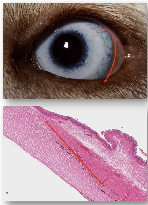
Q: What is the sclera, and how does its structure and function differ from the cornea?
The sclera is the opaque posterior portion of the fibrous tunic that provides structural support and protection to the globe.
Unlike the transparent cornea, which has highly organized collagen, the sclera contains more irregularly arranged collagen, contributing to its opaque appearance.
🔎 What this means:
The sclera forms most of the fibrous tunic and is commonly recognized as the white of the eye.

⭐ Q: What is the uvea, and which structures form its anterior and posterior portions?
The uvea is the vascular tunic of the globe. think of it as a network of blood vessels
The anterior uvea consists of the iris and ciliary body, while the posterior uvea consists of the choroid, which lies between the sclera and retina.
🔎 What this means:
Anterior uvea: Iris and ciliary body.
Posterior uvea: Choroid.
The uvea is especially important for pupillary function, aqueous humor production, and nutritional support of ocular tissues.

⭐ Q: What are the iris sphincter and dilator muscles, and how does their autonomic innervation regulate pupil size?
The iris sphincter muscle contracts under parasympathetic stimulation to constrict the pupil, producing miosis.
The iris dilator muscle contracts under sympathetic stimulation to enlarge the pupil, producing mydriasis.
The balance between these two smooth muscles regulates pupil diameter.
🔎 What this means:
Sphincter — parasympathetic: Constricts the pupil (miosis).
Dilator — sympathetic: Enlarges the pupil (mydriasis).
Loss of sympathetic stimulation impairs pupil dilation and can produce miosis, as described in Horner syndrome.
💡 Think: Parasympathetic makes pupils smaller; sympathetic makes them larger.
🟡 Q: What are the corpora nigra (granula iridica), and in which animals are they commonly present?
Corpora nigra, also called granula iridica, are normal pigmented projections along the pupillary margin of the iris in horses and several other large domestic species.
They are thought to help reduce excessive light entering the eye by acting like a visor.

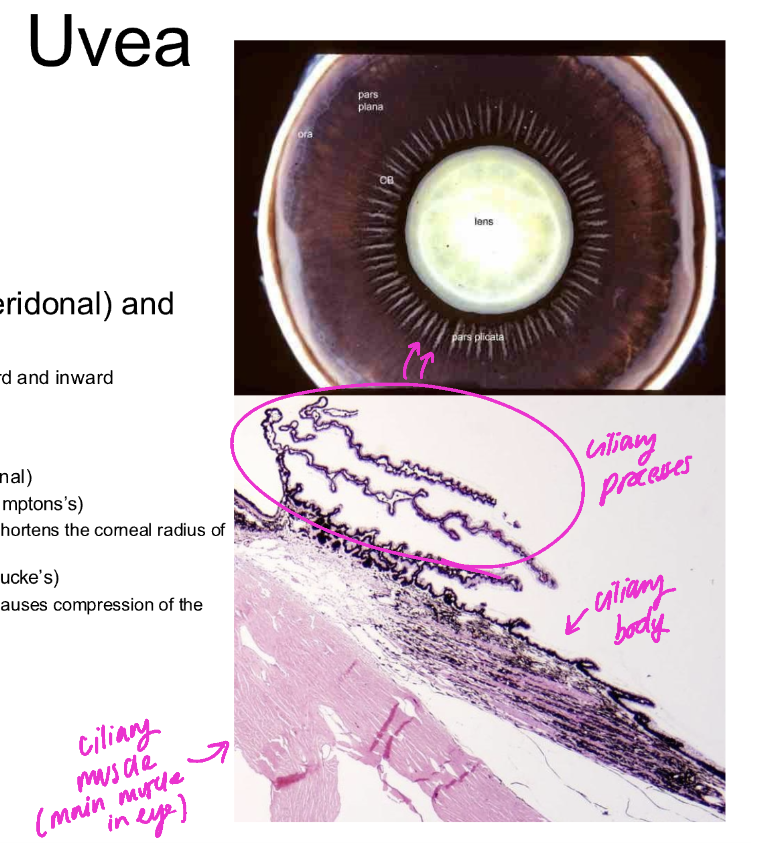
⭐ Q: What are the ciliary processes, and what are their two major functions?
The ciliary processes are vascular ridges extending from the ciliary body behind the iris.
They are involved in aqueous humor production and provide attachment sites for zonular fibers, which help suspend the lens.
🔎 What this means:
Aqueous humor production: Supplies fluid that nourishes avascular ocular structures.
Zonular attachment: Helps support and position the lens.

⭐ Q: What is the ciliary muscle, and how does it contribute to accommodation?
The ciliary muscle is a smooth muscle within the ciliary body that changes the tension and position of structures supporting the lens.
Its contraction and relaxation help alter lens shape and focusing power during accommodation, allowing the eye to adjust between near and distant objects.
🔎 What this means:
Accommodation is the eye's ability to change its optical focusing power.
Dr. Pirie emphasizes that dogs and cats have relatively limited accommodative ability compared with humans.
He also identifies the ciliary muscle as an important source of discomfort during intraocular inflammation.

⭐ Q: What is the choroid, where is it located, and what is its primary function?
The choroid is the vascular posterior portion of the uvea, located between the sclera and retina.
Its primary function is to provide vascular and nutritional support to the retina, and it contains the reflective tapetum lucidum in species that possess one.

Q: What is the tapetum lucidum, where is it located, and how does its structure differ between carnivores and herbivores?
The tapetum lucidum is a reflective structure within the choroid, beneath the retina, that reflects light back through the retina and contributes to improved light sensitivity.
Carnivores have a predominantly cellular tapetum, while herbivores have a predominantly fibrous tapetum.
🔎 What this means:
Cellular tapetum: Found in carnivores such as dogs and cats.
Composed of rectangular cells containing rodlets (no need to know ig?)
Fibrous tapetum: Found in herbivores such as horses and cattle.
Is primarily acellular, it contains few fibrocytes (no need to know ig?)
The tapetum produces the characteristic eyeshine seen when light reflects from an animal's eyes.
⭐ Q: What are the retinal pigment epithelium and neurosensory retina, and how do they differ in function?
The retinal pigment epithelium (RPE) is the outermost layer of the retina and provides support for the photoreceptors rather than directly detecting light.
The neurosensory retina contains the neurons, including photoreceptors, that detect light and transmit visual information toward the brain.

⭐ Q: How is light converted into a neural signal in the retina, and which three major neuron types transmit the information?
Light passes through the inner retinal layers and reaches the photoreceptors (rods and cones), which convert light energy into electrical signals.
These signals pass to bipolar cells and then to ganglion cells, whose axons collectively form the optic nerve (CN II).
🔎 What this means:
Photoreceptors: Detect light and initiate electrical signaling.
Bipolar cells: Relay information from photoreceptors.
Ganglion cells: Send visual information out of the eye through the optic nerve.
PR - photoreceptor layer
Dr. Pirie explicitly emphasizes understanding this three-neuron arrangement rather than memorizing every retinal layer.

Q: What is the difference between nuclear and plexiform layers of the retina?
Nuclear layers contain neuronal cell bodies, while plexiform layers are regions where retinal neurons form synaptic connections.
The outer nuclear layer contains photoreceptor cell bodies, the inner nuclear layer contains bipolar and other neuronal cell bodies, and the ganglion cell layer contains ganglion cell bodies.
🔎 What this means:
Nuclear layers: Where neuronal cell bodies are located.
Plexiform layers: Where neurons communicate through synapses.
This distinction is more important for this lecture than memorizing the names of all ten retinal layers.
Q: What is the difference between rods and cones, and why are most domestic mammals considered to have rod-dominant retinas?
Rods are photoreceptors specialized for low-light vision and sensitivity to movement,
while cones support color vision and greater visual detail under brighter conditions.
Many domestic mammals have rod-dominant retinas, supporting sensitivity in dim lighting rather than the high visual acuity characteristic of cone-rich retinal regions.
Q: Where is the potential space associated with retinal detachment, and why is it clinically important?
The potential space associated with retinal detachment lies between the retinal pigment epithelium (RPE) and the photoreceptor layer.
Fluid accumulation in this space can separate the neurosensory retina from the RPE, producing retinal detachment and disrupting normal visual function.
🔎 What this means:
Dr. Pirie uses systemic hypertension in cats as an example: increased choroidal vascular pressure can cause fluid leakage and subsequent retinal detachment.

Q: What is the area centralis, and how does it differ from the surrounding retina?
The area centralis is a specialized retinal region associated with higher visual acuity and a greater concentration of neurons involved in detailed vision.
It can be distinguished histologically by an increased density of ganglion cells compared with surrounding retinal regions.
⭐ Q: What are the main anatomical components of the lens, and how are they organized?
The lens is a transparent, avascular structure surrounded by anterior and posterior capsules.
A layer of lens epithelial cells lies beneath the anterior capsule and continuously produces lens fibers, which form the bulk of the lens.
The older fibers form the central nucleus, while newer fibers form the peripheral cortex.
🔎 What this means:
Lens capsule: Outer covering.
Lens epithelial cells: Cells that produce new lens fibers.
Lens cortex: Peripheral, newer fibers.
Lens nucleus: Central, older fibers.
The lens fibers are highly organized to minimize light scattering and preserve transparency.
⭐ Q: Why is the lens transparent and avascular, and how does it receive nutrients?
The lens is transparent because its specialized, highly organized lens fibers minimize light scattering, and it lacks blood vessels that would interfere with light transmission.
Because it is avascular, it depends primarily on the surrounding aqueous humor for nutrients.

Q: What are anterior and posterior lens sutures, and how can their orientation help localize a lens opacity?
Lens sutures are patterns formed where lens fibers meet.
In dogs, cats, and horses, the anterior sutures form an upright Y, while posterior sutures form an inverted Y.
Recognizing these patterns can help determine whether a lens opacity lies anteriorly or posteriorly.
🔎 What this means:
Anterior suture: Upright Y.
Posterior suture: Inverted Y.
Dr. Pirie emphasizes this as a practical way to localize certain cataracts.

Q: How do nuclear sclerosis and cataracts differ?
Nuclear sclerosis is an age-related change in the lens nucleus that causes increased hardness and a bluish or hazy appearance.
A cataract is an opacity caused by loss of normal lens transparency and organization.
Nuclear sclerosis is not itself a cataract.
🔎 What this means:
Dr. Pirie describes nuclear sclerosis as resembling a frosted marble, whereas cataracts involve disruption of lens transparency.
⭐ Q: What are lens zonules, and why can damage to them cause lens luxation?
Lens zonules are suspensory fibers extending from the ciliary body to the lens, helping maintain its normal position.
Damage or degeneration of these fibers reduces lens support and can cause lens luxation, in which the lens becomes displaced from its normal position.
🔎 What this means:
Dr. Pirie identifies Jack Russell Terriers as a breed associated with inherited lens luxation.
⭐ Q: How does the lens contribute to accommodation, and why is this process relatively limited in dogs and cats?
The lens contributes to accommodation by changing its optical power as ciliary muscle activity alters tension on the zonular fibers.
Dogs and cats have relatively limited accommodative ability, meaning they cannot adjust their focus between distances as effectively as species with greater accommodation.
Q: What is the vitreous body, where is it located, and what are its primary functions?
The vitreous body is a transparent, gel-like substance filling the vitreous chamber behind the lens and accounting for approximately 80% of globe volume.
It transmits light, helps maintain globe and retinal position, and acts as a diffusion barrier.
🔎 What this means:
Dr. Pirie explains that degeneration of the vitreous can reduce its structural support of the retina and may contribute to retinal detachment.
⭐ Q: What is aqueous humor, where is it produced, and how does it flow through the eye?
Aqueous humor is the fluid produced by the ciliary processes that nourishes avascular ocular structures and helps maintain the eye's internal environment.
It enters the posterior chamber, passes through the pupil into the anterior chamber, and drains through structures at the iridocorneal angle.
🔎 What this means:
Production: Ciliary processes.
Posterior chamber: Receives newly produced aqueous humor.
Pupil: Passage between posterior and anterior chambers.
Anterior chamber: Contains aqueous humor before drainage.
Iridocorneal angle: Major drainage region.
Aqueous humor is different from the tear film, which covers the outside of the eye.
⭐ Q: What is the iridocorneal angle, what structures comprise its drainage apparatus, and why is it clinically important?
The iridocorneal angle is the drainage region located between the peripheral cornea/sclera and the peripheral iris/anterior ciliary body.
Its major structures include the pectinate ligament, trabecular meshwork, and angular aqueous plexus.
Impaired aqueous humor drainage through this region can increase intraocular pressure and contribute to glaucoma.
🔎 What this means:
Pectinate ligament: Structural component of the angle.
Trabecular meshwork: Porous drainage tissue.
Angular aqueous plexus: Part of the aqueous outflow pathway.
💡 Think: Aqueous humor must be produced and drained continuously. If drainage is impaired, pressure can increase.
⭐ Q: What is the pathway for conscious vision from the retina to the visual cortex?
Visual information begins in the retina, travels through the optic nerve (CN II), and reaches the optic chiasm, where a species-dependent proportion of fibers cross to the opposite side.
Information then travels through the optic tracts to the lateral geniculate nucleus (LGN) of the thalamus before reaching the occipital visual cortex through the optic radiations.
🔎 What this means:
Retina: Detects light.
Optic nerve (CN II): Carries visual information.
Optic chiasm: Site where some visual fibers cross.
LGN (thalamus): Relays visual information.
Optic radiations: Carry information toward the cortex.
Occipital cortex: Consciously interprets vision.
⭐ Q: What percentage of optic nerve fibers decussate at the optic chiasm in different species, and how does this relate to visual adaptations?
The proportion of optic nerve fibers undergoing decussation is approximately 50% in primates, 65% in cats, 75% in dogs, 80–90% in herbivores and pigs, and 100% in birds and fish.
Greater decussation is generally associated with more laterally positioned eyes and broader visual fields, while more frontally positioned eyes provide greater binocular overlap and depth perception.
🔎 What this means:
Primates: 50%.
Cats: 65%.
Dogs: 75%.
Herbivores/pigs: 80–90%.
Birds/fish: 100%.
Prey species commonly have laterally positioned eyes to detect threats over a wider area. More frontally positioned eyes provide greater overlap between the two visual fields, supporting depth perception.
⭐ Q: What is the menace response, why is it considered a learned response, and which nervous structures are involved?
The menace response is a learned, conscious response to a threatening visual stimulus that normally produces blinking.
It requires the optic nerve (CN II), visual cortex, cerebellum, and facial nerve (CN VII).
Because the stimulus must undergo conscious cortical processing, the menace response is not a simple reflex.
🔎 What this means:
CN II: Carries the visual stimulus.
Visual cortex: Consciously interprets the stimulus.
Cerebellum: Participates in coordinating the response.
CN VII: Activates the orbicularis oculi to close the eyelid.
⭐ Q: When testing the menace response in one eye, which side of the optic nerve, visual cortex, and facial nerve is primarily evaluated?
Testing the menace response in one eye evaluates the ipsilateral optic nerve (CN II), predominantly the contralateral visual cortex, and the ipsilateral facial nerve (CN VII).
🔎 What this means:
For a menace stimulus presented to the left eye:
Left CN II: Carries the afferent signal.
Right visual cortex: Processes most of the crossing visual information.
Left CN VII: Produces eyelid closure.
This is the simplified pathway emphasized by Dr. Pirie for clinical lesion localization.
Q: How is the menace response evaluated, and why can it be absent in a neurologically normal young animal?
The menace response is evaluated by making a noncontact threatening hand movement toward one eye while covering the opposite eye and avoiding air currents or touching facial hairs.
A normal response includes blinking and may include globe retraction or head movement. It can be absent in young animals because it is a learned response that has not fully developed.
🔎 What this means:
The lecture gives approximate developmental ages:
Foals and calves: 1–2 weeks.
Puppies and kittens: 10–12 weeks.
An absent menace response in an immature animal does not automatically indicate blindness.
⭐ Q: What is the dazzle reflex, which cranial nerves are involved, and how does it differ from the menace response?
The dazzle reflex is an involuntary, subcortical blinking response to a bright light stimulus.
It uses the optic nerve (CN II) as its afferent pathway and the facial nerve (CN VII) to produce eyelid closure, with processing through midbrain structures.
Unlike the menace response, the dazzle reflex does not require conscious visual cortex processing.
🔎 What this means:
Stimulus: Bright light.
Afferent: CN II.
Integration: Subcortical/midbrain.
Efferent: CN VII.
Response: Blink.
💡 Think: Menace involves conscious interpretation; dazzle is an automatic response to bright light.
⭐ Q: What is the pupillary light reflex (PLR), and what is its afferent and efferent pathway?
The pupillary light reflex (PLR) is an involuntary constriction of the pupil in response to light.
Its afferent pathway begins in the retina and travels through the optic nerve (CN II) to the midbrain.
Its efferent pathway uses parasympathetic fibers of the oculomotor nerve (CN III), which synapse in the ciliary ganglion before reaching the iris sphincter through the short ciliary nerves.
🔎 What this means:
CN II: Detects and carries the light stimulus.
Pretectal nuclei: Process the sensory input.
Edinger–Westphal nuclei: Initiate parasympathetic output.
CN III: Carries preganglionic parasympathetic fibers.
Ciliary ganglion: Site of synapse.
Short ciliary nerves: Carry postganglionic fibers to the iris sphincter.
Iris sphincter: Constricts the pupil.
Unlike the menace and dazzle responses, the PLR uses CN III rather than CN VII for its efferent component.
⭐ Q: What is the difference between direct and indirect pupillary light reflexes, and how do these responses differ between mammals and birds?
The direct PLR is constriction of the pupil in the eye receiving the light stimulus,
while the indirect (consensual) PLR is constriction of the opposite pupil.
In mammals, the direct response is generally stronger than the indirect response because of the distribution of crossing visual fibers.
The lecture states that birds do not have a true indirect PLR.
🔎 What this means:
If light is directed into the left eye:
Direct PLR: Left pupil constricts.
Indirect PLR: Right pupil constricts.
The distinction is important because examining both responses helps identify which nerve or pathway may be affected.
⭐ Q: How do the menace response, dazzle reflex, and pupillary light reflex differ in their neural pathways and elicited responses?
The menace response is a learned visual response involving CN II, the visual cortex, cerebellum, and CN VII that produces blinking.
The dazzle reflex is a subcortical light-induced blinking reflex involving CN II and CN VII.
The pupillary light reflex is an involuntary pupil-constriction reflex involving CN II and parasympathetic CN III.
🔎 What this means:
Test | Main structures | Expected response |
|---|---|---|
Menace | CN II, cortex, cerebellum, CN VII | Blink |
Dazzle | CN II, midbrain, CN VII | Blink |
PLR | CN II, midbrain, parasympathetic CN III | Pupil constriction |
💡 Think: Menace and dazzle produce blinking through CN VII; PLR produces pupil constriction through CN III.
⭐ Q: True or false: The pupillary light reflex requires integration in the visual cortex.
False.
The pupillary light reflex is an involuntary response integrated through subcortical pathways involving the midbrain.
It does not require the visual cortex because pupil constriction can occur without conscious visual perception.
🔎 What this means:
An animal can have impaired conscious vision while retaining a pupillary light reflex if the required subcortical pathways remain intact.
⭐ Q: How would a unilateral lesion of the optic nerve (CN II) affect the menace response, dazzle reflex, and pupillary light reflexes?
A unilateral optic nerve (CN II) lesion can eliminate the menace and dazzle responses when the affected eye is stimulated because visual sensory input cannot travel normally to the CNS.
It can also eliminate both the direct and indirect PLRs when light is directed into that eye. However, stimulating the unaffected eye can still produce constriction in both pupils if the efferent CN III pathways are intact.
🔎 What this means:
For a right CN II lesion:
Light in right eye: Neither pupil constricts normally.
Light in left eye: Both pupils can constrict if both CN III pathways are intact.
This pattern identifies an afferent problem because the affected eye cannot transmit the sensory signal.
⭐ Q: How would a unilateral lesion affecting the parasympathetic component of CN III alter the direct and indirect pupillary light reflexes?
A unilateral lesion affecting parasympathetic CN III prevents normal constriction of the pupil on the affected side, regardless of which eye receives light.
The opposite pupil can still constrict normally if its own efferent pathway and the afferent visual pathways are intact.
🔎 What this means:
For a right parasympathetic CN III lesion:
Light in right eye: Right pupil fails to constrict; left pupil can constrict.
Light in left eye: Left pupil constricts; right pupil still fails to constrict.
The problem is efferent because the right pupil cannot carry out the motor response.
⭐ Q: How can a facial nerve (CN VII) lesion affect the menace, dazzle, and pupillary light reflex responses?
A facial nerve (CN VII) lesion can prevent blinking during menace and dazzle testing because CN VII innervates the orbicularis oculi muscle responsible for eyelid closure. The PLR can remain intact because pupil constriction uses parasympathetic CN III rather than CN VII.
⭐ Q: Why can an animal with a visual cortex lesion have an absent menace response but preserved dazzle and pupillary light reflexes?
A visual cortex lesion can impair the menace response because conscious cortical processing is required to interpret the threatening stimulus.
The dazzle and pupillary light reflexes may remain intact because both are mediated by subcortical pathways that do not require conscious visual cortex processing.
🔎 What this means:
Preserved dazzle and PLR responses suggest that the involved sensory and reflex pathways are functioning, even if the animal's conscious visual processing is impaired.
⭐ Q: A dog has no menace response, dazzle reflex, or direct PLR when its right eye is stimulated, and its left pupil also fails to constrict when light is directed into the right eye. However, both pupils constrict when light is directed into the left eye. Which cranial nerve is most likely affected, and why?
The right optic nerve (CN II) is the most likely affected structure because all three responses lose their sensory input when the right eye is stimulated.
The preserved pupil constriction when the left eye is stimulated indicates that the efferent CN III pathways can still function.
🔎 What this means:
This is the clinical localization example Dr. Pirie walks through near the end of the transcript.
Absent right menace: Could involve CN II, CN VII, cortex, or cerebellum.
Absent right dazzle: Narrows the possibilities to CN II or CN VII.
Absent PLRs when stimulating right eye: Points to CN II or the afferent visual pathway.
Preserved bilateral PLRs when stimulating left eye: Supports intact CN III efferent pathways.
The common problem is the right afferent optic pathway, most consistent with a right CN II lesion in this simplified example.