ortho conditions
 Call Kai
 Learn
 Practice Test
 Spaced Repetition
 Match
 Flashcards
 Knowt Play
1/106

There's no tags or description
Looks like no tags are added yet.
107 Terms
What is the SALTER levels used for
epiphysis fxs
describe a 1 on the salter harris
slip/seperated/straight fx across the cartilage of the physis
What is the physis
growth plate
describe a 2 on the salter harris
fx above the physis or away from the joint
describe a 3 on the salter harris
fx below the physis in the epiphysis (lower)
describe a 4 on the salter harris
fx is through the metaphysis, physis, and epiphysis
describe a 5 on the salter harris
physis has been crushed

1
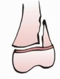
2
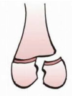
3

4

5
age range for LCP
4-8 y
describe legg-calve perthes (LCP)
avascular event occurring at the femoral head
s/s of LCP
limp
trendelenburg
pain w/ activity
pain in groin/hip/knee
ROM lims for LCP
abd and IR
dx stretegy for LCP
MRI
treatment for LCP
stretching
rest
NSAIDs
bracing?
age range for SCFE
9-16 y
describe slipped capital femoral epiphysis (SCFE)
relative or absolute femoral head displacement
risk factors for SCFE
obesity
nutrition deficiencies
ROM lims for SCFE
hip flexion
abd
IR
s/s of SCFE
limp
pain in groin/hip/knee
posturing in ER
dx strategy for SCFE
XR
treatment for SCFE
surgery
describe developmental dysplasia of the hip (DDH)
wide range of hip disorders that result in increased risk for hip dislocation
when is DDH identified
neonatal screening
What are pre-reqs for normal hip development
normal mm balance
positioning of femoral head in deep acetabulum
How is the acetabulum affected w/ age
depth increases w/ growth
risk factors for DDH
female
breech position
family hx
increased birth weight
special tests for DDH
Barlow’s test
Ortolani sign
describe for barlow’s test
flex and add the hips
apply light pressure on the knee and direct force posteriorly
+ dislocation
describe ortolani’s test
flex hips and knees to 90
thigh gently abd
+ reduce/relocate, may head clunk
what test w/ be positive for more severe DDH
ortolani
What clinical signs may indicate DDH >3 mo
decreased abd
leg length discrepancy
asymmetrical thigh and gluteal folds
dx strategies for DDH
US
XR
What age is the cut off for US when dx DDH
4 mo
if the hip is obviously dislocated, what treatment is done
pavlik harness
What occurs if there is suspected DDH, but none is found
follow up US in 6-8 wks
benefit of pavlik harness
allows ARROM resulting in decreased incidence of avascular necrosis of femoral head
purpose of pavlik harness
restricts ext and add, while allowing abd and flex
how long is a pavlik harness worn
6 wks
what is the best intervention period for pavlik harness
birth to 6 mo
contraindications to pavlik harness
significant m imbalance
significant stiffness in joiints
excessive lig laxity
what is the treatment for DDH from 6 mo to 2 y
closed or open reduction
describe closed reduction
hip reduced while child is under general anesthesia or deep sedation
protocol following closed reduction
hip spica cast for 6 wks, follow up, then another 6 wks
describe open reduction
surgical procedure
protocol following open reduction
hip spica for 12 weeks
potential need for further orthoses (RINO)
what is the treatment for DDH after 2 y
surgery, along w/ femoral shortening or pelvic osteotomy
PT interventions for DDH for infant w/ pavlik harness
screening
PT interventions for DDH after surgery
strengthening
ROM
functional tasks
HEP
name for club foot
congenital talipes equinovarus
resting position for club foot
forefoot add
hindfoot varus
ankle equinus
Why do club feet look smaller
flexible softer heel
causes of club foot
male
smoking during pregnancy
early amniocentesis
viral infections during pregnancy
neurogenic conditions
goals of clubfoot intervention
correct deformity
retain mobility/strength
What are the different approaches for clubfoot treatment
extrinsic and intrinsic
When is extrinsic club foot intervention performed
soft tissue deformity
When is intrinsic club foot intervention performed
rigid deformity
What type of clubfoot deformity is easier to treat
soft tissue
What are the extrinsic club foot interventions
ponsetti method
bracing
sleep braces
describe the ponsetti method
casting to correct
correct cavus
foot from under talus
correct equinus
how long is bracing performed after ponsetti
3 mo
how long is sleep bracing used
2-4 ys
What type of brace is used for sleeping when using the extrinsic club foot intervention method
boots and bar
how long is the ponsetti method
4-6 wk
What are the intrinsic club foot interventions
surgical intervention at 6-9 mo old
types of surgical intervention for clubfoot
release of cords/joint capsule
placing pins
casting
when are clubfoot pinss removed
after 4-6 wks
how long is the clubfoot casted after surgery
6-12 wk
clubfoot interventions after bracing/surgery
ankle stab/strength
LE strengthening
ankle stretches
balance
shoe modifications (smaller size)
define idiopatjic scoliosis
3 dimensional curvature of the spine, with >10 degrees in the coronal plane
how is scoliosis measured
cobb method
what are the 2 types of scoliosis curves
structural
nonstructural
define structural curve
cannot be voluntarily/passively/forcibly corrected
define non-structural curve
full corrects clinically and radiographically
does not have a rotational component
What typically causes non-structural curves
leg length discrepancy
potential causes of scoliosis
growth timing
spine slenderness
genetics
asymmetrical bone loading due to positioning
vestibular dysfunction
purpose the cobb methos
measurement of the curve angle on XR
procedure for cobb method
most cephalic and caudal vert w/ max tilt toward convexity
lines extend outward to create an angle to be measured
how is scoliosis named
for the convexity
what are the 3 subtypes of idiopathic scoliosis
infantile
juvenile
adolescent
age range for infantile scoliosis
0-3
age range for juvenile scoliosis
4- puberty
age range for adolecent scoliosis
puberty (12 F, 14 M)
what type of scoliosis tends to spontaneously resolve
infantile
saying prognosis for surgery to correct scoliosis
>30 degree curve, w onset before puberty = 100% need for surgery
What gender tends to have more severe curvature
female
how fast does adolecent scoliosis progress
1 degree/day of no treatment
what is used to measure skeletal maturity
risser sign
What risser grades are at risk for scoliosis cure progression
0-3
risser sign grade 0
no fusion at the ilium
risser sign grade 1
25% fusion at ilium
risser sign grade 2
50% fusion at ilium
risser sign grade 3
75% fusion at ilium
risser sign grade 4
100% fusion at ilium
risser sign grade 5
full fusion of iliac epiphysis
medical management of <25 degree scoliosis
monitor every 3 mo
medical management of 25-40 degree scoliosis
spinal orthosis