BIOLOGY 1 1st Sem Midterms Summary
 Call Kai
 Learn
 Practice Test
 Spaced Repetition
 Match
 Flashcards
 Knowt Play
1/148

There's no tags or description
Looks like no tags are added yet.
149 Terms
COMPOUND MICROSCOPE
Combination of lenses that produces a highly magnified image of microscopic living entities and other details; has more than one lens and its own light source

MICROGRAPH
Image produced by a microscope
MAGNIFICATION
Measure of an optical instrument for an object to appear larger
RESOLUTION
Clarity of an image
VIDEO MICROSCOPY
Using image processing to view or photograph microscope images on a TV or computer screen
COMPOUND MICROSCOPE (KIND)
Commonly used in schools, can magnify objects up to 2,000x, used to examine cells and thin sections of tissues, uses sunlight or artificial light as its light source

PHASE-CONTRAST MICROSCOPE
Used to examine transparent specimens, such as unstained cells
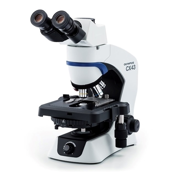
STEREOSCOPIC / DISSECTING MICROSCOPE
Has 2 eyepieces (binocular head), providing a three-dimensional (3D) view, used for biological dissections and entomology

ELECTRON MICROSCOPE
Uses beams of electrons instead of light, can magnify objects up to 10,000,000x, electron micrographs are usually black and white

TRANSMISSION ELECTRON MICROSCOPE (TEM)
Used to study internal structures of cells, usually cut into very thin sections
SCANNING ELECTRON MICROSCOPE (SEM)
Used to examine three-dimensional surface structures and shapes of specimens
ELECTRON MICROSCOPE TYPES
Scanning Electron Microscope (SEM) and Transmission Electron Microscope (TEM)
FLUORESCENCE MICROSCOPE
Uses fluorescent dyes that glow when exposed to specific wavelengths of light, used to study the location of specific molecules or organelles inside cells

CONFOCAL SCANNING MICROSCOPE
Used to examine three-dimensional structure of cells or organelles without physically cutting the specimen into sections, uses laser beams to scan across the specimen, produces detailed sections that can be combined to create a 3D image

FOOT or BASE
Supports the entire weight of the microscope
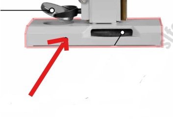
STAGE CONTROL
Stands on the base and supports the stage, and moves the stage clips in horizontal and vertical positions

ARM
A curved, strong structure used to handle the microscope

MECHANICAL STAGE
Flat, rectangular plate connected to the lower end of the arm and the specimen is placed here

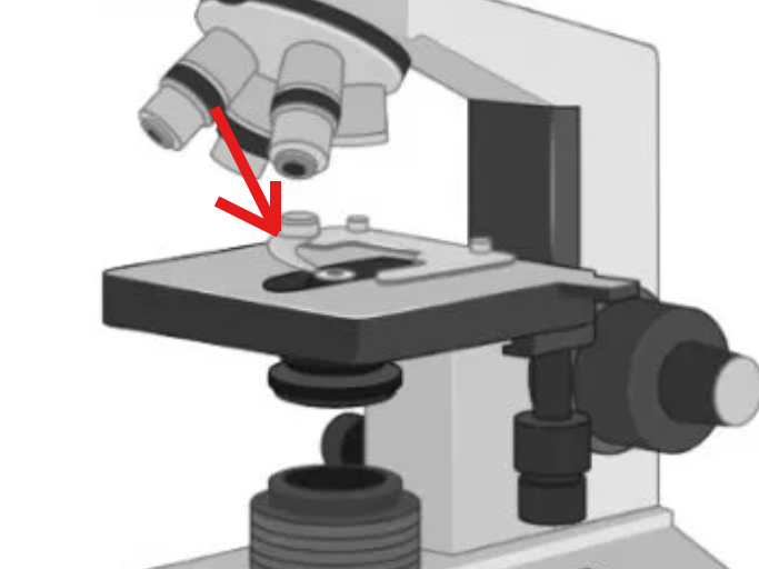
STAGE CLIP
Holds the slide securely in its position and keeps it stable
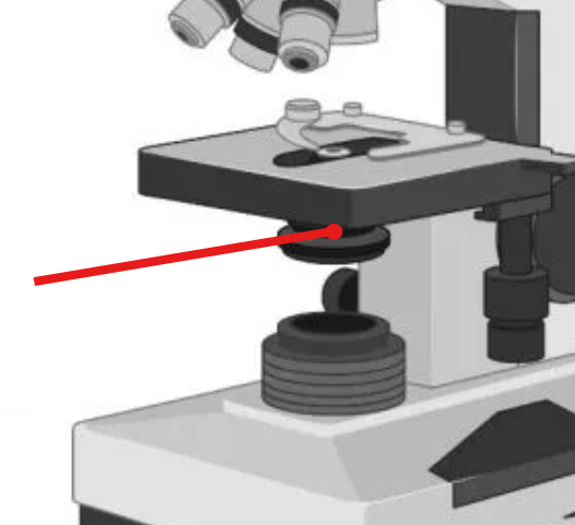
DIAPHRAGM
Controls and adjusts the intensity of light that passes

IRIS DIAPHRAGM
Controls the light with an adjustable opening

DISC DIAPHRAGM
Controls light using different-sized holes

CONDENSER
Gathers and focuses light from the illuminator onto the specimen
NOSEPIECE
Rotating turret that holds multiple objective lenses

BODY TUBE or HEAD
Upper part of the arm comprises a hollow and tubular structure

FINE ADJUSTMENT KNOB
Smaller knob that makes sharp and small focus adjustments (1)

COARSE ADJUSTMENT KNOB
Larger knob that moves the tube/stage up and down and large movement/rough focusing (2)

CONDENSER FOCUS KNOB
A knob that moves the condenser vertically to control the focal concentration of light.

DIOPTER ADJUSTMENT
Adjusts focus for differences between your eyes

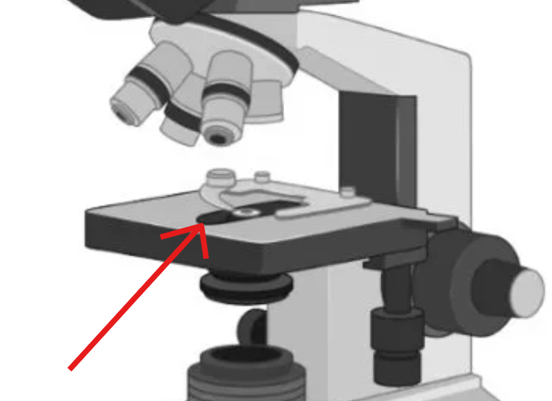
APERTURE
Hole in the middle of the stage that allows light to reach the specimen
POWER SWITCH
Turns the illuminator on and off
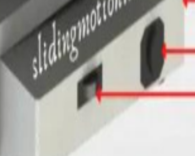
LIGHT INTENSITY CONTROL
Changes the intensity of the light source

INCLINATION JOINT
Pivoting mechanism that allows the upper assembly to tilt backwards

RACK STOP
Safety feature that limits how high the stage rises to prevent the objective from hitting the slide
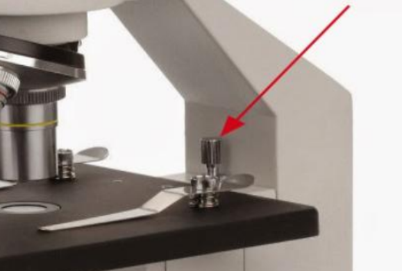
THUMB SCREW
Attaches the microscope head to the arm
EYEPIECE or OCULAR
Located at the top of the body tube; the lens you look through to view the specimen (Typically 10x power, though 15x & 20x exist)

ILLUMINATOR
Light source of the microscope that provides light to illuminate the specimen (Older microscopes may use a mirror to reflect light)
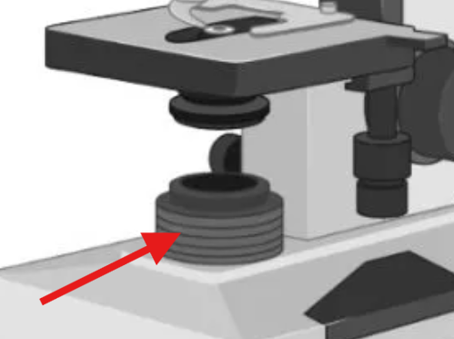
OBJECTIVE LENS
Lenses located at the nosepiece that magnify the specimen at different magnification levels (4x, 10x, 40x, 100x)

4X (SCANNING OBJECTIVE)
Shortest and lowest, used to locate the specimen
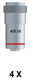
10X (LOW POWER OBJECTIVE)
Middle sized and magnification, used for general overview

40X (HIGH POWER OBJECTIVE)
Longest and highest magnification power, used for detail structure viewing

100X (OIL IMMERSION OBJECTIVE)
Requires a drop of oil to be placed for optical performance by reducing light refraction, used for tiny specimens like bacteria

What happens when magnification is higher?
Higher magnification results in a greater amount of light and detail, but a smaller field of view
TOTAL MAGNIFICATION FORMULA
Eyepiece lens magnification × Objective lens magnification
MAGNIFICATION OF AN IMAGE FORMULA
Total magnification power × Size of the specimen
MICROSCOPE SLIDES
Thin sheet of glass used to hold the specimen in place
COVER GLASS
Avoids contact of the specimen with the lens and holds it in place
SPECIMEN (MICROSCOPY)
Piece or portion of a sample selected for examination
CELL
Basic biological unit of all known living organisms
ROBERT HOOKE
Devised one of the earliest microscopes using a thin slice of cork; called porous structures "cellulae" (Latin for "small room"); first to be credited with publishing the first description of a cell in 1666
Robert Hooke was first to be credited with publishing the first description of a cell in this year
1666
"cellulae"
Latin for "small room"
ANTONIE VAN LEEUWENHOEK
First to study magnified cells; devised his own simple microscope with one lens; observed tiny living organisms in pond water called "animalcules"
"animalcules"
tiny living organisms in pond water
Leeuwenhoek’s achievement
First to study magnified cells
MATTHIAS JACOB SCHLEIDEN
Botanist who focused his interests on the study of plant cells
THEODOR SCHWANN
Examined animal cells and found that all bodies of living organisms are made up of cells
RUDOLF CARL VIRCHOW
Proposed the third tenet in the cell theory, explaining where new cells originate
FIELD OF VIEW (FoV)
How much of the specimen we can be able to see through a microscope
FIELD OF VIEW (mm) FORMULA
Field number of eyepiece / Objective magnification
FIELD NUMBER (FN)
Diameter of the image area that is engraved on the eyepiece
PROKARYOTIC CELL
First kind of cell to develop (~3.5 billion years ago); simple organisms possessing cells that are smaller in size with simple internal structures; reproduce very fast; examples are bacteria, blue-green algae, and archaeans
PLASMA MEMBRANE (PROKARYOTIC)
Semipermeable membrane that allows waste products out while preventing essential products from leaving
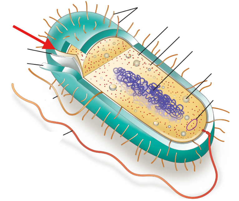
CELL WALL (PROKARYOTIC)
Rigid wall that maintains cell shape, especially when exposed to water influx

CAPSULE
Protective layer made up of polysaccharides lying outside the cell wall

NUCLEOID REGION
Irregular-shaped region without a distinct nucleus where DNA is stored
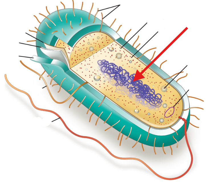
CYTOPLASM (PROKARYOTIC)
Lacks distinct interior compartments, and organelles are spaced

RIBOSOME (PROKARYOTIC)
Small structure where proteins are made
PLASMID
Small, circular DNA molecule found mainly in bacteria that carries additional genetic information

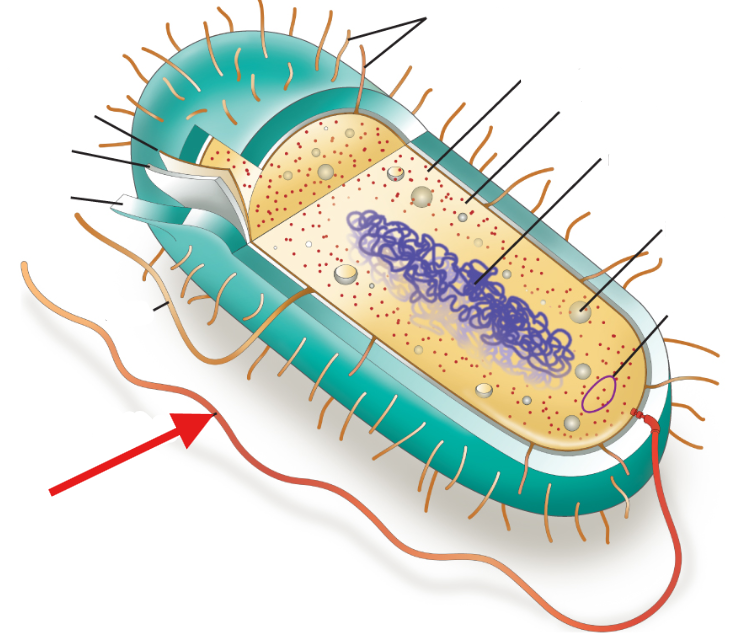
FLAGELLUM (PROKARYOTIC)
Long, whip-like structure that helps a cell move (allows locomotion)
FIMBRIAE
Small, bristle-like fibers that help bacteria attach to surfaces

CONJUGATION PILI
Tubular structures that function for cell-to-cell communication (some bacteria develop drug resistance across generations)
EUKARYOTIC CELL
Evolved about 1.8 billion years ago; larger size and more complex structural components; genetic material (DNA) encased within a distinct nucleus; many membrane-bound interior compartments; examples are fungi, plants, animals, and protists
CELL/PLASMA MEMBRANE (EUKARYOTIC)
Thin barrier that separates the cell from the external environment and acts as a gatekeeper for regulating what enters and leaves
CELL WALL (EUKARYOTIC)
Rigid layer that gives protection, support, and shape to the cell and is made up of the polysaccharide cellulose
CYTOPLASM (EUKARYOTIC)
Jelly-like substance that fills the cell and holds the organelles in place
CYTOSOL
Fluid portion of the cytoplasm, mainly made of water and surrounding the organelles
NUCLEUS (EUKARYOTIC)
Storehouse of genetic information in the form of DNA and protects it from damage
DNA
Long chain of molecules divided into genes that contain instructions for making proteins
CHROMATIN
DNA packaged with histone proteins
NUCLEUS ENVELOPE
Encloses the DNA and contains holes called nuclear pores that allow large molecules to pass between the nucleus and cytoplasm
NUCLEAR PORES
Holes in the nuclear envelope allowing large molecules to pass between nucleus and cytoplasm
NUCLEOLUS
Dense region inside the nucleus where ribosomes are produced
RIBOSOME (EUKARYOTIC)
Small structures that make protein; enter through nuclear pores into cytoplasm where protein synthesis occurs
ENDOPLASMIC RETICULUM (ER)
Interconnected network of thin and folded membranes forming a maze of enclosed spaces in the cytoplasm
LUMEN
Interior of the ER maze where processes like protein and lipid production occur
ROUGH ER
Covered with ribosomes; makes proteins for membranes and secretion
SMOOTH ER
Lacks ribosomes; makes lipids and helps break down drugs and alcohol
GOLGI APPARATUS
Layered stack of membranes that modifies, sorts, and delivers proteins
VESICLES
Small membrane-bound sacs that transport proteins from the ER to the Golgi apparatus
VACUOLES
Membrane-bound sacs that store water, nutrients, wastes, and other substances
LYSOSOMES
Organelles containing digestive enzymes that break down wastes and worn-out cell parts
PEROXISOMES
Organelles surrounded by a single membrane, containing digestive enzymes like catalase for breaking down toxic materials
MITOCHONDRIA
Supply energy to the cell; round/oval shaped organelles with two membranes
PLASTIDS (CHLOROPLASTS)
Help plants convert solar energy into chemical energy and are involved in photosynthesis

THYLAKOIDS
Outer and inner membrane sacs inside chloroplasts containing chlorophyll
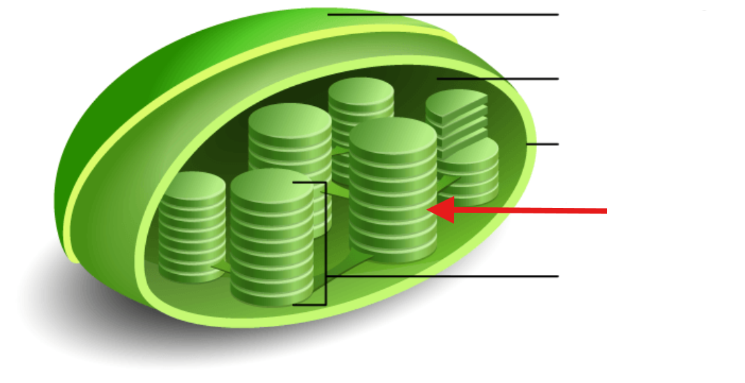
GRANUM
A stack of thylakoids

STROMA
Liquid portion of chloroplasts

CENTROSOME
Small, dense region of cytoplasm and serves as the main microtubule organizing center (MTOC)

CENTRIOLES
Cylinder-shaped organelles made of nine triplets of microtubules arranged in a ring
