Week 2: Acute Cardiac Conditions
 Call Kai
 Learn
Learn Practice Test
 Spaced Repetition
 Match
Match Flashcards
 Knowt Play
1/71

There's no tags or description
Looks like no tags are added yet.
Name | Mastery | Learn | Test | Matching | Spaced | Call with Kai | Chat |
|---|
No analytics yet
Send a link to your students to track their progress
72 Terms
Coronary Artery Disease (CAD)
chronic stable angina and acute coronary syndromes
waxy substances (plaque) builds up inside the coronary arteries
leading to atherosclerosis
IS preventable
How is CAD diagnosed?
cardiac cath
angiogram
ECG and Echo are helpful
CAD Non-Modified Risk Factors
family history
increasing age
>45 males
>55 females
↑ incidence in African Americans
premature menopause and pregancy-associated disorders
CAD Modifiable Risk Factors
hyperlipidema
tobacco use
damages epithelium
HTN
Diabetes
Metabolic Syndrome
Physical Inactivity
Chronic inflammatory conditions
Rheumatoid arthritis, lupus, HIV
CAD: Hyperlipidemia Risk Factor
elevated cholesterol levels
forms plaques
statins can be used to lower bad cholesterol
Tobacco Use: Hyperlipidemia Risk Factor
smoking impacts heart health
damages epithelium
cessation strategies
HTN: Hyperlipidemia Risk Factor
endothelial damage (injury to vessels)
blood pressure management
Diabetes: Hyperlipidemia Risk Factor
chronic ↑ blood sugar → endothelial damage
lifestyle modifications are needed
Metabolic Syndrome: Hyperlipidemia Risk Factor
assess waist size → adipose tissue in abdomen is more active → release cytokines that induce inflammatory activity in vessels
inflammation can:
damage endothelial damage
promote plaque formation
increase insulin resistance
↑ risk of HTN, HD, and stroke
Chronic Inflammation Conditions: Hyperlipidemia Risk Factor
Aortic aneurysm → main cause is atherosclerosis
All vessels are linked → disease in one area of vessels may be indicative of disease in other areas
Excess circulating lipids
Chronic kidney disease
Impact on cardiovascular health
slows down the body’s ability to clear out fats
Peripheral Venous Disease (PVD)
Dysfunction or damage to one-way venous valves in the lower extremities.
Prevents efficient blood return to the right side of the heart, leading to venous stasis
(pooling in legs).
Manifests as lower-extremity edema
increases the risk of DVT
Atherosclerosis
Abnormal accumulation of lipid deposits and fibrous tissue within arterial walls and lumen
Atherosclerosis Pathogenesis
damaged endothelium
fatty streak and lipid core formation
lipids accumulate
fibrous plaque
raised plaques are visible
blood flow is reduced
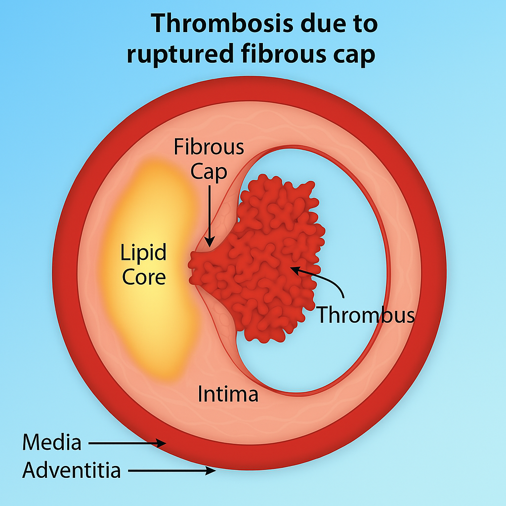
complicated lesion: artery blockage that has burst open and formed a blood clot
When does Classic (Stable) Angina Pectoris occur?
occurs with stress or exertion
Classic (Stable) Angina Pectoris
chest pain or discomfort
chest pain resolves in 15 mins
relieved with nitroglycerin
Unstable (Pre-infarction) Angina Pectoris
occurs frequently with progressive severity
does NOT get better with rest
unrelated to activity (unpredictable)
Why does Stable and Unstable Angina Pectoris occur?
artery narrowing
partial occlusion
Variant Angina Pectoris
occurs DURING rest
caused by vasospasm
can be caused by nicotine or cocaine
Myocardial Infarction (MI)
Prolonged ischemia of the heart muscles caused by an imbalance between oxygen supply and demand
increased workload of the heart due to the plaque buildup
What are the causes of MIs?
plaque rupture
protective cap over an atherosclerotic plaque ruptures → blood clot forms to try to heal it →occludes the entire lumen
spasm
muscle severely constricts → blood cannot pass through
inflammation
damage
Ischemia
blood flow and oxygen to a part of the body are reduced or blocked
EKG Change: Ischemia
inverted T wave
EKG Change: Injury
ST elevated
EKG Change: Infarction
wide Q wave
delay in depolarization of ventricles → widened Q wave is a sign of irreversible damage to the ventricles
MI Assessment
chest pain
HTN (early)
HoTN (shock)
↑ HR
↓ HR (damage)
S3 and S4 heart sounds
N/V
Chest Pain in MI
“squeezing, choking, smothering”
chest pain not relieved by rest or nitro
women: discomfort in the arms, back, neck, jaw, or stomach
MI: Hypertensive Response
happens early
body is responding to pain, anxiety, and decreased O2
sympathetic nervous system (fight or flight)
release of epinephrine and norepinephrine → constricts blood vessels → ↑ BP
HoTN during MI
cardiogenic shock
damaged heart cannot pump enough blood/O2 to maintain adequate circulation
due to blockages
often MORE concerning
Tachycardia in MI
The body senses that the heart muscle is not getting enough oxygen and activates the sympathetic nervous system.
Release of epinephrine and norepinephrine → ↑HR
Bradycardia in MI
Damage to the heart’s electrical conduction system
SA/AV node ischemia → HR ^*
3rd degree heart block
S3 and S4 Heart Sounds in MI
S3 and S4 are extra heart sounds
S1 and S2 are normal
S3 Heart Sounds in MI
Volume overload, poor ventricular pumping. Damaged ventricle.
“The ventricle is failing”
MI Relation: shows heart failure after MI
S1-S2-S3 (“ken-tuc-ky”)
S4 Heart Sounds in MI
Atria contract to force blood into a stiff, non-compliant ventricle
“The ventricle is stiff”
MI Relation: acute ischemia or recent MI
S4-S1-S2 (“ten-nes-see”)
Diagnostic Labs: MI
Troponin T and Troponin I
Creatine Kinase-MB (CK-MB)
Myoglobin
CBC
12-lead EKG
cardiac cath.
chest x-ray
Troponin T and Troponin I
GOLD STANDARD for MI marker
released from heart when there is strain
NSTEMI = elevated troponin but no ST changes
How long does Troponin T and Troponin I stay elevated for?
elevated within a few hours of acute MI
remain elevated for 3 to 6 days
Creatine Kinase-MB (CK-MB) in MI
creatine kinase isoenzyme found mainly in heart
begins to increase within a few hours of chest pain
peaks within about 24 hours
Why is CBC needed in MI?
anemia can cause MI
checks for bleeding
checks coagulation
Emergency Management of the Patient with ACS/MI
Assess ABCs (Defibrilate)
O2 sat > 95%
ECG within 10 mins
Assess pain
RRT (rapid response team)
Vascular access needed
Aspirin
Why is morphine used during MIs?
used for pain
reduces preload
workload of heart
*can cause respiratory depression
Oxygen Saturation with MI
keep > 95%
supernormal O2 can be bad
MI: Collaborative Management
Reperfusion Therapy: Fibrinolytics ("clot-busting" drugs)
Door-to-needle goal of 30 minutes
Door-to-balloon time is 90 minutes
Tissue Plasminogen Activator (t-PA)
What is reperfusion therapy (fibrinolytics)?
Fibrinolytics dissolves thrombi in coronary arteries and restores myocardial blood flow
What is Tissue Plasminogen Activator (t-PA)?
Powerful "clot-busting" medication
*Carries a severe risk of dangerous bleeding
MI: Ongoing Management
continuous EKG
O2
pain relief
prevent complications
glycemic control
MI: Complications
dysrhythmias
HF
cardiogenic shock
papillary muscle rupture
wall rupture
acute kidney injury
hyperglycemia
What dysrhythmias occur with MI?
Vtach, Vfib
Vtach with pulse → synch cardioversion
Why does HF occur with MI?
arteries and veins become more sensitive to vasopressors when people age
the loss of blood flow during an MI deprives the heart muscle (myocardium) of oxygen
Papillary Muscle Rupture in Acute MI: Pathophysiology & Management
Mitral valve damaged → blood back flows → pressure in lungs, low blood flow to body → SOB, hypotensive → emergent valve replacement (surgery)
Why does blood glucose increase with MI?
stress of MI can increase cortisol
high cortisol → increases blood sugar
What is reperfusion therapy?
critical medical treatment that restores blood flow to blocked blood vessels
gold-standard reperfusion therapy: Percutaneous Coronary Intervention (PCI)
threading a catheter to the blockage
inflating a tiny balloon to open the artery, and placing a stent

Reperfusion Therapy Timing
door-to-needle = 30 minutes
start catheter
door-to-balloon = 90 minutes
balloon to open vessel
Reperfusion Therapy: Collaborative Management
advice pt to stay still
feeling warm/hot is normal when dye is infused
HOLD anticoagulants 3 days before cath.
dual antiplatelet therapy
What is dual antiplatelet therapy used in post reperfusion therapy?
Aspirin + Clopidogrel (Plavix)
use for 1 year
prevents re-stenosis (re-narrowing)
What is Coronary Artery Bypass Graft (CABG)?
surgeons take a healthy blood vessel from another part of the body and use it to create a detour around blocked or narrowed coronary arteries

The Bypass Machine
temporarily takes over your heart and lung functions, circulating and oxygenating blood while the surgeon temporarily stops your heart to perform the grafts safely
What is the goal of The Bypass Machine
maintain cardiac and pulmonary function
Risks of The Bypass Machine
coagulopathy
increased bleeding risk
inflammatory response
due to contact with artificial surfaces in bypass machine
Coronary Artery Bypass Graft (CABG): Collaborative Management (before)
skin prep: clip vs shave
shaving can increase infection risk
medication: surgical antibiotic prophylaxis
Inflammatory & Structural Heart Disease: Regurgitation
valve does NOT CLOSE properly and blood backflows through the valve
causes high volume problem
ex. mitral valve not closing → back flow to left atrium → into lungs
Inflammatory & Structural Heart Disease: Stenosis
valve does NOT OPEN completely (narrowing) and blood flow through the value is reduced → murmur
valve is calcified and stiff

Valve Prolapse
stretching of the valve leaflet into the atrium during systole
mitral has two leaflets
valve become floppy, weirdly shaped
Causes of Valvular Heart Disease?
Rheumatic heart disease (RHD)
Endocarditis
Marfan’s syndrome (tall, thin)
Can cause valve prolapse
Congenital
Age-related valve calcification (aortic stenosis)
Need valve replacement to maintain CO
ACS/MI
Valvular Heart Disease: Nursing Priority Problems
Decreased cardiac output related to valvular incompetence
Excess fluid volume related to heart failure
Activity intolerance related to decreased cardiac output
HF is a concern
Check wt, medication management
What is Endocarditis
An infection of the endocardial surface including valves, caused by bacterial, fungal or viral agents
Endocartditis: Possible Ports of Entry
Oral cavity
Skin rash/lesion/abscess
Surgery or invasive procedure
Endocarditis: Clinical Manifestations
fever
new heart murmur
Osler nodes, Janeway lesions, Roth spots, and splinter hemorrhages in nailbeds
Endocarditis: Osler’s Nodes
Painful, erythematous or purple, pea-like nodules
Most commonly found on pads of fingers & toes

Endocarditis: Janeway Lesions
Nontender, erythematous & nodular or lesions
Most commonly found on palms & soles

Endocarditis: Roth’s Spots
Oval-shaped, white-centered hemorrhages
Present on retina during fundoscopic exam

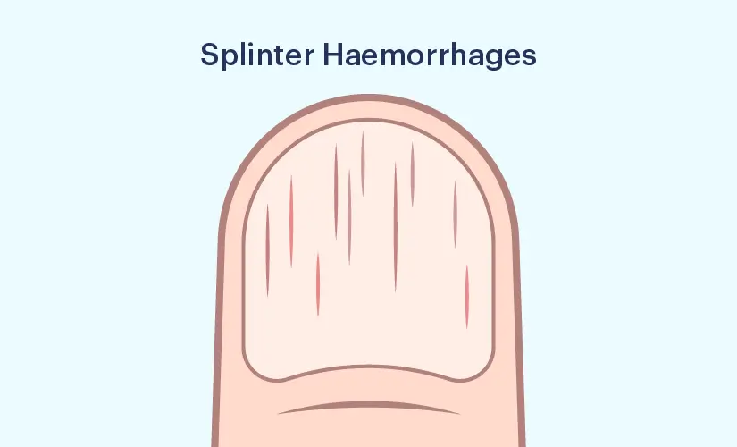
Endocarditis: Splinter Hemorrhages
Small, linear hemorrhages under the nails that are usually asymptomatic
Infective Endocarditis: Collaborative Management
A serious, life-threatening infection of the inner lining of the heart chambers and valves
Obtain blood cultures BEFORE starting antibiotics.
Specific type of endocarditis