Gastrointestinal Tract
 Call Kai
 Learn
 Practice Test
 Spaced Repetition
 Match
 Flashcards
 Knowt Play
1/64

There's no tags or description
Looks like no tags are added yet.
65 Terms
What are the major activities of the digestive system?
Ingestion
Secretion - release of water, acid, ions, enzymes, mucus, bile, and hormones to support digestion
Motility - movement of material via smooth-muscle activity
Digestion - mechanical + chemical breakdown
Absorption - transfer of digestive products from GI lumen into blood or lymph
Elimination/defecation
Where does absorption primarily occur?
In the small intestine, specifically the jejunum and duodenum
Small units from digestion, water, vitamins, and electrolytes are transferred
Anything water-soluble: absorbed into capillaries in intestinal villi
Anything lipid soluble: absorbed into lymph vessels in intestinal villi
Why is the GI tract considered part of the external environment?
Its lumen is continuous with the outside world from mouth to anus
Material in the lumen is not truly inside the body until it crosses the intestinal epithelium

What is the peritoneal cavity?
A closed sac in the abdominal cavity into which organs descend during embryologic development
The peritoneum covers or supports abdominal organs

What is the mesentery?
A peritoneal attachment that anchors much of the intestine dorsally while still allowing mobility
It also carries blood vessels, lymphatics, and nerves

What are the four major histologic layers of the GI wall?
Mucosa, submucosa, muscularis externa, and serosa.

What are the three layers of the mucosa?
Mucosal epithelium (innermost), lamina propria, and muscularis mucosae (outermost).
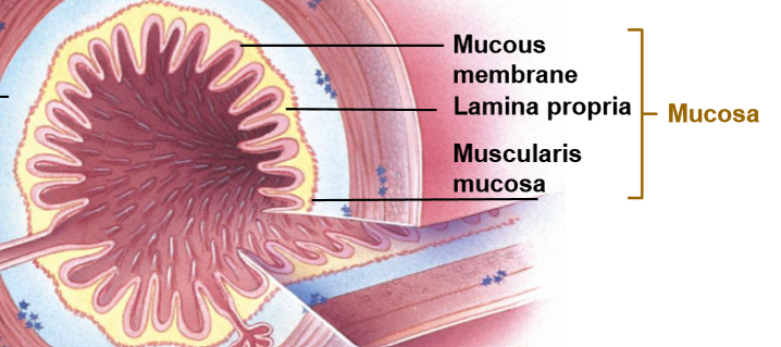
What is the function of the mucosal epithelium?
It forms the luminal surface and is specialized for secretion, digestion, absorption, or protection depending on GI location.
Contains exocrine gland cells, endocrine gland cells, and epithelial cells

What is the lamina propria?
A loose connective-tissue layer in the mucosa that contains capillaries, lymphatic vessels, immune cells, and sometimes glands.
Houses gut-associated lymphoid tissue (GALT)

What is the muscularis mucosae?
A thin layer of smooth muscle that creates local movement of the mucosa and increases contact with luminal contents.

What is the submucosa?
A thick connective-tissue layer that supports the gut wall
Provides distensibility and elasticity
It contains larger blood/lymph vessels and the submucosal nerve plexus.

What is the submucosal plexus?
An intrinsic layer of nerves running in the wall of the intestine that helps regulate secretion, blood flow, and local GI function.
Overall how the GI tract can govern its own motility

What is the muscularis externa?
The main smooth-muscle coat of the GI tract, usually with an inner circular layer and outer longitudinal layer.
What happens when the circular layer vs longitudinal layer of muscularis externa contracts?
Circular/inner layer: contraction decreases the diameter of the GI lumen
Longitudinal/outer layer: contraction shortens the length of that GI segment
What is the myenteric plexus?
The nerve plexus between the circular and longitudinal muscle layers of the muscularis externa
Coordinates GI motility
What is peristalsis? What about segmentation?
Peristalsis: coordinated wave of contraction that propels GI contents forward through the digestive tract
Segmentation: Local rhythmic contraction that mixes luminal contents

What is the serosa?
The outer covering of much of the GI tract - the visceral peritoneum
Secretes serous fluid
Lubricates/reduces friction b/w digestion organs and viscera
Continuous with the mesentery throughout most of the tract
Attachment provides fixation
Supports organs in proper place

What is the stomach?
A J-shaped ac-like chamber that’s divided into the fundus, body and antrum
Stores ingested food
Secretes HCl and enzymes to being protein digestion
Mixes to pulverize food into chyme
Pyloric sphincter provides barrier to regulate chyme flow from stomach to S.I.

What protects the stomach from digesting itself?
The gastric mucosal barrier: surface mucus and bicarbonate protect the epithelium from HCl and pepsin.
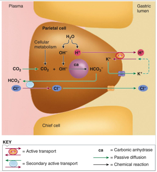
What are the major functions of HCl in the stomach?
Activates pepsinogen to pepsin and provides the acid medium (low pH) for optimal pepsin activity
Aids breakdown of connective tissue and muscle fibers
Unfolds/denatures proteins
Helps kill microorganisms ingested with food
What do chief cells secrete?
Pepsinogen, an inactive precursor that is converted to the protein-digesting enzyme pepsin in acidic conditions.
Secreting the inactive form protects the cells and stomach wall from autodigestion.

What is the pancreas’s endocrine role?
Releases hormones into the blood to regulation nutrient metabolism
Insulin and glucagon
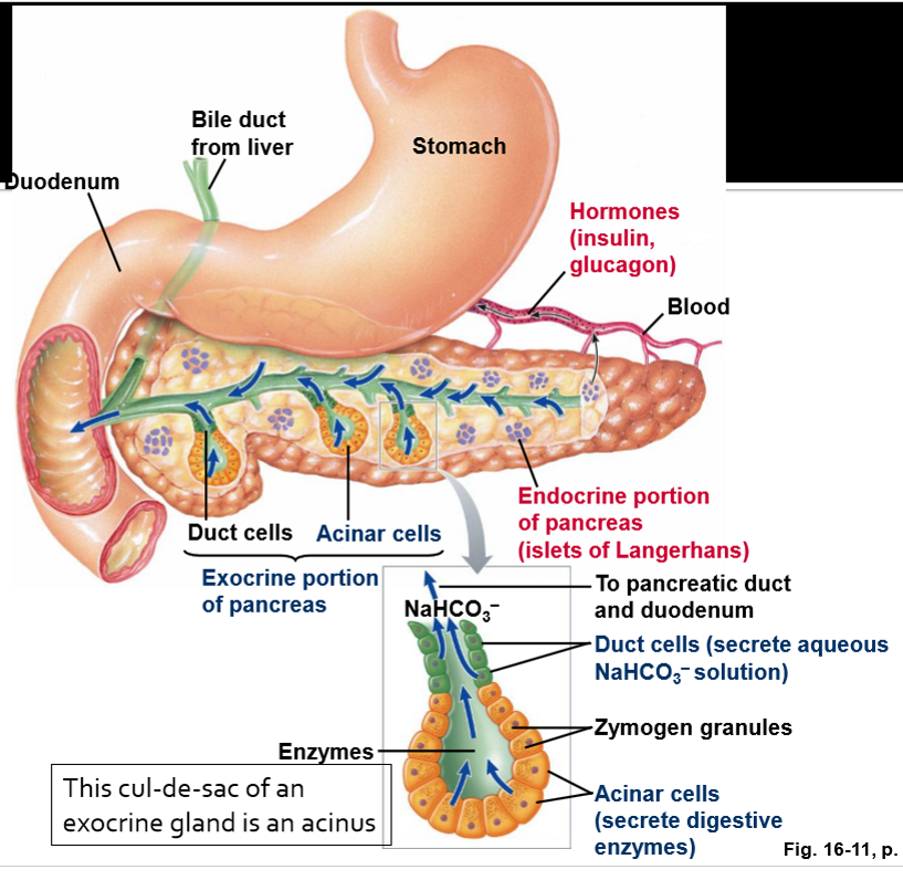
What is the pancreas's exocrine role?
Delivers digestive enzymes and bicarbonate-rich fluid into the duodenum
Pancreatic enzymes are secreted by acinar cells
Aqueous alkaline solution is secreted by duct cells (bicarbonate)
What stimulates the pancreas to release secretions?
Secretin from duodenal mucosa stimulates the duct cells
CCK from duodenal mucosa stimulates the acinar cells
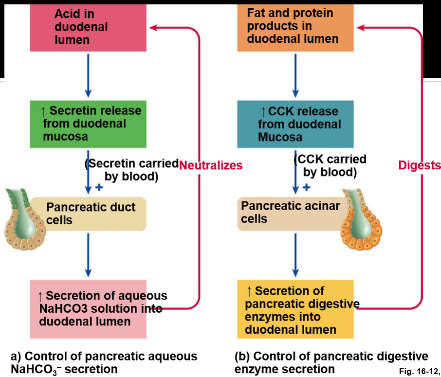
How do secretin and CCK coordinate pancreatic secretion in the duodenum?
Acidic chyme in the duodenum stimulates secretin release from duodenal mucosa
Secretin travels in blood to pancreatic duct cells, which secrete aqueous NaHCO₃ to neutralize acid
Fat and protein products stimulate CCK release
CCK travels in blood to pancreatic acinar cells, which secrete digestive enzymes into the duodenum.

What is the basic structural unit of the liver?
The hepatic lobule, organized around a central vein
Lobules are bounded at each of the six outer corns by portal triads (groups of vessels)

What is the portal triad?
A branch of the hepatic portal vein, a branch of the hepatic artery, and a bile duct.

What are the two blood supplies to the liver?
Receives blood from the hepatic artery (~25%)
Perfuses liver tissue/brings oxygen + nutrients
Receives blood from portal vein (~75%)
Drains the stomach and intestine; contains materials absorbed from GI tract
Liver detoxifies/subjects portal blood to immune surveillance

Compare the dual blood supplies to the liver
Hepatic arterial blood functions to nourish
Portal blood flows into the liver for conditioning before it’s allowed to flow into systemic veins

How does blood flow through and drain from the liver?
Blood from branches of the hepatic artery and hepatic portal vein enters hepatic sinusoids
Flows toward the central vein of each lobule, then drains into hepatic veins → caudal vena cava → heart.

What is the direction of bile flow in a hepatic lobule?
Bile flows from hepatocytes toward bile canaliculi and bile ducts, generally opposite the direction of blood flow.

What are hepatic sinusoids?
Specialized capillary channels where blood from the hepatic artery and portal vein mixes and exchanges substances with hepatocytes
Very leaky
Allows hepatocytes to take or secrete nutrients from sinusoids while also secreting bile salts into the canaliculi

What are Kupffer cells?
Resident macrophages in liver sinusoids that remove microbes, debris, and aged cells from blood.
Why does blood from the GI tract travel to the liver before entering systemic circulation?
The liver processes absorbed nutrients, stores/modifies substances, detoxifies compounds, and performs immune surveillance before blood reaches the systemic circulation.
Liver will take the first “hit” from any disease that is affecting the GI tract

How can cirrhosis lead to portal hypertension and ascites?
Fibrosis/scarring increases resistance to portal blood flow
Scar tissue replaces hepatocytes → liver loses function
Increased portal pressure promotes fluid accumulation in the abdominal cavity = ascites

What is the major digestive role of the liver?
The liver produces bile, which is delivered to the small intestine to aid digestion and absorption of fats.

What are bile salts?
Amphipathic components of bile that emulsify fats and help form micelles for lipid digestion and absorption
They get recycled through the enterohepatic circulation
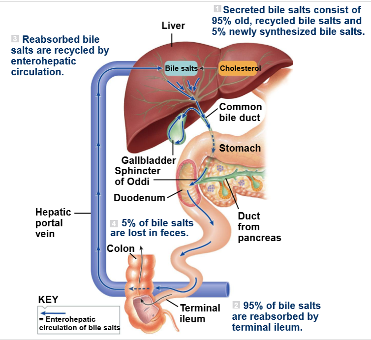
Describe enterohepatic circulation of bile salts
Liver makes bile salts and secretes them as part of bile
Bile then enters the duodenum via common bile duct to aid fat digestion
~95% of bile salts are reabsorbed in the terminal ileum and returned to the liver via hepatic portal vein for reuse
~5% are lost in feces

What is emulsification of fat?
The physical breakup of large lipid droplets into smaller droplets, increasing surface area available to digestive enzymes.
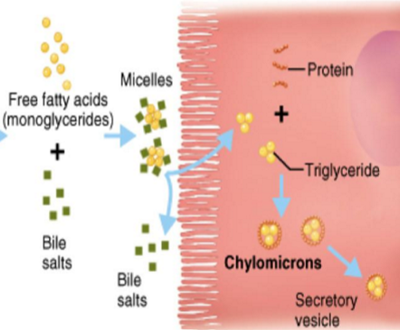
What is a micelle?
A small bile-salt-containing structure that carries lipid digestion products through the watery intestinal lumen to the epithelial surface.
What is the small intestine's major role?
It is the primary site of enzymatic digestion and nutrient absorption
Juice secreted by the S.I. contains mucous and aqueous salts, not digestive enzymes
Rather the digestive enzymes are part of the brush border (they’re proteins within the membrane of border epithelial cells)
Enteropeptidase
Disaccharidases
Aminopeptidases
What occurs in the duodenum, jejunum, and ileum?
Duodenum: Acidic chyme mixes with pancreatic secretions and bile
Much digestion begins or continues here
Jejunum: major absorptive of nutrients, water, electrolytes
Ileum: reabsorption of bile salts and absorption of vitamin B12 bound to intrinsic factor

What are circular folds?
Large folds of the small-intestinal mucosa and submucosa that increase surface area and slow movement of chyme.

What are villi?
Fingerlike projections of small-intestinal mucosa that greatly increase absorptive surface area.

What are microvilli?
Tiny projections on the apical surface of intestinal epithelial cells that form the brush border and dramatically increase surface area.

What is the brush border?
The dense microvillus-covered apical surface of intestinal epithelial cells
Contains brush-border enzymes and transport proteins important for final digestion and absorption

Why are villi well suited for absorption?
Each villus contains blood capillaries and a lymphatic vessel, allowing rapid removal of absorbed nutrients.
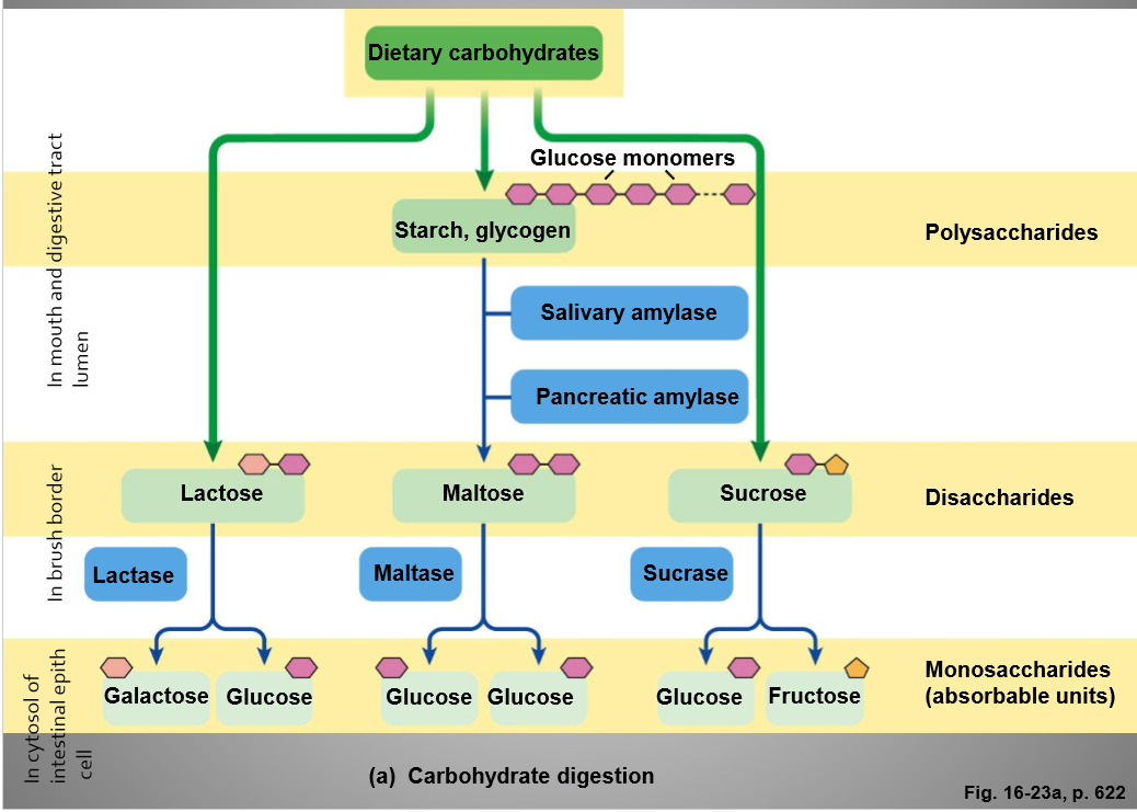
What are the end products of carbohydrate digestion?
Monosaccharides, mainly glucose, galactose, and fructose.

How are glucose and galactose absorbed from the intestinal lumen?
They enter intestinal epithelial cells across the apical membrane through SGLT1, a Na⁺-glucose cotransporter
They leave across the basolateral membrane through GLUT2 into the blood

How is fructose absorbed from the intestinal lumen?
Fructose enters intestinal epithelial cells across the apical membrane by facilitated diffusion through GLUT5
It leaves across the basolateral membrane through GLUT2 into the blood
What is the role of the basolateral Na⁺/K⁺ pump in nutrient absorption?
It uses ATP to pump Na⁺ out of the epithelial cell and K⁺ in, keeping intracellular Na⁺ low
This Na⁺ gradient powers SGLT1-mediated uptake of glucose and galactose from the intestinal lumen

What are the end products of protein digestion?
Amino acids plus small peptides.

How are amino acids absorbed from the intestinal lumen?
Amino acids enter intestinal epithelial cells across the apical membrane through Na⁺-dependent amino acid cotransporters
They then leave across the basolateral membrane through amino acid transporters and enter blood capillaries
The Na⁺/K⁺ pump maintains the Na⁺ gradient that powers apical uptake
How are small peptides absorbed from the intestinal lumen?
Small peptides enter intestinal epithelial cells across the apical membrane through PepT1, an H⁺-dependent peptide cotransporter
Intracellular peptidases break them into amino acids, which leave the basolateral membrane through amino acid transporters and enter blood

How are fats digested and absorbed?
Bile salts emulsify fat
Pancreatic lipase digests triglycerides
Products enter micelles → diffuse into epithelial cells → repackaged → leave as chylomicrons through lymph
What is a lacteal and how does it relate to fat absorption?
A lymphatic vessel inside an intestinal villus
It absorbs chylomicrons containing dietary triglycerides and carries them into the lymphatic system