Bio Dr P: Animal Transport
 Call Kai
 Learn
 Practice Test
 Spaced Repetition
 Match
 Flashcards
 Knowt Play
1/32

Earn XP
Description and Tags
33 Terms
The need for a transport system depends on 3 factors
Size
Metabolic activity
SA:V
Invertebrate circulatory systems diagram
(need to be able to label the aorta and heart in neat notes pg 11)
Invertebrate circulatory system
Some invertebrates (insects) have an open circulatory system which are not enclosed in blood vessels
The haemolymph (fluid) flows freely through the body cavity
How does the circulatory system work in invertebrates?
Invertebrate hearts are segmented
They contract in a wave from the back of the abdomen to the front called peristalsis
The heart pumps haemolymph into a single main artery (aorta) which opens up into body cavity and allows fluid to flow around the insects organs supplying nutrients
The fluid then returns into heart segments through a series of valves to be recirculated again
Vertebrate circulatory systems
All vertebrates have a closed circulatory system so blood is enclosed in blood vessels
Fish circulatory system diagram

Fish circulatory system
Fish have a single circulatory system
As blood passes through the gills to body blood pressure decreases
So blood flows slowly and rate at which oxygen and nutrients are delivered to respiring tissues and carbon dioxide and urea are removed is limited
Why is this type of ciculatory system sufficient for fish?
Because they are not as metabolically active as mammals
Mammalian circulatory system diagram

Mammalian circulatory system
Mammals have a double circulatory system consisting of:
Pulmonary circuit- the right side of the heart pumps blood to the lungs which then returns to the left side
Systemic circuit- the left side of the heart pumps blood to the body which then returns to the right side
What does the double circulatory system in metablically active mammals allow?
Allows the heart to increase pressure so the rate of flow is fast
Pressure can be controlled (kept low in pulmonary circuit and high in systemic circuit)
Role of arteries
Arteries carry blood away from the heart at high pressure to the lungs or the rest of the body
Arteries diagram
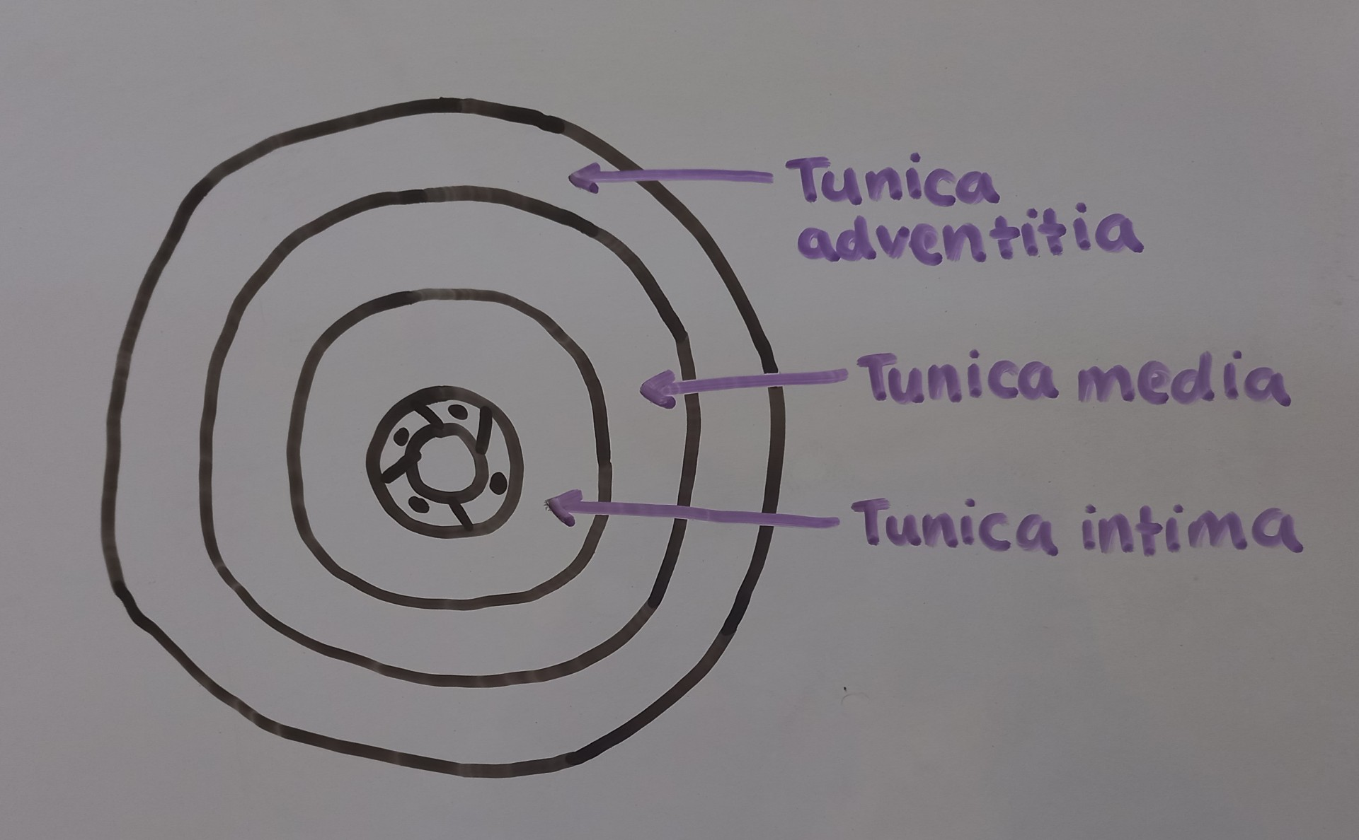
3 different layers in the arteries and their function
Tunica intima- has a layer of smooth endothelial cells to reduce friction + a thin layer of elastic tissue to allow stretch and recoil to maintain blood pressure
Tunica media- has a thick layer of smooth muscle which contracts to constrict the artery narrowing the lumen so blood pressure can be increased and maintained
Tunica adventitia- has a thick layer of collagen and elastic tissue to provide mechanical strength to withstand pressure + to recoil to maintain pressure
Role of veins
Veins carry blood to the heart under low pressure
Veins diagram

Key features of veins
Large lumen to ease blood flow. The larger the diameter of the vessel the less resistance there is to blood flow
Thinner layers of collagen, smooth muscle and elastic tissue as they don't need to stretch and recoil
Have valves to prevent the backflow of blood. Movement of blood is controlled by contraction of skeletal muscle
Role of capillaries
Involved in carrying out the exchange of substances between the blood and the tissues
Capillaries diagram

Key features of capillaries
Consist of a single layer of endothelial cells
Very narrow lumen so red blood cells are squeezed against the walls helping oxygen transport
Fenestrations (gaps in the walls) allow movement of dissolved substances
Role of arterioles
They occur between arteries and capillaries
They are smaller vessels that have a layer of smooth muscle
Contstriction of the arteriole walls can divert blood
Role of venules
They collect blood from the capillary bed and have layers similar to veins
Heart diagram

Structure of the heart
The heart is divided into 4 chambers
It consists of cardiac muscle and adipose tissue on the outside for external protection
The septum separates the ventricles from each other ensuring that oxygenated blood in the left side and deoxygenated blood in the right side of the heart are kept separate
Coronary arteries supply the heart with oxygen
How is pressure in the heart generated?
It is generated by the cardiac muscle contracting
Atria and ventricles
Atria are thinner than ventricles as they don't need to generate much pressure
Walls of right ventricle are thinner than left ventricle as they only pump deoxygenated blood to the lungs
If blood pressure is too high in right ventricle then alveoli will be damaged
Insufficient supply of oxygen to the heart can lead to
Angina or myocardial infarction (heart attack)
Explain each stage of the cardiac cycle
Atrial and ventricular diastole- both atria and both ventricles are relaxed, blood passively enters atria then flows through AV valves into both ventricles
Atrial systole- both atria contract decreasing volume + increasing pressure in atria, more glood flows through AV valves into ventricles increasing ventricular volume and ventricular pressure
Ventricular systole- both ventricles contract decreasing volume and increasing the pressure in the ventricles, AV valves close + SL valve opens, blood flows into pulmonary arteries and aorta
At the end of ventricular systole atria relax + blood flows into atria (beginning of atrial diastole)

AV valves
During diastole when ventricles relax pressure falls as atria are passively filling with blood causing atrial pressure to rise above ventricular pressure and AV valves open allowing blood to flow through
When atria relax and ventricles begin to contract ventricular pressure rises above atrial pressure and blood flows up forcing AV valve to shut

SL valves
During ventricular systole ventricular pressure rises above pressure in major arteries which opens the SL valves and allows blood to flow through
During diastole as ventricles relax ventricular pressure falls below arterial pressure + blood flows down forcing the valve to shut
