lower extremity flashcards
 Call Kai
 Learn
 Practice Test
 Spaced Repetition
 Match
 Flashcards
 Knowt Play
1/86

There's no tags or description
Looks like no tags are added yet.
87 Terms
label the joints of the foot in # order
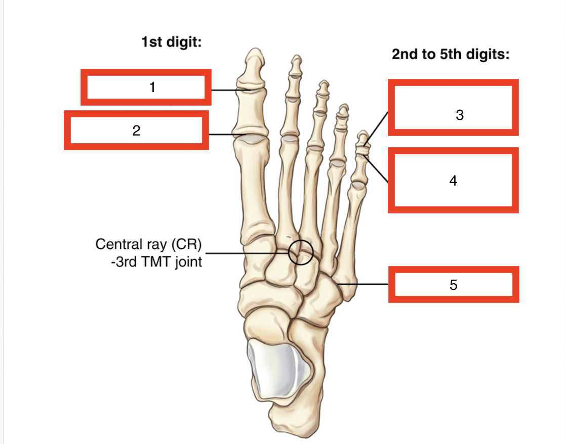
interphalangeal joint
metatarsophalangeal joint
distal interphalangeal joint
proximal interphalangeal joint
tarsometatarsal joint
label the following

tarsometatarsal joint
metatarsophalangeal joint
proximal interphalangeal joint
distal interphalangeal joint
interphalangeal joint
sesamoid bones
in the lower limbs sesamoid bones tend to be ____ and more significant radiographically
larger
the largest sesamoid in the body is the ___
patella
sesamoid bones are almost always present in the ____or the ___ surface , at the head of the 1st metatarsal near the 1st _____ joint
posterior, plantar, metatarsophalangeal
the sesamoid bone on the medial side of the lower limb is called the ____ sesamoid and the lateral is the ___ sesamoid bone
tibial, fibular
how many tarsals, metatarsals, and phalanges are in the foot
7 tarsals, 5 metatarsals, 14 phalanges
how many bones are in the foot
26
the LAT cuneiform sits on top of what metatarsal
3rd metatarsal
the intermediate cuneiform sits on top of what metatarsal
2nd metatarsal
the medial cuneiform sits on top of what metatarsal?
1st metatarsal
which toe has only a proximal and distal phalanx?
big toe
the calcaneus/ cuboid are on top of which metatarsals and what side of the foot?
the 5th and 4th metatarsal. lateral side
the base of the 5th metatarsal expands ___ forming a prominent ___ which provides attachment of a tendon
laterally, tuberosity
what tendon is attached to the 5th metatarsal tuberosity
peroneus brevis and
the proximal portion of the 5th metatarsal and its tuberosity is a common ____
trauma site
what are 2 common fractures of the 5th metatarsal tuberosity?
avulsion and jones
a fracture caued by sudden rolling/pulling of the ankle causing the peroneus brevis to pull a small piece of bone from the tuberosity is known as _____ fracture
avulsion

what kind of fracture is this?
jones fracture
a jones fracture is slow and difficult to heal compared to other foot fractures because the 5th metetarsal naturally has poor _____
blood supply
the centering point for AP/oblique projections of the foot is
the 3rd metatarsal or tarsometatarsal joint
the tarsal of the foot are ____ and less ____ compare to the carpals of the hand
larger, mobile
the largest and strongest bone of the foot is the
calcaneus
the tuberosity of the calcaneus is a common site for ___ which are sharp outgrowths of the bone that can be painful on weight bearing
bone spurs
the calcaneus articulates with 2 bones, one anteriorly and the other superiorly, which are they?
anterior: cuboid
superior: talus
what are the 3 facets of the subtalar (talocalcaneal ) joint
posterior, anterior, middle

label the following in # order
posterior facet
middle facet
anterior facet
sustentaculum tali
subtalar joint
what is the opening in the middle of the subtalar joint that allows ligaments to pass through
sinus tarsi
the navicular articulates with ___ bones, which are they?
5, talus, cuboid, 3 cuneiforms
the biggest cuneiform is the ___ and the smallest is the ___
medial cuneiform , intermediate cuneiform

What is A on the lateral foot radiograph?
Tibia

What is B on the lateral foot radiograph?
Calcaneus

What is C on the lateral foot radiograph?
Tuberosity of the calcaneus (calcaneal tuberosity)

What is D on the lateral foot radiograph?
Cuboid

What is E on the lateral foot radiograph?
fifth metatarsal tuberosity
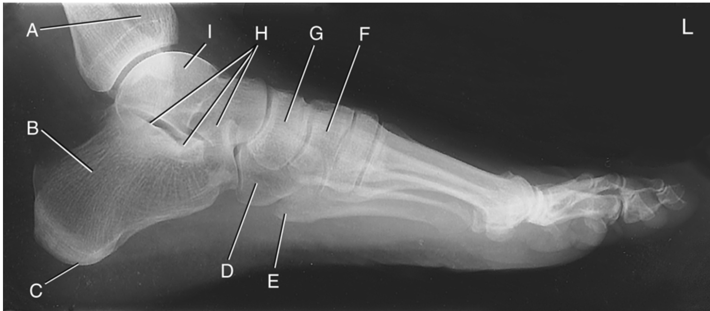
What is F on the lateral foot radiograph?
Cuneiforms

What is G on the lateral foot radiograph?
Navicular

What is H on the lateral foot radiograph?
Subtalar joint

What is I on the lateral foot radiograph?
Talus

What is A on the AP oblique foot radiograph?
Interphalangeal joint of the first digit

What is B on the AP oblique foot radiograph?
Proximal phalanx of the first digit

What is C on the AP oblique foot radiograph?
First metatarsophalangeal joint

What is D on the AP oblique foot radiograph?
Head of the first metatarsal

What is E on the AP oblique foot radiograph?
Body of the first metatarsal
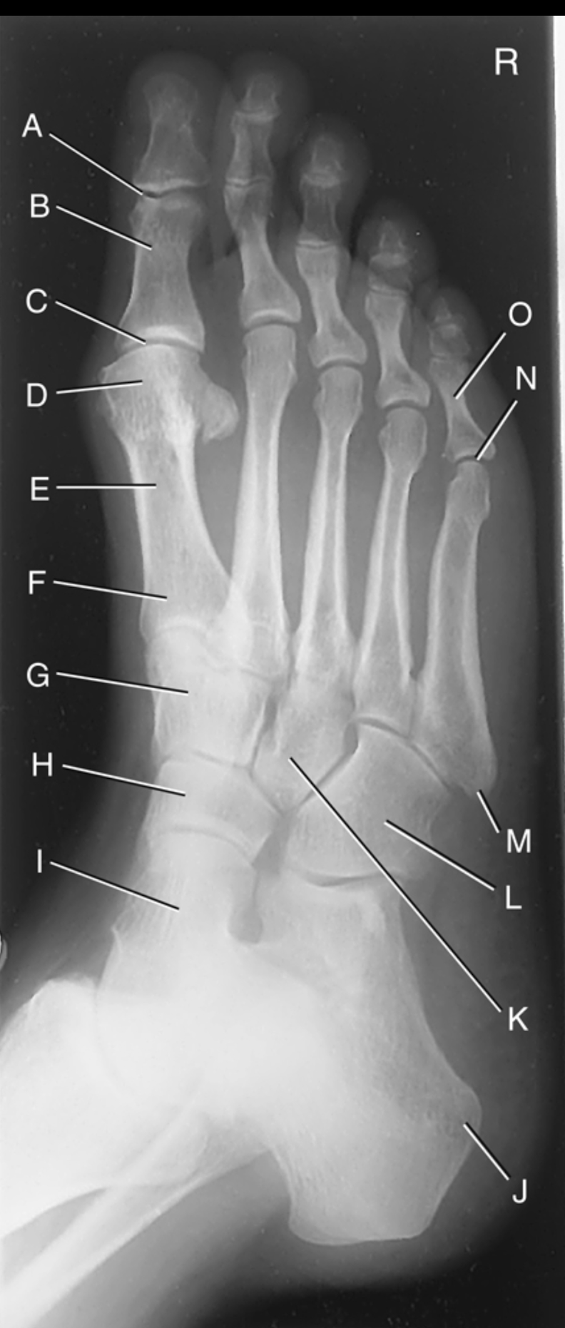
What is F on the AP oblique foot radiograph?
Base of the first metatarsal

What is G on the AP oblique foot radiograph?
Medial cuneiform

What is H on the AP oblique foot radiograph?
Navicular

What is I on the AP oblique foot radiograph?
Talus

What is J on the AP oblique foot radiograph?
Tuberosity of calcaneus

What is K on the AP oblique foot radiograph?
Lateral cuneiform
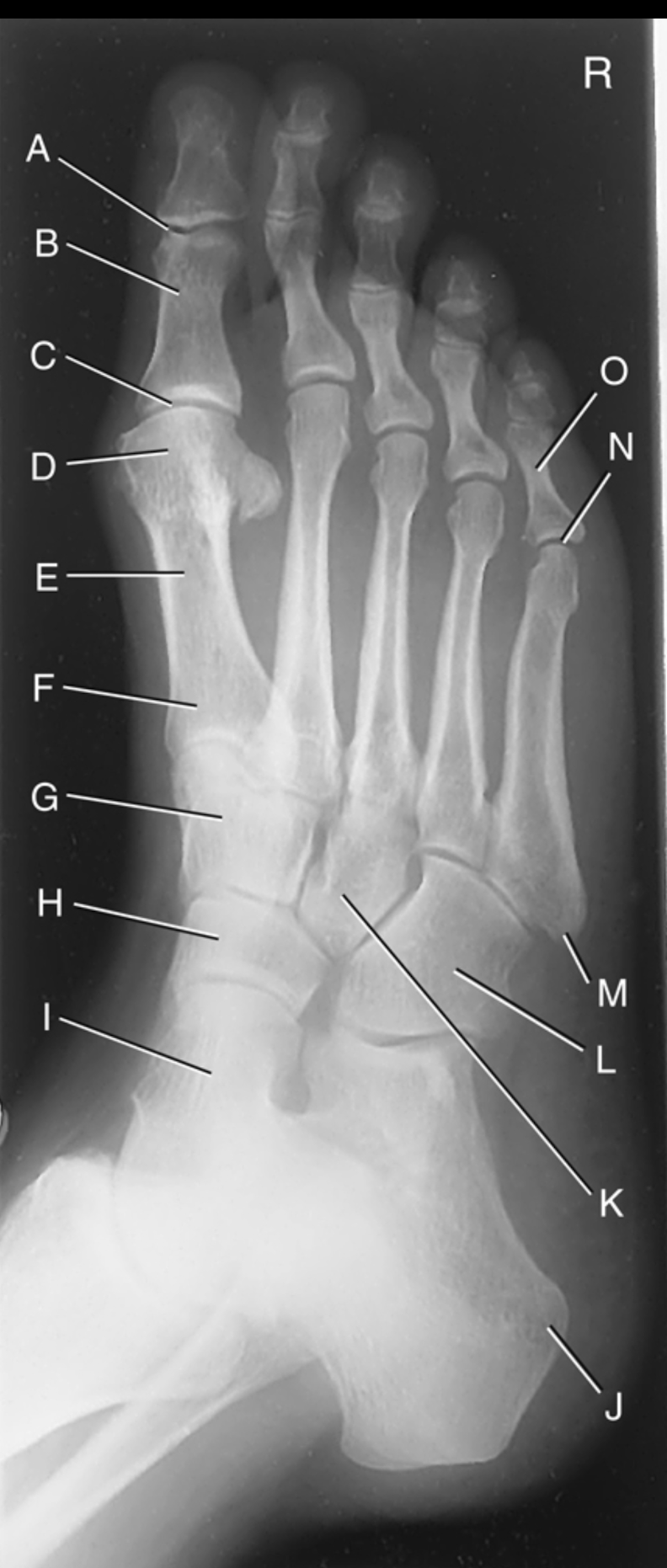
What is L on the AP oblique foot radiograph?
Cuboid

What is M on the AP oblique foot radiograph?
Tuberosity of the fifth metatarsal

What is N on the AP oblique foot radiograph?
Fifth metatarsophalangeal joint

What is O on the AP oblique foot radiograph?
Proximal phalanx of the fifth digit


What is A on the AP mortise ankle radiograph?
Fibula

What is B on the AP mortise ankle radiograph?
Lateral malleolus

What is C on the AP mortise ankle radiograph?
Mortise joint

What is D on the AP mortise ankle radiograph?
Talus
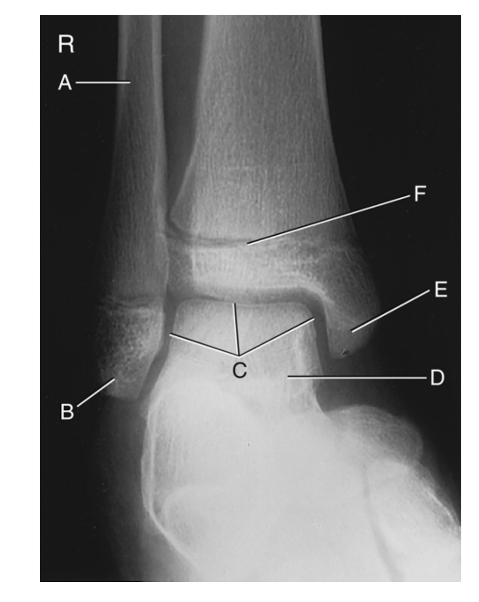
What is E on the AP mortise ankle radiograph?
Medial malleolus

What is F on the AP mortise ankle radiograph?
Epiphyseal plate (epiphyseal line)

What is A on the lateral ankle radiograph?
Fibula

What is B on the lateral ankle radiograph?
Calcaneus

What is C on the lateral ankle radiograph?
Cuboid

What is D on the lateral ankle radiograph?
Tuberosity of the fifth metatarsal

What is E on the lateral ankle radiograph?
navicular

What is F on the lateral ankle radiograph?
talus

What is G on the lateral ankle radiograph?
Sinus tarsi
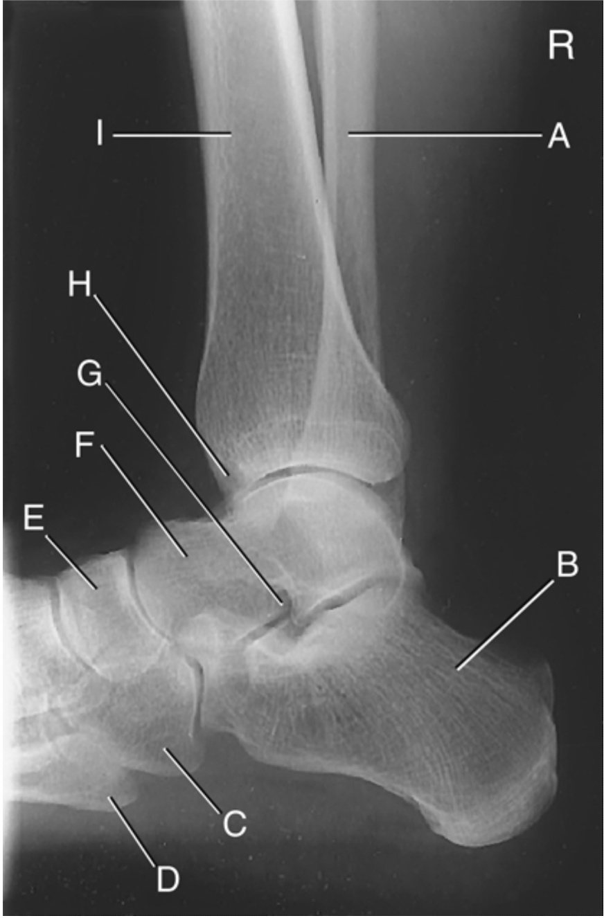
What is H on the lateral ankle radiograph?
anterior tubercle

What is I on the lateral ankle radiograph?
Tibia
the articular facets making up the tibial plateau slope posteriorly from __ to ____ degrees in relation to the long axis of the tibia
10 , 20
when an AP knee is positioned, the CR must be angled in relation to the IR / the tabletop to be ___ to the tibial plateau.
parallel
A CR angle on AP knee is essential to show an __ ___ space
open joint
Generalized decreased bone density, cortical thinning,
and possible Looser zones (pseudofractures) indicates what pathology ?
osteomalacia
punched-out erosions with overhanging edges, soft-tissue swelling, and sometimes tophi indicate what pathology
gout
Uneven joint-space narrowing, osteophytes (bone spurs), subchondral sclerosis, and possible subchondral cysts is indicative of what pathology ?
osteoarthritis
Bone enlargement, cortical thickening, coarse trabeculae, and mixed areas of sclerosis and bone destruction are indicative of what pathology
paget disease
increased bone density (whiter bones), reduced distinction between cortex and medulla, and sometimes a bone-within-bone appearance are indicative of what pathology
osteopetrosis
Acute: early radiographs may be normal; later, bone
destruction and periosteal reaction.
Chronic: sclerosis,
cortical thickening, and possible sequestrum (dead
bone).
osteomyelitis
for an AP knee the CR angle depends on ASIS to table top measurement, if the table top measurement is less than 19cm what is the angle
3-5 caudad
for an AP knee the CR angle depends on ASIS to table top measurement, if the table top measurement is greater than 24cm what is the angle
3-5 cephalad
for an AP knee the CR angle depends on ASIS to table top measurement, if the table top measurement is 19- 24cm what is the angle
0 (perpendicular)