Exam 3 Sturcutre and Function
 Call Kai
 Learn
 Practice Test
 Spaced Repetition
 Match
 Flashcards
 Knowt Play
1/178

There's no tags or description
Looks like no tags are added yet.
179 Terms
What is the primary role of a neuron?
To receive, process, and transmit information through electrical and chemical signaling.
How do neurons communicate?
Through electrical signals and chemical signals called neurotransmitters.
What are the key characteristics of neurons?
They are electrically excitable cells that communicate through electrical and chemical signaling. They have a high metabolic rate, are long-lived, and generally do not divide.
What are the general functions of neuroglia (glial cells)?
Support, protection, growth, and communication.
What are the functions of astrocytes?
Anchor neurons to blood vessels, regulate the extracellular environment, help form the blood-brain barrier, repair damaged tissue, provide glucose to neurons, secrete nerve growth factor, and regulate blood vessel diameter.
What is the function of oligodendrocytes?
Myelinate certain axons in the CNS.
What is the function of microglial cells?
Act as phagocytes that remove unwanted material and cellular debris.
What are the functions of ependymal cells?
Line cavities in the brain and spinal cord, produce cerebrospinal fluid (CSF), and use cilia to circulate the fluid.
What is the function of Schwann cells?
Myelinate certain axons in the PNS.
What is the function of satellite cells?
Surround and support neuron cell bodies in the PNS.
FRONT: What are the components of the CNS?
The brain and spinal cord.
What are the components of the PNS?
Cranial nerves, spinal nerves, and ganglia.
Where do cranial nerves originate?
From the brain or brainstem.
Where do spinal nerves originate?
From the spinal cord.
What is the general function of the PNS?
To carry information to and from the CNS.
What is the difference between cranial nerves and spinal nerves?
Cranial nerves originate from the brain or brainstem, while spinal nerves originate from the spinal cord.
What are ganglia and nuclei?
Collections of neuron cell bodies.
What is the difference between ganglia and nuclei?
Ganglia are located in the PNS, while nuclei are located in the CNS.
What are nerves?
Bundles of axons in the PNS surrounded by connective tissue that carry information to or from the CNS.
What are tracts?
Bundles of axons in the CNS that carry information within the CNS, such as between brain regions or up and down the spinal cord.
What is the difference between nerves and tracts?
Nerves are bundles of axons in the PNS surrounded by connective tissue. Tracts are bundles of axons in the CNS without connective tissue wrappings
What factors affect the conduction velocity of an action potential?
Axon diameter and whether the axon is myelinated or unmyelinated.
How does axon diameter affect conduction velocity?
Larger-diameter axons conduct impulses faster.
How does myelination affect conduction velocity?
Myelinated axons conduct impulses faster than unmyelinated axons.
What is the significance of an action potential?
It allows electrical signals to travel long distances along an axon without decreasing in strength.
What cells in the nervous system generate action potentials?
Electrically excitable cells, especially neurons.
What causes voltage-gated ion channels to open?
Changes in membrane potential.
Where are voltage-gated ion channels located in a neuron?
Along the axon, with important concentrations in the trigger zone and at the nodes of Ranvier in myelinated axons.
What types of ion channels can be opened by a stimulus?
Chemically gated, mechanically gated, light-gated, and temperature-sensitive channels.
What causes chemically gated ion channels to open?
A neurotransmitter binds to its receptor.
Where are chemically gated ion channels commonly located?
On the dendrites and cell body.
What is the resting membrane potential of a neuron?
Approximately -70 mV.
What does the resting membrane potential indicate?
The inside of the neuron is more negative than the outside.
What happens between the initial stimulation of a neuron and the events at the axon hillock?
A stimulus opens ion channels on the receptive regions, producing a local potential. The local potential spreads toward the trigger zone, where it may bring the membrane to threshold.
What is a depolarizing local potential?
A local change in membrane potential that makes the inside less negative, bringing the neuron closer to threshol
What is a hyperpolarizing local potential?
A local change in membrane potential that makes the inside more negative, moving the neuron farther from threshold.
How do depolarizing and hyperpolarizing local potentials affect the likelihood of an action potential?
Depolarizing local potentials increase the likelihood of reaching threshold. Hyperpolarizing local potentials decrease the likelihood.
What is threshold?
The critical membrane voltage required to trigger an action potential, approximately -55 mV.
What does it mean when a neuron reaches threshold?
The membrane has reached the critical voltage needed to open voltage-gated channels and initiate an action potential.
Where is the trigger zone for an action potential?
At the axon hillock and initial segment, rather than the main cell body.
What happens when a neuron reaches threshold?
An all-or-none action potential is initiated.
How does a postsynaptic neuron respond when excitatory and inhibitory presynaptic neurons are both signaling it?
The postsynaptic neuron integrates the excitatory and inhibitory local potentials through summation. Their combined effect determines whether threshold is reached.
What is a local potential?
A graded change in membrane potential caused by a stimulus. It can be depolarizing or hyperpolarizing.
What are the main characteristics of a local potential?
Graded (a bigger stimulus opens more ion channels producing a bigger local potential. )
Decremental (it fades as it travels away from the stimulus.)
Reversible (it stops when the stimulus ends)
What are the main characteristics of an action potential?
All-or-none (it either happens completely or not at all once threshold is reached)
Non-decremental (it does not fade as it travels)
Irreversible (once triggered, it cannot be stopped.)
What is the difference between a local potential and an action potential?
A local potential is graded, decremental, and reversible. An action potential is all-or-none, non-decremental, and irreversible.
What effect does an excitatory neurotransmitter have on a neuron?
It produces a depolarizing local potential, bringing the membrane closer to threshold and increasing the likelihood of an action potential.
What effect does an inhibitory neurotransmitter have on a neuron?
It produces a hyperpolarizing local potential, moving the membrane farther from threshold and decreasing the likelihood of an action potential.
During depolarization, how does the charge inside the neuron change?
The inside becomes less negative and may become positive relative to the outside.
During depolarization, which ions move, and in which direction?
Sodium (Na+) ions move into the cell.
During repolarization, how does the charge inside the neuron change?
The inside returns toward a negative charge relative to the outside.
During repolarization, which ions move, and in which direction?
Potassium (K+) ions move out of the cell.
Why do sodium and potassium ions move when their voltage-gated channels open?
They move down their electrochemical gradients. Sodium is more concentrated outside the cell, while potassium is more concentrated inside the cell.
What happens during the resting phase of an action potential graph
The membrane is at its resting potential, approximately -70 mV, with the inside more negative than the outside.
What happens during the depolarization phase of an action potential graph?
Voltage-gated sodium channels open, sodium enters the cell, and the membrane potential becomes more positive.
What happens at the peak of an action potential graph?
The membrane reaches its most positive voltage as sodium channels inactivate and potassium channels contribute to the beginning of repolarization.
What happens during the repolarization phase of an action potential graph?
Potassium leaves the cell, causing the membrane potential to become more negative.
What happens during the hyperpolarization phase of an action potential graph?
The membrane becomes temporarily more negative than its resting potential because potassium channels remain open briefly.
What happens after hyperpolarization?
The membrane returns toward its resting potential as the ion channels return to their resting states.
What is the electrical status of a neuron at rest?
The inside is negative relative to the outside, approximately -70 mV.
What is the electrical status of a neuron during depolarization?
The inside becomes less negative and may temporarily become positive relative to the outside.
The inside becomes less negative and may temporarily become positive relative to the outside.
The inside returns toward a negative charge relative to the outside
What does the sodium-potassium pump do?
It uses ATP to move three sodium ions out of the cell and two potassium ions into the cell.
Why is the sodium-potassium pump important?
It maintains the sodium and potassium concentration gradients needed for normal neuronal function. Repolarization restores the electrical conditions but not the original ionic conditions.
What is the refractory period?
The period following an action potential during which another action potential cannot occur or requires a stronger stimulus.
What is the difference between the absolute and relative refractory periods?
During the absolute refractory period, another action potential cannot occur. During the relative refractory period, another action potential can occur if a stronger stimulus is applied.
Why is the refractory period important?
It helps prevent backward conduction of an action potential and limits the frequency at which a neuron can fire.
What happens after an action potential reaches the axon terminal?
Voltage-gated calcium channels open, calcium enters the terminal, and neurotransmitter vesicles release their contents into the synaptic cleft.
How does an excitatory postsynaptic potential (EPSP) affect the postsynaptic membrane?
It causes partial depolarization, commonly through sodium or calcium entering the cell, making an action potential more likely.
How does an inhibitory postsynaptic potential (IPSP) affect the postsynaptic membrane?
It causes hyperpolarization, commonly through potassium leaving the cell or chloride entering it, making an action potential less likely.
What is an example of an excitatory neurotransmitter?
Glutamate.
What is an example of an inhibitory neurotransmitter?
GABA (gamma-aminobutyric acid).
How is a neurotransmitter signal stopped?
Through diffusion, degradation, or reuptake.
What blood potassium and calcium ranges are listed in the PowerPoint?
Potassium: 3.8 to 4.5 mEq/L. Calcium: 9.2 to 10.4 mEq/L, as written on the slide.
How do changes in blood potassium levels affect action potentials and neuromuscular activity?
They alter the resting membrane potential and cellular excitability, which can interfere with normal nerve and muscle activity.
How do changes in blood calcium levels affect action potentials and neuromuscular activity?
Low calcium increases neuromuscular excitability, while high calcium generally decreases it.
How does an action potential travel down an unmyelinated axon?
Through contiguous conduction, where adjacent sections of the membrane depolarize continuously along the axon.
How does an action potential travel down a myelinated axon?
Through saltatory conduction, where the action potential travels from one node of Ranvier to the next.
What is the difference between contiguous conduction and saltatory conduction?
Contiguous conduction occurs along the membrane of unmyelinated axons. Saltatory conduction occurs in myelinated axons, with the action potential regenerating at the nodes of Ranvier.
How does lidocaine block pain signals?
It blocks voltage-gated sodium channels, preventing sensory neurons from generating or conducting action potentials that transmit pain signals.
How many pairs of spinal nerves are there?
31 pairs.
Are spinal nerves sensory, motor, or mixed?
Mixed, they carry both sensory and motor signals.
Are spinal nerves part of the CNS or PNS?
PNS
What are the holes called where spinal nerves leave the vertebral canal?
Intervertebral foramina.
How many pairs of cranial nerves are there?
12 pairs
Are cranial nerves sensory, motor, or mixed?
They can be sensory, motor, or mixed, depending on the nerve.
Are cranial nerves part of the CNS or PNS?
PNS
Where do most cranial nerves serve structures?
The head and neck.
How is the vagus nerve different from most other cranial nerves?
It extends beyond the head and neck to supply organs in the chest and abdomen.
The spinal cord lies within what structure of the vertebral column?
The vertebral canal.
Where does the spinal cord begin?
At the foramen magnum, where it continues from the medulla oblongata of the brainstem.
Where does the spinal cord end in an adult
Approximately at the L1 to L2 vertebral level.
What is the conus medullaris?
The tapered inferior end of the spinal cord.
What is the cauda equina?
The collection of spinal nerve roots extending below the end of the spinal cord.
What are the three spinal meninges, from superficial to deep?
Dura mater, arachnoid mater, and pia mater.

Where is the epidural space located, and what does it contain?
Outside the dura mater but inside the vertebral canal. It contains fat and blood vessels.
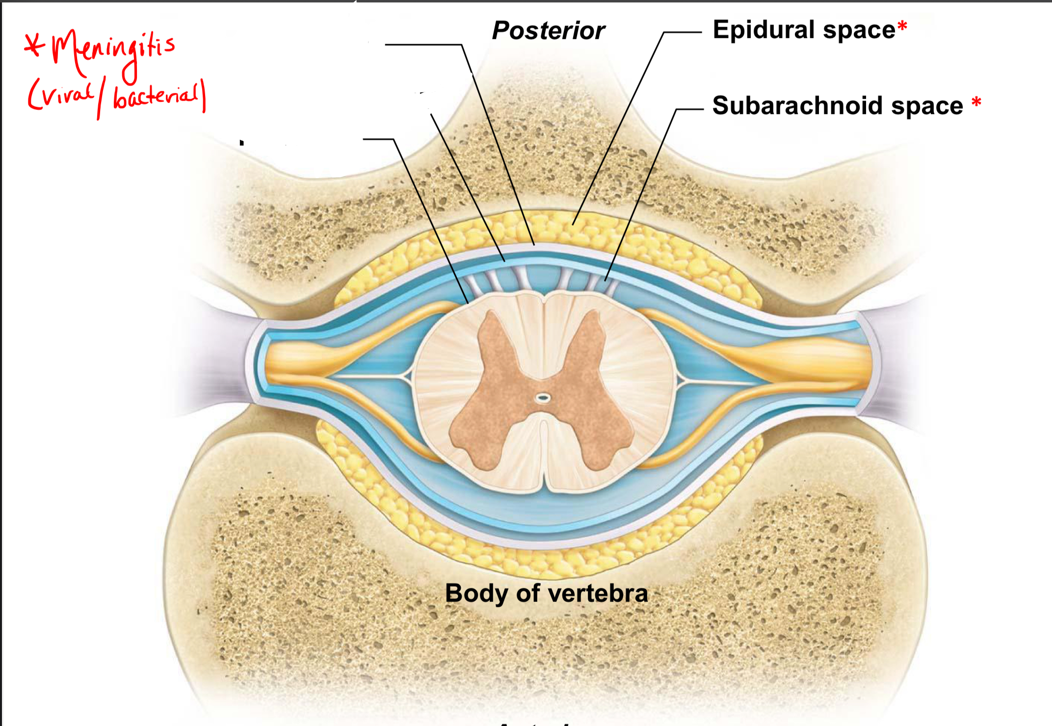
Where is the subarachnoid space located, and what does it contain?
Between the arachnoid mater and pia mater. It contains cerebrospinal fluid (CSF).

Where is a lumbar puncture typically performed?
At the L3 to L4 or L4 to L5 level, with the needle entering the subarachnoid space.
What is meningitis, and what causes it?
Inflammation of the meninges, commonly caused by a viral or bacterial infection.
What is spina bifida, and what causes it?
A neural tube defect in which the vertebral arches fail to close completely during development.