Exam 2 (Anatomy)
 Call Kai
 Learn
Learn Practice Test
 Spaced Repetition
 Match
Match Flashcards
 Knowt Play
1/118

There's no tags or description
Looks like no tags are added yet.
Name | Mastery | Learn | Test | Matching | Spaced | Call with Kai | Chat |
|---|
No analytics yet
Send a link to your students to track their progress
119 Terms
Knee Joint Muscles
Extensors: __________ muscle group
Flexors: ____________ muscle group + others
Knee Joint Muscles
Extensors: Quadricep muscle group (quad = four muscles)
Flexors: Hamstring muscle group + others
What makes up the Pes Anserinus
Gracilis, Sart, Semi T
Origin = ____________
Insertion = ________________
Innervation = _______________
Origin = (where it starts)
Insertion = (where it ends)
Innervation = (what nerve communicates with it)

Vastus Lateralis
Origin:
Insertion:
Innervation:
Action:
Notes:
Origin: Linea Aspera and greater trochanter of femur
Insertion: Tibial tuberosity via patella and patellar tendon.
Innervation: Femoral
Action: Knee extension
Notes: Largest quadricep muscle, uniarticular


Vastus Intermedius
Origin:
Insertion:
Innervation:
Action:
Notes:
Origin: Anterior surface of femur
Insertion: Tibial tuberosity via patella
Innervation: Femoral
Action: Knee extension
Notes: Under Rectus Femoris (layer deeper), uniarticular. Smallest quad muscle


Vastus Medialis
Origin:
Insertion:
Innervation:
Action:
Notes:
Origin: Linea aspera of femur
Insertion: Tibial tuberosity via patella
Innervation: Femoral
Action: Knee Extension
Notes: uniarticular, VMO

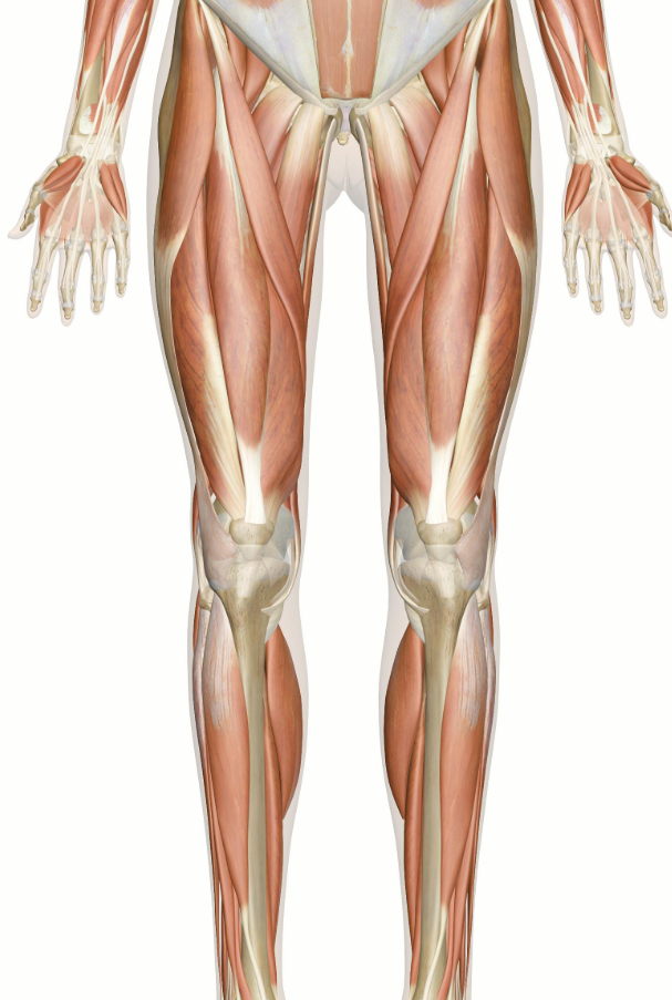
Rectus Femoris
Origin:
Insertion:
Innervation:
Action:
Notes:
Origin: Anterior Inferior iliac spine (AIIS) and superior rim of acetabulum
Insertion: Tibial tuberosity via patella
Innervation: Femoral
Action: Knee extension & Hip flexion (FABER)
Notes: Biarticular / over vastus intermedius
![<p><span style="background-color: transparent;">[NAME]</span></p><ul><li><p><span style="background-color: transparent;">Origin:</span></p></li><li><p><span style="background-color: transparent;">Insertion:</span></p></li><li><p><span style="background-color: transparent;">Innervation:</span></p></li><li><p><span style="background-color: transparent;">Action:</span></p></li><li><p><span style="background-color: transparent;">Notes:</span></p></li></ul><p></p>](https://assets.knowt.com/user-attachments/3168a957-30df-45ee-a896-f097f46f224a.png)
[NAME]
Origin:
Insertion:
Innervation:
Action:
Notes:
Semitendinosus
Origin: Ischial tuberosity
Insertion: Proximal, medial tibia below condyle
Innervation: Sciatic (tibial division)
Action: Knee flexion, medial rotation of tibia, & Hip extension & hyperextension
Notes: medially located; one of three muscles that make up the pes anserinus

![<p><span style="background-color: transparent;">[NAME]</span></p><ul><li><p><span style="background-color: transparent;">Origin:</span></p></li><li><p><span style="background-color: transparent;">Insertion:</span></p></li><li><p><span style="background-color: transparent;">Innervation:</span></p></li><li><p><span style="background-color: transparent;">Action:</span></p></li><li><p><span style="background-color: transparent;">Notes:</span></p></li></ul><p></p>](https://assets.knowt.com/user-attachments/63fc279f-8229-4c9b-9743-3eb02b560fd2.png)
[NAME]
Origin:
Insertion:
Innervation:
Action:
Notes:
Semimembranosus
Origin: Ischial tuberosity
Insertion: posterior surface of medial condyle of tibia
Innervation: Sciatic (tibial division)
Action: Knee flexion, Knee medial rotation of tibia, & hip extension
Notes: medially oriented

![<p><span style="background-color: transparent;">[NAME]</span></p><ul><li><p><span style="background-color: transparent;">Origin:</span></p></li><li><p><span style="background-color: transparent;">Insertion:</span></p></li><li><p><span style="background-color: transparent;">Innervation:</span></p></li><li><p><span style="background-color: transparent;">Action:</span></p></li><li><p><span style="background-color: transparent;">Notes:</span></p></li></ul><p></p>](https://assets.knowt.com/user-attachments/2abc9aac-c7e9-4719-8576-232f7ba826e5.png)
[NAME]
Origin:
Insertion:
Innervation:
Action:
Notes:
Biceps Femoris
Origin:
Long Head: Ischial tuberosity
Controls action of hip extension
Short Head: linea aspera
Insertion: Head of fibula & lateral condyle of tibia
Innervation: sciatic and common peroneal
Action: Knee flexion, knee lateral rotation, and Hip extension (long head only)
Notes: lateral muscle

![<p><span style="background-color: transparent;">[NAME]</span></p><ul><li><p><span style="background-color: transparent;">Origin:</span></p></li><li><p><span style="background-color: transparent;">Insertion:</span></p></li><li><p><span style="background-color: transparent;">Innervation:</span></p></li><li><p><span style="background-color: transparent;">Action:</span></p></li><li><p><span style="background-color: transparent;">Notes:</span></p></li></ul><p></p>](https://assets.knowt.com/user-attachments/c3b04c3f-baa6-48c0-8720-a693b9ff5883.png)
[NAME]
Origin:
Insertion:
Innervation:
Action:
Notes:
Gastrocnemius
Origin: posterior surface of femoral condyles above medial and lateral joint surfaces
Insertion: calcaneus via achilles (calcanean) tendon
Innervation: Tibial
Action: Knee flexion & Ankle plantarflexion (pointing foot like ballerina)
Notes: 2 heads, medial head is more distal

![<p><span style="background-color: transparent;">[NAME]</span></p><ul><li><p><span style="background-color: transparent;">Origin:</span></p></li><li><p><span style="background-color: transparent;">Insertion:</span></p></li><li><p><span style="background-color: transparent;">Innervation:</span></p></li><li><p><span style="background-color: transparent;">Action:</span></p></li><li><p><span style="background-color: transparent;">Notes:</span></p></li></ul><p></p>](https://assets.knowt.com/user-attachments/03376b20-7326-4f3b-8e6f-efd63c23d406.png)
[NAME]
Origin:
Insertion:
Innervation:
Action:
Notes:
Plantaris
Origin: Lateral supracondylar ridge
Insertion: Calcaneus
Innervation: Tibial
Action: Knee flexion & Ankle plantarflexion
Notes: Posterior to the gastrocnemius
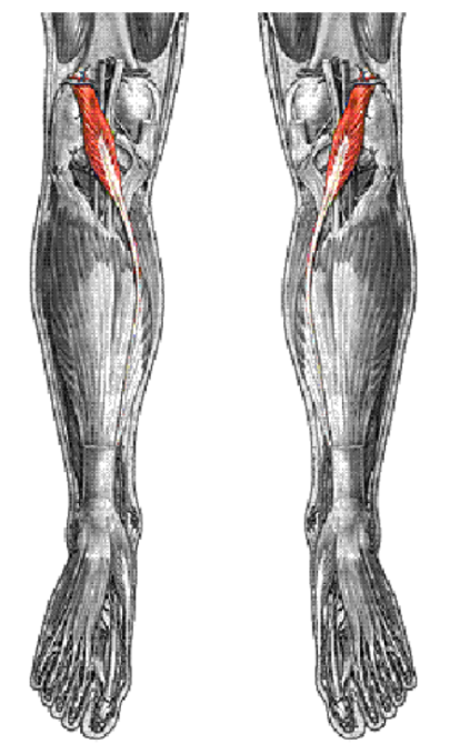

Popliteus
Origin:
Insertion:
Innervation:
Action:
Notes:
Origin: Lateral condyle of femur
Insertion: Posterior surface of proximal tibial shaft
Innervation: Tibial
Action: Laterally rotates Femur or medially rotates tibia
Think about what each end would do if pulling (like twisting a jar open by rotating lid and jar opposite directions)
Notes: This is the most posterior in the knee


Sartorius
Origin:
Insertion:
Innervation:
Action:
Notes:
Origin: Anterior superior iliac spine (ASIS)
Insertion: medial surface of tibia near tibial tuberosity
Innervation: Femoral
Action: Knee flexion, knee medial rotation, hip flexion, hip lateral rotation of the femur
Notes: One of three muscles that make up the pes anserinus

![<p><span style="background-color: transparent;">[NAME]</span></p><ul><li><p><span style="background-color: transparent;">Origin:</span></p></li><li><p><span style="background-color: transparent;">Insertion:</span></p></li><li><p><span style="background-color: transparent;">Innervation:</span></p></li><li><p><span style="background-color: transparent;">Action:</span></p></li><li><p><span style="background-color: transparent;">Notes:</span></p></li></ul><p></p>](https://assets.knowt.com/user-attachments/ce33671b-83e8-4019-b636-16eb2a32b14c.png)
[NAME]
Origin:
Insertion:
Innervation:
Action:
Notes:
Gracilis
Origin: Inferior ramus of pubis (body of pubis)
Insertion: Medial surface of tibia inferior to medial condyle
Innervation: Obturator
Action: (FAdIr) Knee flexion, knee medial rotation of tibia, hip adduction, hip medial rotation
Notes: One of three muscles that make up the pes anserinus
(pic is a medial view)


Popliteal Fossa
Vessels and nerves pass through the Popliteal Fossa


Cross Section view of proximal thigh
Sartorius: Anterior
Gracillis: Medial compartment
BF(LH), ST, SM: Posterior

FASCIA OF THE THIGH - Intermuscular Septa (IMS)
Medial IMS
Divides the ______ & ______ Compartments
Lateral IMS
Divides the ______ & ______ Compartments
Posterior IMS
Divides the ______ & ______ Compartments
FASCIA OF THE THIGH - Intermuscular Septa (IMS) (thin walls of tissue)
Medial IMS: Divides the Anterior & Medial Compartments
Lateral IMS: Divides the Anterior & Posterior Compartments
Posterior IMS: Divides the Medial & Posterior Compartments


Nerves in the thigh
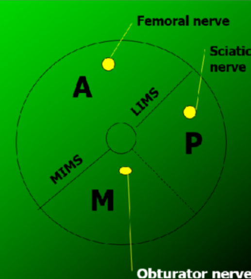

Femur & Innominate

Flexors of the Knee
Hamstring muscles
(3)
______________
______________
______________
______________
______________
Flexors of the Knee (posterior because it makes the knee bend)
Hamstring muscles
Semitendinosus
Semimembranosus
Biceps femoris
Gastrocnemius
Plantaris
Popliteus
Sartorius
Gracilis
3 NERVES OF THE LOWER EXTREMITY
Femoral nerve: Anterior compartment
Obturator nerve: Medial compartment
Sciatic nerve: Posterior compartment
Anastemoses of the Knee
If you’re not getting enough flow from a vessel for whatever reason, an anastomosis is bypassing that vessel and taking an alternate route through the knee


Course of Blood Vessels


Branches of the sciatic nerve
Superficial peroneal
Piriformis
Common Peroneal
Deep peroneal
Tibial
Sciatic
Medial plantar (foot)
Lateral plantar (foot)
Piriformis: Triangular muscle deep in the butt, just above the sciatic nerve
Common Peroneal (lateral compartment) (Peroneal = fibular)
Superficial peroneal (lateral compartment)
Deep peroneal (anterior compartment)
Tibial (posterior compartment)
Medial plantar (foot)
Lateral plantar (foot)

Joints of the leg (Shank) and Foot
3 Bones: ________________
Connective Point: ________________
Joints of the leg (Shank) and Foot
3 Bones: Tibia, Fibula, Talus (foot)
Connective Point: Medial/lateral malleolus (The bottom of the tibia and the outer fibula reach down to create a socket around the top of the talus)
____________ support joints
Ligaments support joints
Tibiofibular Joints:
Joint One: ____________
Movement type: ____________
2 Ligamentous supports: ____________
Joint Two: ____________
Movement type: ____________
Ligamentous supports: ____________
Tibiofibular Joints:
Joint One: Proximal Tibiofibular
Movement type: Synovial, gliding
Ligamentous supports:
Anterior proximal tibiofibular ligament
Posterior proximal tibiofibular ligament
Joint Two: Distal Tibiofibular
Movement type: Synovial, gliding
Ligamentous supports:
Anterior distal tibiofibular ligament
Posterior distal tibiofibular ligament
Interosseous (tibiofibular) ligament

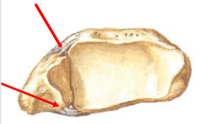
2 of 3 Distal Tibiofibular Joint Ligamentous supports
Distal Tibiofibular Joint Ligamentous supports

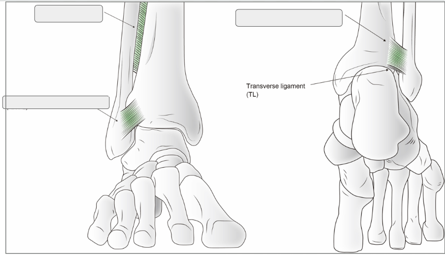
Anterior distal/inferior tibiofibular ligament
Posterior distal/inferior tibiofibular ligament
Interosseous (tibiofibular) ligament
Anterior distal/inferior tibiofibular ligament
Posterior distal/inferior tibiofibular ligament
Interosseous (tibiofibular) ligament

Middle Tibiofibular Joint
Cartilaginous, synovial, or fibrous joint: _____________
Attachments: _____________
Functions: _____________
Middle Tibiofibular Joint
Cartilaginous, synovial, or fibrous joint: Fibrous
Attachments: Connects the interosseous (“between bone”) borders of the tibia and fibula
Functions: Separates the anterior compartment from the posterior compartment and serves as sites for muscular attachments
Ankle Joint
Ankle Joint fancy name: _________
Bones: _____________________
Movement type: ___________
Ankle Joint
Ankle Joint fancy name: Talocrural
Bones: Tibia, fibula, talus (trochlear surface)
Movement type: Synovial – Hinge Joint
Ankle Joint Movements
Sagittal Plane (two types):
_________________
_________________
(#) degrees clinically is treated as zero (neutral position)
Ankle Joint Movements
Sagittal Plane (two types):
Dorsiflexion (DF): Anterior angle decreasing or less than 90 degrees
Plantarflexion (PF): Anterior angle increasing or greater than 90 degrees
90 degrees clinically is treated as zero (neutral position)

Intertarsal Joints (tarsal = _______________)
Joint Type: _____________
7 Tarsal Bones:
Talus (trochlea)
Calcaneus
Navicular
Cuboid
Medial Cuneiform
Intermediate Cuneiform
Lateral Cuneiform
Intertarsal Joints (tarsal = ankle and rear part of the foot)
Joint Type: Synovial – gliding
7 Tarsal Bones:
Talus (trochlea)
Calcaneus
Navicular
Cuboid
Medial (#1) Cuneiform
Intermediate (#2) Cuneiform
Lateral (#3) Cuneiform
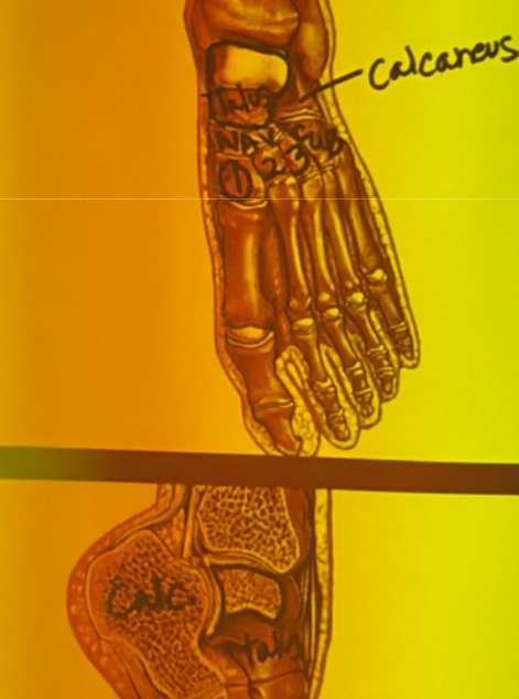
Intertarsal Movement
________ Plane (mostly)
Two movement types:
Inversion: __________,__________,__________
Eversion: __________,__________,__________
Intertarsal Movement
Frontal Plane (mostly)
Two movement types:
Inversion: Supination (sole inward), Adduction, Varus
Eversion: Pronation, Abduction, Valgus

Intertarsal Joints
Most Movement occurs at the:
________ joint: (________ ligament)
________ joint
Known as: ________ motion
Intertarsal Joints
Most Movement occurs at the:
Subtalar joint: (Talocalcaneo ligament)
Talonavicular joint
Known as: Rearfoot motion

Inversion occurs with Plantarflexion (pointing foot down)
The (medial/lateral malleolus) extends more inferior and posterior than the (medial/lateral malleolus). This forces the foot inward as it points downward.
The axis of rotation for the intertarsal/subtalar joints is angled (acute/oblique). Plantarflexion automatically couples with inversion along this axis.
In plantarflexion, the (medial/lateral ligaments) become taut, pulling and stabilizing the ankle into inversion.
Inversion occurs with Plantarflexion (pointing foot down)
The lateral malleolus extends more inferior and posterior than the medial malleolus. This forces the foot inward as it points downward.
The axis of rotation for the intertarsal/subtalar joints is angled (oblique). Plantarflexion automatically couples with inversion along this axis.
In plantarflexion, the lateral ligaments become taut, pulling and stabilizing the ankle into inversion.

Category: Major Lateral Ankle and Intertarsal Ligaments
Anterior talofibular (connects ____ & ____)
Calcaneofibular (connects ____ & ____)
Posterior talofibular (connects ____ & ____)
Prevents: ___________________
Category: Major Lateral Ankle and Intertarsal Ligaments
Anterior talofibular (connects talus & fibula)
Calcaneofibular (connects calcaneus & fibula)
Posterior talofibular (connects talus & fibula)
Prevents: Excessive Inversion


Category: __________________________
Category: Major Medial Ankle Ligaments

Category: __________________
Type of Joint: ______________
Bones Involved: ____________________
Movement: _______________________
Function: ______________________
Category: Tarsometatarsal Joints
Type of Joint: Synovial, gliding
Bones Involved: Cuneiforms (three wedge-shaped bones) and the cuboid bone connect with the bases of the metatarsals (the long bones of the foot).
Movement: No named movement (no flexing or extending, only slight shifting)
Function: Tight joint capsules and strong ligaments designed to stabilize the foot and hold the arch position


Category: ______________________
Joint Type: ______________________
Movements:
Sagittal: ______________________
Transverse: ______________________
Category: Metatarsophalangeal (MP) Joints
Joint Type: Synovial – condyloid
Movements:
Sagittal: Flexion, extension, hyperextension
Transverse: Abduction, Adduction


Category: Interphalangeal (IP) Joints
Joint Type: ___________________________
Movements:
Sagittal: ___________________________
LABEL:
PIP = Proximal IP joints
DIP = Distal IP joints
Category: Interphalangeal (IP) Joints
Joint Type: Synovial, condyloid (but function as hinge joint due to tight lateral ligaments)
Movements:
Sagittal: Flexion, extension, hyperextension

The Three Foot Arches
Medial Longitudinal
Lateral Longitudinal
Transverse

Functional Segments of Foot (3)
Rearfoot
Midfoot
Forefoot: Metatarsals & phallanges


Frontal Plane Analysis


Medial longitudinal arch bones:
(Flexible/Inflexible)
Medial longitudinal arch bones
Calcaneus
Talus
Navicular
Cuneiforms
1st three metatarsals
Flexible

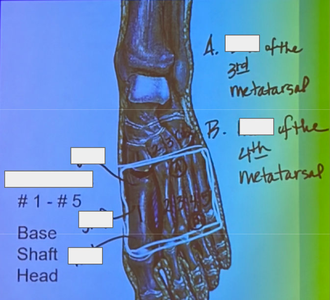
Transverse arch in mid-foot bones:
Transverse arch in forefoot bones:
Transverse arch in mid-foot bones
Cuboid
Cuneiforms
Metatarsals (Base)
Transverse arch in forefoot bones
Metatarsals (Head)

Ligaments that support the arches
Plantar aponeurosis
Long Plantar- lateral
Spring- medial
Bony attachment site: Sustentaculum Tali & Navicular

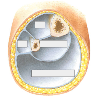
Lower leg
Lower leg
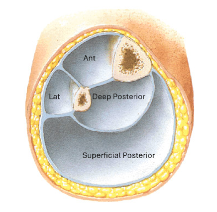

Anterior
Attaches to ___________________
Separates ________ from ________ compartments
Posterior
Attaches to ___________________
Separates the ________ from the ________ compartments
Deep transverse fascia
Extends from the ___________ to the ______________
Deep transverse fascia
Extends from the medial tibial border to the posterior fibular border


Deep transverse fascia
Deep transverse fascia
–Extends from the medial tibial border to the posterior fibular border

Myology: ______________
(#) compartments in the leg that each have an associated nerve: ________________
Myology: Study of Muscles
Three Compartments in the leg that each have an associated nerve:
Anterior: Deep Peroneal
Lateral: Superficial Peroneal
Posterior: Tibial
5 Nerves in the Leg
•Sciatic n.
•Common peroneal n.
•Tibial nerve n.
•Deep peroneal n.
•Superficial peroneal nerve
Muscles, common innervation, and common action in the Posterior Superficial & Posterior Deep Compartments
Superficial: (S = Space)
Gastrocnemius
Plantaris
Soleus
Deep: Pop TDH
Popliteus
Posterior Tibialis
Flexor Digitorum Longus
Flexor Hallucis Longus
Common Innervation: Tibial
Common Action: Ankle plantarflexion
Muscles, common nerve, and common action in the Lateral & Anterior Leg Compartments
Anterior
Anterior Tibialis
Extensor Hallucis Longus
Extensor Digitorum Longus
Peroneus Tertius*
Common Innervation: Deep peroneal nerve
Common Action: Ankle dorsiflexion
Lateral (peroneus means lateral)
Peroneus Longus
Peroneus Brevis
Common Innervation: Superficial peroneal nerve
Common Action: Eversion

Gastrocnemius: Origin, insertion, innervation, action
•Origin: posterior surface of femoral condyles
•Insertion: calcaneus via achilles tendon
•Innervation: tibial
•Action: Ankle plantarflexion (pointing foot like ballerina), knee flexion.
•Notes: 2 heads, medial head is more distal


Plantaris (Origin, insertion, innervation, action)
•Origin: lateral supracondylar ridge of femur
•Insertion: calcaneus
•Innervation: tibial
•Action: Ankle plantarflexion, Knee flexion
•Notes: Posterior to the gastrocnemius
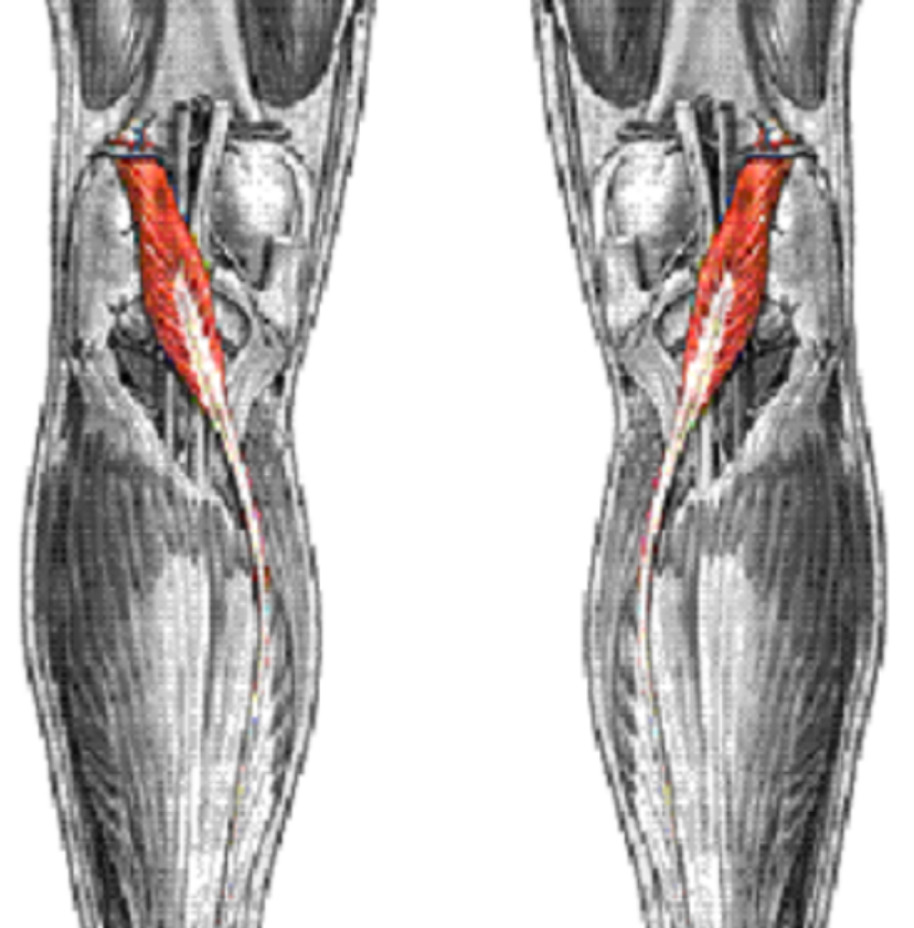

Soleus (Origin, insertion, innervation, action)
•Origin: Head and proximal fibula & posterior tibia
•Insertion: calcaneus via achilles tendon
•Innervation: tibial
•Action: Ankle plantarflexion


Posterior Tibialis (Origin, insertion, innervation, action)
•Origin: Interosseus membrane and tibial and fibular shafts
•Insertion: Tarsals and metatarsals (navicular)
•Innervation: tibial
•Action: Ankle plantarflexion, Inversion (supination)
•Notes: Deep muscle


Flexor Digitorum Longus (Origin, insertion, innervation, action)
•Origin: Posteromedial surface of tibia
•Insertion: Inferior surfaces of distal phalanges of toes 2-5
•Innervation: tibial
•Action: Ankle plantarflexion, Intertarsal inversion, MP & IP flexion for toes 2-5


Flexor Hallucis Longus (Origin, insertion, innervation, action)
•Origin: Posterior surface of fibula
•Insertion: Inferior surface of distal phalanx of # 1.
•Innervation: tibial
•Action: Ankle plantarflexion, Intertarsal inversion, MP & IP flexion of #1.

![<p>[NAME]</p><ul><li><p>Origin</p></li><li><p>Insertion</p></li><li><p>Innervation</p></li><li><p>Action</p></li></ul><p></p>](https://assets.knowt.com/user-attachments/997f1652-d6a6-4e1f-959e-794ac51948da.png)
[NAME]
Origin
Insertion
Innervation
Action
Peroneus Longus
•Origin: lateral proximal surface of fibula & tibial condyle
•Insertion: Base of 1st metatarsal and medial cuneiform***
•Innervation: superficial peroneal
•Action: Ankle plantarflexion, eversion

![<p>[NAME] (Origin, insertion, innervation, action)</p>](https://assets.knowt.com/user-attachments/195e151f-b208-4947-a90d-33adff64230f.png)
[NAME] (Origin, insertion, innervation, action)
Peroneus Brevis
•Origin: Lateral mid shaft of fibula
•Insertion: Base of 5th metatarsal
•Innervation: superficial peroneal
•Action: Ankle plantarflexion, Eversion
(Underneath peroneus longus)


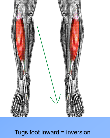
Tibialis Anterior (Origin, insertion, innervation, action)
•Origin: Lateral condyle and shaft of tibia
•Insertion: Base of 1st metatarsal and medial cuneiform
•Innervation: Deep peroneal
•Action: Ankle dorsiflexion, intertarsal inversion
Notes: Feel the tendon
![<p>[NAME] (Origin, insertion, innervation, action)</p>](https://assets.knowt.com/user-attachments/29fafc4d-04dc-451a-954f-107170c39642.png)
[NAME] (Origin, insertion, innervation, action)
Peroneus Tertius
•Origin: Distal anterior shaft of fibula
•Insertion: Base of the 5th metatarsal
•Innervation: Deep Peroneal
•Action: Ankle dorsiflexion, Intertarsal eversion
•Notes: Part of EDL muscle, separate tendon

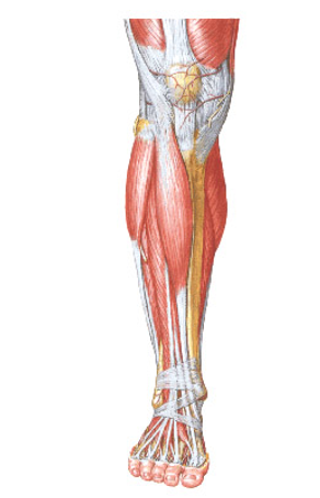
Extensor Digitorum Longus (Origin, insertion, innervation, action)
•Origin: Lateral condyle of tibia, anterior surface of fibula
•Insertion: superior surfaces of phalanges 2-5
•Innervation: deep peroneal
•Action: Ankle dorsiflexion, extension of MP & IP joints. Toes 2-5
Centered over ankle joint at crossing point = no inversion/eversion (Digitorum = toes 2-5)


Extensor Hallucis Longus (Origin, insertion, innervation, action)
•Origin: Anterior surface of fibula
•Insertion: Superior surface of distal phalanx of #1
•Innervation: deep peroneal
•Action: Ankle dorsiflexion, intertarsal inversion, MP & IP extension of # 1.

Name for Supporting CT of leg:
Retinaculum

Extensor Inferior Retinaculum (Attachments & Holds)
Attachments
–Stem (lat. Part) attaches to the upper surface of the calcaneous
–Prox. Band – reaches the medial malleolus
–Distal band – extends inferomedially to the plantar aponeurosis
Holds: TA, EHL, EDL, PT
(Extensor = holds down muscles that extends)


Extensor Superior Retinaculum (Attachments & Holds)
•Attachments
–Laterally to the distal anterior border of the fibula
–Medially to the distal anterior border of the tibia
Holds: TA, EHL, EDL, PT
(Extensor = holds down muscles that extends)

![<p>[NAME] (attachments & holds)</p>](https://assets.knowt.com/user-attachments/21707bb5-fcb8-4062-917c-208e3a09efd0.png)
[NAME] (attachments & holds)
Flexor Retinaculum
Attachments
Anteriorly to the tip of the medial malleolus
Continues posteriorly to the medial calcaneal process and plantar aponeurosis
Holds: TP, FDL, & FHL


Superior Peroneal Retinaculum (attachments & holds)
•Attachments
–Back of the lateral malleolus
–To lateral calcaneal surface
•Holds: PL, PB


Inferior Peroneal Retinaculum (attachments & holds)
•Attachments
–Continues anteriorly with the inferior extensor retinaculum
–To lateral calcaneal surface
•Holds: PL, PB


Foot Muscle Layer 1 Identifications & Actions
Flexor Digitorum Brevis: middle and proximal phalanges of the lateral four toes
Abductor Hallucis: Abducts #1
Abductor Digiti Minimi: Abducts #5

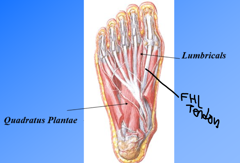
Foot Muscle Layer 2 Identifications & Actions
Quadratus Plantae: MP & IP flexion of 2-5
Lumbricals: MP flexion, IP extension
Tendons of FDL / Tendon of FHL (tubes on the medial side of foot- FDL on top)

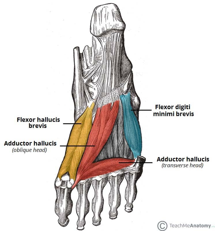
Foot Muscle Layer 3 Identifications & Actions
Flexor Hallucis Brevis: 1st MP and IP flexion
Adductor Hallucis (Transverse and Oblique Heads): Adduct # 1
Flexor Digiti Minimi Brevis: MP # 5 flexion

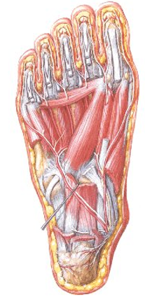
Foot Muscle Layer 4 Identifications & Actions
Plantar Interossei: Adduct toes - PAD (3 of them)
Dorsal Interossei: Abducts toes - DAB (4 of them)


2 Muscles on dorsal aspect of foot (identification & action)
Extensor Digitorum Brevis: MP 2-5 extension & hyperextension
Extensor Hallucis Brevis: MP # 1 extension & Hyperextension

Tibial N. divides into ________ and ________ Nerves in the foot
Tibial N. divides into Medial and Lateral Plantar Nerves in the foot

Anterior Tibial Artery turns into the ___________ once it crosses the ankle joint
Anterior Tibial Artery turns into the Dorsalis Pedis once it crosses the ankle joint

Posterior Tibial Artery splits into the ____________ and ____________
Posterior Tibial Artery splits into the Medial Plantar A. and Lateral Plantar A.

Fancy name for The Hip Joint:
Fancy name for The Hip Joint: COXA

FEMUR (_________)
•Head of Femur
•Greater Trochanter
•Lesser Trochanter
•Intertrochanteric line
FEMUR (Ball in COXA)
•Head of Femur
•Greater Trochanter
•Lesser Trochanter
•Intertrochanteric line


Ilium
Iliac crest
Anterior Superior Iliac Spine
Anterior Inferior Iliac Spine
Posterior Superior Iliac Spine
Posterior Inferior Iliac Spine
Acetabulum (socket)
Lunate surface
Acetabular fossa
Acetabular notch
Ilium
Iliac crest
Anterior Superior Iliac Spine
Anterior Inferior Iliac Spine
Posterior Superior Iliac Spine
Posterior Inferior Iliac Spine
Acetabulum (socket)
Lunate surface
Acetabular fossa
Acetabular notch


INNOMINATE BONE vs Pelvis
Left innominate bone + right innominate bone = 1 pelvis


Ischium
Body
Ischial tuberosity
Ischial ramus (superior and inferior)
Pubis
Body
Ramus (superior and inferior)

CLASSIFICATION OF HIP JOINT
Joint type:
(One axis/Multiaxial)
(Simple/Complex)
(Stable/Unstable)
CLASSIFICATION OF HIP JOINT
Joint type: Synovial / Ball and Socket
Multiaxial
Complex
Stable
Hip Joint Motion: (Femur/pelvis) moving and (femur/pelvis) stationary
Sagittal Plane:
Flexion: _____________
Extension: _____________
Hyperextension: _____________
Frontal Plane:
Abduction: _____________
Adduction: _____________
Hip Joint Motion: Femur moving and pelvis stationary
Sagittal Plane:
Flexion: kick leg up
Extension: bring leg back to ana. pos.
Hyperextension: kick backwards past AP
Frontal Plane:
Abduction: kick leg out
Adduction: bring leg in
Hip Joint Motion: (Femur/pelvis) moving and (femur/pelvis) stationary
Transverse Plane:
External rotation: _____________
Internal rotation: _____________
Horizontal abduction: _____________
Horizontal adduction: _____________
Circumduction: _____________
Hip Joint Motion: Femur moving and pelvis stationary
Transverse Plane:
External rotation: rotate out (lateral)
Internal rotation: rotate in (medial)
Horizontal abduction: hip flexed + abduction
Horizontal adduction: hip flexed + adduction
Circumduction: circling leg

ANGLE OF INCLINATION (COXA)
Angle of inclination for: Normal, Coxa Valga, and Coxa Vera

COXA VALGA

COXA VARA

HIP JOINT LIGAMENTS: Each ligament is a regional ________ of the ________
List them: ______________________
HIP JOINT LIGAMENTS: Each ligament is a regional thickening of the capsule
List them:
•Iliofemoral
•Ishiofemoral
•Pubofemoral
•Transverse acetabular
•Ligamentum teres
![<p>[NAME]</p><ul><li><p>Proximal Attachment:</p></li><li><p>Distal Attachment:</p></li><li><p>Extrinsic or capsular:</p></li><li><p>Function:</p></li></ul><p></p>](https://assets.knowt.com/user-attachments/c15138e7-fe1e-4cbc-9fa7-bdb623c55410.png)
[NAME]
Proximal Attachment:
Distal Attachment:
Extrinsic or capsular:
Function:
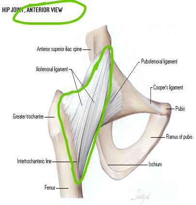
ILIOFEMORAL LIGAMENT
•PA: Below AIIS, Ilial rim of acetabulum
•DA: Intertrochanteric Line
•Extrinsic
•FUNCTION:
•Checks Hip hyperEXT
•Checks Hip ABD
(stopping hip from pulling away)
![<p>[NAME]</p><ul><li><p>Proximal Attachment:</p></li><li><p>Distal Attachment:</p></li><li><p>Extrinsic or capsular:</p></li><li><p>Function:</p></li></ul><p></p>](https://assets.knowt.com/user-attachments/446f5470-aa82-4737-8a88-401a5e1cfc40.png)
[NAME]
Proximal Attachment:
Distal Attachment:
Extrinsic or capsular:
Function:
PUBOFEMORAL LIGAMENT
•PA: Pubic rim of acetabulum
•DA: Blends with Iliofemoral Ligament at intertrochanteric line
•Capsular
•FUNCTION: Checks Hip hyperEXT and ABD

![<p>[NAME]</p><ul><li><p>Proximal Attachment:</p></li><li><p>Distal Attachment:</p></li><li><p>Extrinsic or capsular:</p></li><li><p>Function:</p></li></ul><p></p>](https://assets.knowt.com/user-attachments/084638a7-d0d9-46b9-8ef9-e423b69d15d3.png)
[NAME]
Proximal Attachment:
Distal Attachment:
Extrinsic or capsular:
Function:
ISCHIOFEMORAL LIGAMENT
•PA: Ischial rim of acetabulum
•DA: Intertrochanteric crest and medial surface of GT
•Capsular
•FUNCTION: Oblique Fibers check hip hyperEXT and Int. Rot.

![<p>[NAME]</p><ul><li><p>Attachments: </p></li><li><p>Extrinsic or capsular:</p></li><li><p>Function:</p></li></ul><p></p>](https://assets.knowt.com/user-attachments/9d270415-0c5a-4936-b424-e51b532ec559.png)
[NAME]
Attachments:
Extrinsic or capsular:
Function:
TRANSVERSE ACETABULAR LIGAMENT
•Attaches adjacent inferior ends of the acetabular notch
•Extrinsic
•FUNCTION:
Completes round articular surface
Protects neurovascular supply of the head of the femur

![<p>[NAME]</p><ul><li><p>Proximal Attachment:</p></li><li><p>Distal Attachment:</p></li><li><p>Function:</p></li></ul><p></p>](https://assets.knowt.com/user-attachments/aba64580-1273-46ea-8c1a-c6cd6967c57d.png)
[NAME]
Proximal Attachment:
Distal Attachment:
Function:
LIGAMENTUM TERES
PA: Around margins of acetabular notch
DA: Fovea capitis of femur (indentation on the head of the femur)
Function: Limits distraction (pulling leg out) and adduction + serves as a pipe

![<p>[NAME]</p><p><span>•<strong>Origin</strong>: </span></p><p><span>•<strong>Insertion</strong>: </span></p><p><span>•<strong>Innervation</strong>: </span></p><p><span>•<strong>Action</strong>: </span></p><p></p>](https://assets.knowt.com/user-attachments/d4db39af-5445-4ebe-a947-3eae92e34e60.png)
[NAME]
•Origin:
•Insertion:
•Innervation:
•Action:
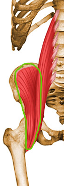
Iliacus - PART of the Iliopsoas muscle
•Origin: Iliac fossa of ilium
•Insertion: Lesser trochanter of femur
•Innervation: Femoral
•Action: Hip flexion, lateral rotation of femur
![<p>[NAME]</p><p>•<strong>Origin</strong>: </p><p>•<strong>Insertion</strong>: </p><p>•<strong>Innervation</strong>: </p><p>•<strong>Action</strong>: </p>](https://assets.knowt.com/user-attachments/cd2d83ed-f625-41cc-a89e-12541ffa355a.png)
[NAME]
•Origin:
•Insertion:
•Innervation:
•Action:
Psoas Major - PART of the Iliopsoas muscle
•Origin: Anterior surface & transverse processes of T12-L5
•Insertion: Lesser trochanter of femur
•Innervation: Lumbar plexus L2-L3
•Action: Hip flexion, lateral rotation of femur.
