light microscopy
 Call Kai
 Learn
 Practice Test
 Spaced Repetition
 Match
 Flashcards
 Knowt Play
1/48

There's no tags or description
Looks like no tags are added yet.
49 Terms
why are things visible?
source of illumination emits photons that act on objects
eye lense focuses light emitted by / bouncing off from objects
photoreceptor cells in retina activated by photons of light with certian wavelengths
6 ways light reacts with matter
transmission
reflection
refraction
diffraction
absorption
scattering
why are things not visible? (6)
not enough photons - eg turn lights off
wrong kind of photons - only ultraviolet to infared
objects too small for our array of photoreceptors to resolve -
objects do not interact with or emit visible light
objects interact with light the same as surrounding material
materials or lenses bend light to cloak the object
as an object gets further away from the detector, the angle between the object’s top and bottom…
decreases
convex lenses
refract / bend light, increasing the angle of the top and bottom of the object
spreads out the light hitting detector, making object appear larger
magnifying lens
refract light coming from an object
object appears larger and further way
compound lens parts and roles (5)
light from specimen hits detector
eyepiece (ocular) lens magnifies the image from the objective lens
objective lens magnifies light from specimen
condenser lens focuses light onto the specimen
virtual image appears to observer as enlarged specimen
magnification equation
product of objective lens x ocular lens
‘not enough photons” fix on microscope
use condenser lens to focus light on sample
“wrong kind of photon” fix on microscope
use detectors or cameras sensitive to other wavelengths
“object too small for array of photoreceptors to resolve” fix on microscope
use compound lenses to magnify sample
resolution
smallest distance at which two objects can be distinguished from one object
what determines resolution? (3)
how much light is lost between specimen and lenses
how much light is gathered by lenses
wavelength used for iillumination
smaller / shorter wavelength…
higher resolution
mis match between elements / refractive indexes in light path result in..
loss of signal between sample, lenses and detector
match refractive index =
higher resolution
refraction at interface between glass and air =
less light enters objective lens
immersion oil role
refractive index = glass
prevents loss from refraction and gathers all light
angular aperture of the objective lens
how much of the light is collected by the objective lens
cone of light coming from sample
higher angle means more light gathered
numerical aperture
0 number that describes the light gathering ability and resolving power of an objective lens
light is information
more information, better resolution so:
limited refraction between specimens and lens
shorter wavelength of illumination (more energy)
limit of resolutions between microscopes
~200nm for visible light microscopes (ideal, ~500nm without super-resolution techniques)
~2 nm for electron microscopes
brightfield or transmitted light
some structures within cells are visible because they interact with light differently from surrounding medium (water, air)
many things in cells are hard to see without stains or labels
Phase contrast
boosts contrast resulting from differences in refractivity between cells and cellular structures
DIC (differential interference contrast)
uses polarised light to boost contrast from diffraction of light within the sample
2 typs of enhanced contrast in label-free microscopy
dic (differential interference contrast)
phase-contrast
“objects interact with light the same as surrounding medium`’ fix on microscope
use optics that boost contrast between components with different material properties
“objects do not interact with or emit any visible light”
use stains or labels that can be detected by eyes or camera
fluorophores
molecules that emit energy in the form of photons of a longer wavelength when hit with specific shorter wavelength photons.
e.g. Ub to blue, green to red, red to infared
attach fluorescent molecules to molecules or proteins that label cellular structures
quinine
fluorescent molecule, excited by UV light, emitting blue light
mechanism of fluorescence (3)
photons of a particular wavelength carrying a particular amount of energy excite the electrons in the fluorophore
as electrons go back to ground state (unexcited) , energy is released as emitted photon
less energy is released as light than as absorbed, so emitted photons have longer wavelengths
fluorescence microscopy
light passes through the objective lens to and from sample
goes through excitation and emission filters
dichroic (2-colour) mirror: shorter wavelengths reflected onto sample, longer wavelengths pass through detector

A visible light
B excitation filter
C ocular lens
D emission filter
E dichroic mirror
F objective lens
Commonly used fluorescent labels
DAPI anf Hoechst - small molecules that bind to DNA, UV/ blue
Fluorophore-conjugated molecules, e.g. phalloidin, from toxic mushrooms, binds to F-actin
fluorescent molecules that localises to cells or organelles
GFP - green fluorescent protein (2)
derived from jellyfish A. victoria
encodes fluorescent protein that can be incorporated into plasmids and genomes
how to use a fluorescent protein sequence (2)
FP sequence at beginning or end of another protein to make a hybrid tagged protein
FP gene after promoter to monitor gene expression. green = promoter active
how to increase resolution in fluorescence microscopy (2)
reduce out-of-focus fluorophore excitation
reduce out-of-focus emitted light collection
confocal fluorescence microscopy (3)
improves resolution by removing out of focus light
excitation source = laser
pinholes focus light in x,y,z direction onto specimen

two-photon confocal microscopy (2)
improves resolution in z by only exciting fluorophore where beams of light / photons cross paths
2 longer photon beams energy adds up to that of one shorter photon
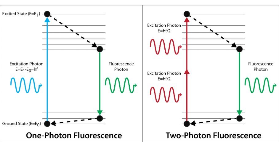
digital deconvolution
mathematically improve images using information about the diffraction of light, removing artefacts
super resolution microscopy (4)
technical and mathematical methods
resolution better than the diffraction limit
200 nm in x,y
500 in z
epitope
the part of the antigen that the antibody recognises
monoclonal antibodies
bind to one epitope
polyclonal antibodies
mixture of antibodies that recognise epitopes on the same antigen
antibodies
defences against pathogens
bind tightly to target molecules through their variable regions
direct and indirect immuno-labelling
direct: primary Ab binds to antigen epitope, with label attached
indirect: primary Ab doesn’t have label, secondary AB binds to first AB.
2nd Ab’s antigen is the primary’s constant region

immunohistochemistry
antibou + enzyme → colour change in sample
immunofluorescence

multiplexed fluorescent labelling
combining multiple dyes / antibodies
multiple fluorescent labels in one image
labelled in sequence, then put together in composite image