Oxygenation & Perfusion (40)
 Call Kai
 Learn
 Practice Test
 Spaced Repetition
 Match
 Flashcards
 Knowt Play
1/80

There's no tags or description
Looks like no tags are added yet.
81 Terms
Adventitious Breath Sounds
abnormal breath sounds heard over the lungs
Alveoli
small air sacs at the end of the terminal bronchioles.
they are the site of gas exchange.
Angina
a temporary imbalance.
between the amount of oxygen needed by the heart and the amount delivered to the heart muscles.
Arrhythmia
abnormal heart rhythm.
caused by the heart beating too fast, too slow, or irregularly.
Arterial Blood Gas (ABG)
a diagnostic test examining arterial blood.
used to determine the pressure exerted by oxygen and carbon dioxide in the blood.
Atelectasis
incomplete expansion or collapse of a part of the lungs

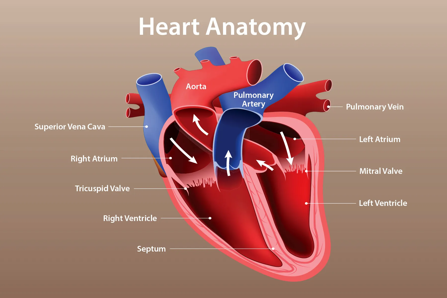
Atria
upper chambers of the heart

Atrioventricular (AV) Node
a node of specialized heart muscle.
located in the septal wall of the right atrium.
receives impulses from the sinoatrial node.
transmits them to the atrioventricular bundle.

Atrioventricular (AV) Bundle
a bundle of modified heart muscle.
Purpose: it transmits the cardiac impulse from the atrioventricular (AV) node to the ventricles.
causing them to contract.
Bradypnea
slow breathing
Bronchial Breath Sounds
Loud, high pitched, harsh “blowing” breathing sounds.
normally heard over the larynx and trachea.
sound on expiration is longer than inspiration.
Bronchodilators
medication that relaxes contractions of smooth muscles of the bronchioles
Bronchovesicular Breath Sounds
normal breath sounds heard over the mainstem bronchus.
they are moderate blowing sounds.
inspiration is equal to expiration.
Capnography
a method to monitor ventilation and, indirectly, blood flow through the lungs

Cardiopulmonary
relating to the heart and the lungs
Cardiovascular
relating to the heart and blood vessels
Cilia
Tiny, hair-like structures.
moves mucus, dust, and germs out of the airways.
Circulation
the continuous one-way circuit of blood through the blood vessels, with the heart as the pump
Crackles
fine, crackling sounds.
made when air moves through thin wet secretions in the lungs.
Diffusion
the tendency of solutes to move freely throughout a solvent.
from an area of higher concentration to an area of lower concentration.
until equilibrium is established.
in respirations, it is the movement of oxygen and carbon dioxide between the air (in the alveoli) and the blood (in the capillaries).
Any change in the surface area available for diffusion hinders diffusion.

Dyspnea
difficulty breathing
Dysrhythmia
an abnormal cardiac rhythm
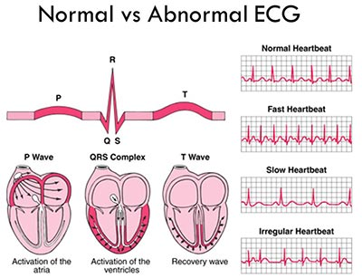
Electrocardiogram (ECG/EKG)
A test that records the electrical activity of the heart to check for abnormal heart rhythms and other heart problems.
such as myocardial ischemia and infarction, heart damage, rhythm and conduction disturbances, chamber enlargement, electrolyte imbalances, and drug toxicity.
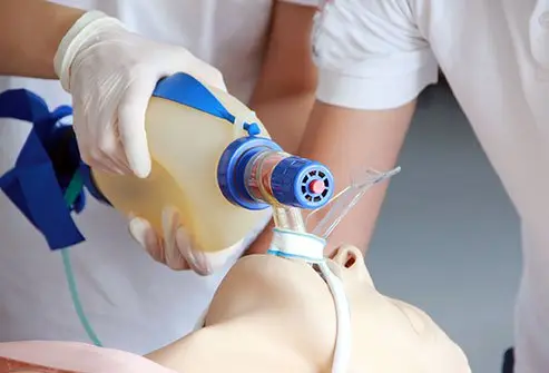
Endotracheal Tube
A tube (polyvinyl chloride) placed through the mouth or nose into the trachea.
to help a patient breathe.
Expiration
the act of breathing out
aka: exhalation
Heart Failure
occurs when the heart is unable to pump a sufficient blood supply.
results in inadequate perfusion and oxygenation of tissues.
heart conditions may include chronic hypertension, coronary artery disease, and disease of the heart valves.
Symptoms include shortness of breath, edema (swelling), and fatigue.
Hyperventilation
condition where more than the normal amount of air enters and leaves the lungs.
Breathing too fast or too deeply.
This causes the body to lose too much carbon dioxide (CO₂).
Hypoventilation
decreased rate or depth of air movement into the lungs
Hypoxemia
deficient (lack of) oxygenation of blood

Hypoxia
inadequate amount of oxygen available to the cells
Inspiration
the act of breathing in
aka: inhalation
Internal Respiration
the exchange of oxygen and carbon dioxide.
occurs between the circulating blood and tissue cells.
Ischemia
deficiency of blood and oxygen in a particular area
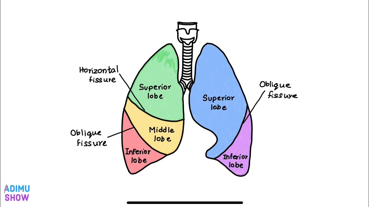
Lungs
they are composed of elastic tissue that can stretch or recoil.
They extend from the base at the level of the diaphragm to the apex (top), which is above the first rib.
The heart lies between the right and left lungs.
Each lung is divided into lobes.
Right lung: three lobes.
Left lung: two lobes.
Each lobe is subdivided into segments or lobules.
Myocardial Infarction
one type of acute coronary syndrome.
characterized by the death of heart tissue due to lack of oxygen.
aka: a heart attack

Oxygenation
the process of providing cells with life-sustaining oxygen

Perfusion
when oxygenated blood passes through body tissues
depends on an adequate blood supply and proper cardiovascular functioning to carry oxygen & carbon dioxide to and from the lungs
Pulmonary Ventilation
movement of air into and out of the lungs
Pulse Oximetry
noninvasive technique that measures the oxygen saturation (SpO2) of arterial blood

Respiration
the gas exchange between the atmospheric air in the alveoli and blood in the capillaries.
The process of breathing in oxygen and breathing out carbon dioxide.
Sinoatrial (SA) Node
a mass of tissue in the upper right atrium.
just below the opening of the superior vena cava.
Purpose: it initiates the transmission of electrical impulses.
contracts the heart at regular intervals.
aka: the pacemaker

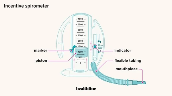
Spirometer
the instrument used to measure lung capacities and volumes.
one type is used to encourage deep breathing (incentive spirometry)
Sputum
respiratory secretion expelled by coughing or clearing the throat
Subcutaneous Emphysema
Air trapped under the skin.
usually around the chest or neck.
causes swelling or a crackling feeling when touched.
Surfactant
A detergent-like phospholipid (or substance) in the lungs.
Purpose: to keep the alveoli (air sacs) from collapsing.
helps make breathing easier.
reduces the surface tension of the fluid lining the alveoli.
Tachypnea
fast breathing
Tracheostomy
A surgical opening in the neck into the trachea to help a person breathe.
Tracheostomy Tube
A curved tube.
placed through an artificial opening in the neck into the trachea (windpipe).
purpose: to help a person breathe.

Ventricles
lower chambers of the heart
Vesicular Breath Sounds
normal sound of respirations.
heard on auscultation over peripheral lung areas.
Wheezes
continuous musical sounds.
produced as air passes through constricted airways by swelling, narrowing, secretions, or tumors.
often heard in patients with asthma, tumors, or a buildup of secretions.
low pitched sound, snoring quality.
air travels through thick secretions.
Stridor
high-pitched sound.
cuased by blockage in the upper airway.
can be heard across the room.
dangerous & emergency.
requires immediate intervention.
DO NOT NEED A STETHOSCOPE TO HEAR IT!!!!
a serous membrane that lines the lungs and thoracic cavity.
The visceral pleura covers the lungs.
the parietal pleura lines the thoracic cavity.
Pleural fluid between the membranes acts as a lubricant and an adhesive agent.
purpose: to hold the lungs in an expanded position.
Atherosclerosis
A condition where fatty deposits (plaque) build up inside the arteries
causing them to narrow and making it harder for blood to flow.
Kyphosis
An excessive outward curve of the upper spine that causes a rounded or hunched back.
Nasal Cannula
Low flow ( 1 - 6 LPM)
1–2 L/min = 24–28%
3–5 L/min = 32–40%
6 L/min = 44%
Simple Mask
Low flow
5–8 L/min = 40–60%
Nonrebreather Mask
Low flow
10–15 L/min = 80–95%
The bag is for the air you breathe in
the nurse should remember that an oxygen concentration of more than 50% given for longer than 48 hours can cause oxygen toxicity in the client.
Venturi Mask
High flow
4–6 L/min = 24–40%
the only mask that gives a specific amount of O₂ to a patient
Tent
aka: Shovel Mask
Flow: 6 to 10 LPM
it is VERY noisy & uncomfortable!
Bag-Valve Mask
aka: Ambue
Flow: 15 LPM
Used on people with respiratory arrest
administer 1 breath every 3-5 sec
check to see if patient’s chest is rising and falling
Peak Expiratory Flow Rate
the point of highest flow during forced expiration.
Measures how fast a person can blow air out of their lungs after taking a deep breath.
PEFR reflects changes in the size of pulmonary airways and is measured using a peak flow meter.
It is routinely used for patients with moderate or severe asthma to measure the severity of the disease and degree of disease management.
With the patient standing or sitting with the back positioned as straight as possible, the patient takes a deep breath and places the peak flow meter in the mouth, closing the lips tightly around the mouthpiece.
The patient forcibly exhales into the peak flow meter, and an indicator on the meter rises to a number.
The patient is asked to repeat this three times, and the highest number is recorded.

Pulmonary Function Tests
Tests that measure how well the lungs move air in and out and how well they transfer oxygen.

Thoracentesis
the procedure of puncturing the chest wall and inserting a needle into the pleural space to aspirate pleural fluid, air, or both
A thoracentesis may be performed to obtain a specimen for diagnostic purposes, to remove fluid or air that has accumulated in the pleural cavity and is causing respiratory difficulty and discomfort, or to instill medications
The physician or other advanced practice professional can perform a thoracentesis at the bedside with the nurse assisting, or it can be performed in a procedure room, in the radiology department, or in an outpatient setting
The patient is required to sign a permit for this procedure.
Because the cavity being entered is sterile, surgical asepsis is required.
Standard precautions also are used; transmission-based precautions are implemented as indicated.

Holter Monitor
A portable device that continuously records the heart’s electrical activity, usually for 24–48 hours, to detect abnormal heart rhythms. patient can NOT get this wet

Lung Scan
An imaging test that checks the lungs for problems, such as blood clots or poor blood flow.
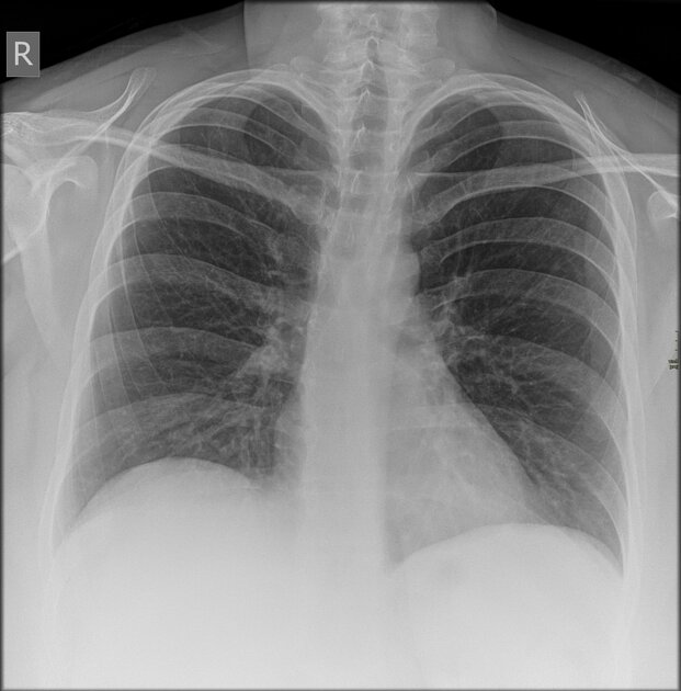
Radiography
The use of X-rays to create images of the inside of the body to help diagnose medical conditions.

Oropharyngeal Airway
a device placed through the mouth to help keep a patient's airway open.
patient is unconscious
it is important to get the right size
the device is not secure
Nasopharyngeal Airway
a tube inserted through the nose to help keep the airway open.
must use lubrication when inserting
patient is alert
requires frequent suctioning
Cardiac Coronary Catheterization
A procedure in which a thin tube is inserted into a blood vessel and guided to the heart to check for blockages in the coronary arteries.
insertion site: wrist, grion, arm, neck or collarbone
ex: A doctor performs cardiac catheterization to find a blockage that may be causing chest pain.
Cardiac Exercise Stress Testing
A test that monitors the heart’s activity while a patient exercises to see how well the heart works under stress.
ex: A patient walks on a treadmill while their heart rate, blood pressure, and heart rhythm are monitored.
Echocardiogram
An ultrasound test that uses sound waves to create pictures of the heart and show how well it works.
Transesophageal Echocardiogram (TEE)
A test that uses an ultrasound probe inserted into the esophagus to get detailed pictures of the heart.
Complete Blood Count (CBC) Test
provides a broad overview of a patient's health by evaluating the three cellular components of blood: red blood cells, white blood cells, and platelets
The test indicates the body’s response to illness, and may take 5 to 10 minutes to collect. The results of the CBC may indicate that further testing is needed. There may be some discomfort experienced during the venipuncture
Food and fluid do NOT need to be withheld before this test.
Flow Meter
a medical device used to measure and regulate the exact rate at which a gas or liquid is delivered to a patient
Oxygen Concentrator
a medical device that draws in room air, filters out the nitrogen, and delivers concentrated, purified oxygen to a patient.
Unlike heavy oxygen tanks, an oxygen concentrator never runs out of oxygen because it makes its own on demand, as long as it has electricity or battery power. It is most commonly used for patients requiring long-term oxygen therapy at home, such as those with Chronic Obstructive Pulmonary Disease (COPD)
It eliminates the need for a central reservoir of pipe oxygen and it is an economical choice. It may increase the client's electric bill. These can be used in homes, not long-term care facilities.
Continuous Positive Airway Pressure (CPAP) Mask
delivers a constant stream of pressurized room air to keep your upper airway open during sleep
used for patients with sleep apnea
Pleural Effusion
occurs when an abnormal, excessive amount of fluid accumulates in the pleural space
Airway Resistance
Airway resistance is the result of any impediment or obstruction that air meets as it moves through the airway. Any process that changes the bronchial diameter or width causes airway resistance. Obstruction in any part of the normal passageways impedes respiration. Obstruction can be caused by a foreign substance, such as a piece of food, a coin, or a toy, or by liquids, as in the case of a drowning victim. Obstruction can also result from secretions (e.g., excessive or thickened secretions) or tissues (e.g., tumors or edema of the respiratory tract). A decrease in the size of air passages resulting from constriction or poor neck positioning can also impede respiration.
Fremitus
the vibration felt on a patient's chest or back when they speak or breathe.