lung cancer
 Call Kai
 Learn
 Practice Test
 Spaced Repetition
 Match
 Flashcards
 Knowt Play
1/46

There's no tags or description
Looks like no tags are added yet.
47 Terms
upper, middle, lower
lobes of the right lung
(2 lobes): upper, lower + lingula
lobes of the left lung
horizontal and oblique
fissures in the right lung
oblique only
fissures in the left lung
Right mainstem = wider, shorter, more vertical → aspiration risk
main difference between the right and left mainstem bronchi
Right: Pulmonary artery Anterior
Left: Pulmonary artery Superior
what is the RALS mnemonic?
gas exchange
purpose of type I pneumocytes
surfactant
purpose of type II pneumocytes
total forced exhalation
meaning of FVC
↓ FEV1, ↓ FEV1/FVC
obstructive pattern PFTs
↓ FEV1, ↓ FVC, normal/high ratio
restrictive pattern PFTs
V/Q mismatch (most common)
Shunt
Diffusion impairment
common causes of hypoxia in relation to gas exchange
emphysema, fibrosis
DLCO is decreased in
lung cancer
number one cause of cancer death worldwide
adenocarcinoma
more common lung cancer type in women
Age 50-80 years
≥20 pack-year smoking history
Current smoker OR quit within 15 years
criteria for annual low dose chest CT screening
peripheral
location of adenocarcinoma of the lungs
adenocarcinoma
lung cancer type more common in nonsmokers, women
central
location of squamous cell lung carcinoma
keratin pearls, cavitation
histologic features of squamous cell lung cancer

adenocarcinoma of the lung
Peripheral
More common in Women
Most common in nonsmokers and overall
Bronchioalveolar subtype = lepidic, with hazy infiltrates similar to pneumonia

squamous cell
lung cancer type associated with PThrP---> hypercalemia
"oat cells", neuroendocrine
histology features of small cell carcinoma

associated with rapid growth and early mets
prognosis of small cell carcinoma
SIADH (↑ ADH)
Cushing (ACTH)
Lambert-Eaton syndrome
paraneoplastic syndromes associated with small cell
esophageal compression
cause of dysphagia associated with lung cancer
facial swelling
distended neck veins
signs of superior vena cava syndrome associated with lung cancer
hypercalcemia
paraneoplastic syndrome associated with squamous cell
SIADH
paraneoplastic syndrome most associated with small cell
adenocarcinoma
digital clubbing is most associated with this lung cancer type
contrast chest CT including upper abdomen/adrenals
inital imaging workup of lung cancer
TNM staging
staging of non-squmous lung cancer
limited stage: confined to one hemithorax
extensive stage: goes beyond that
staging of squamous cell lung carcinoma
metastatic disease (advanced stage).
pleural/pericardial effusion in the setting of lung cancer indicates
treated primarily with systemic therapy ± radiation, not surgery
treatment for most cases of small cell lung cancer
surveillance imaging is generally recommended every 6 months for 2 years, then annually to detect recurrence or a second primary lung cancer
recommendation for screening after definitive curative intent therapy for lung cancer
-age 50-80
-20 pack-year smoking history
currently smoke or quit within the last 15 years. -Stop screening once the person has not smoked for 15 years or develops a health problem that substantially limits life expectancy or ability/willingness to undergo curative lung surgery.
USPSTF recommendation for annual low dose CT screening
pneumothorax
most common complication of percutaneous lung biopsy
Classic sign:
visible visceral pleural line
no lung markings peripheral to that line.
signs of pneumothorax on upright chest XR
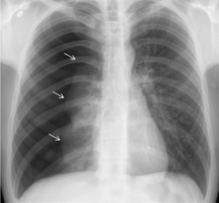
deep sulcus sign: abnormally lucent, deep lateral costophrenic angle
basilar hyperlucency
unusually sharp diaphragmatic or mediastinal contours.
signs of pneumothorax on supine chest XR
mediastinal shift away from affected side
depressed hemidiaphragm
radiographic signs of tension pneumothorax
obstructive shock
compresses the affected lung
shifts/compresses the mediastinum
reduces venous return to the heart
what type of shock may be caused by tension pneumothorax
Immediate needle/finger decompression followed by chest tube
treatment for tension pneumothorax
mediastinal shift
abnormal movement of the structures within the mediastinum to one side of the chest cavity

increased volume
-think massive plaural effusion
mediastinal shift away from the opaque side indicates
volume loss
complete lung collapse/atelectasis from mainstem obstruction
prior pneumonectomy
mediastinal shift toward the opaque side indicates
Away = push (effusion pushes mediastinum away)
Toward = pull (collapse pulls mediastinum toward it)
main causes of mediastinal shift