Quiz 10
 Call Kai
 Learn
 Practice Test
 Spaced Repetition
 Match
 Flashcards
 Knowt Play
1/143

There's no tags or description
Looks like no tags are added yet.
144 Terms

Cell division
the process in which one parent cell divides to produce two daughter cells
parent cell
You start with one cell, called the _
daughter cells
The parent cell divides into two new cells, called _. The _ receive genetic information from the parent cell.
binary fission
a form of asexual reproduction and cell division used primarily by prokaryotic organisms, such as bacteria and Archaea. During this process, a single parent cell duplicates its genetic material and splits into two identical daughter cells. Process:
DNA replication begins at a specific location called the origin of replication.
Replication produces two origins.
As the chromosome continues to replicate, one origin moves toward the opposite end of the cell.
The cell elongates while replication occurs.
When replication is complete, the bacterium has reached about twice its original size.
Proteins cause the plasma membrane to pinch inward.
The parent cell separates into two daughter cells.
Each daughter cell inherits a complete genome.
does not involve mitosis
In bacteria, most genes are carried on a single bacterial chromosome. This chromosome consists of:
One circular DNA molecule.
Associated proteins: proteins help organize bacterial division by moving bacterial chromosomes (similar to actin in eukaryotic cells) and helps pinch the plasma membrane inward (similar to tubulin in eukaryotic cells)
It is highly coiled and folded so it can fit inside the cell.
Reproduction in unicellular organisms
First major role of cell division.
A unicellular organism is an organism made up of only ONE cell. For these organisms, cell division is actually reproduction because the organism itself is just one cell. When that cell divides into two, it creates two separate living organisms.
Ex: bacteria, amoebas, certain types of algae.
Growth and development in multicellular organisms
2nd major role of cell division.
multicellular organisms start off as one cell.
Humans start off as a fertilized egg, also known as a zygote. A fertilized egg forms when a sperm cell and an egg cell combine. That single cell then repeatedly divides, creating more and more cells. The process looks something like this:
One fertilized egg exists.
It divides into two cells.
Those two cells divide into four.
Four divide into eight.
The number of cells continues increasing.
Eventually, a multicellular organism develops.
In multicellular organisms, cell division allows growth and development.

Repair and replacement
3rd major role of cell division.
Cell division doesn't stop once an organism becomes fully grown. Bodies constantly loses cells. Some cells die naturally because of normal wear and tear. Others are damaged through accidents or everyday activities. Bodies need to replace them. Cell division makes this possible.
Cell division allows organisms to repair damaged tissue and replace cells that die.
Ex: bone marrow, skin cells being replaced, cells in your digestive tract being replaced, damaged tissue being repaired
Bone marrow
the soft tissue inside certain bones that produces blood cells. Cells in your _ continuously divide to produce new blood cells.
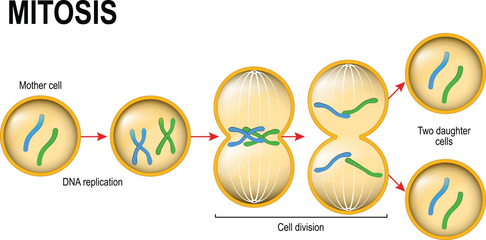
mitosis
division of the nucleus that produces two nuclei with the same chromosome number as the original nucleus.
the process that separates duplicated chromosomes into two groups so that each future nucleus receives an identical set of chromosomes.
a type of cell division in eukaryotic cells where a single parent cell replicates its chromosomes and divides them equally, producing two genetically identical daughter cells
Basic steps of cell reproduction:
DNA replication: One parent cell divides. The cell makes an identical copy of its DNA.
DNA distribution: The two copies (duplicated chromosomes) are separated and moved to opposite sides of the cell.
Cell division: The cell physically separates into two daughter cells via cytokinesis.
The 2 daughter cells have generally have the same chromosome number and nearly identical genetic information as the parent cell.
For human cell: 46 → 46 + 46
Each daughter cell receives 46 chromosomes. _ is important for growth, development, and tissue repair.
cell reproduction
Basic steps of _:
before replication: the parent cell has its original DNA
DNA replication: the DNA is copied, producing 2 identical copies
Divisions: each daughter cell receives one complete copy
The result is two daughter cells with genetically identical DNA.
This is the foundation of cell division.
DNA replication
The process of making an identical copy of DNA
Genetically identical
Having the same genetic information.
genome
the complete collection of an organism's genetic information.
Every cell needs the appropriate genetic information to perform its functions. Before a cell divides, it must make a copy of its _ so that both daughter cells receive a complete _
It is the entire instruction manual, consisting of many chromosomes
chromosomes
typical human cell contains approximately 2 meters of DNA. The DNA is extremely long, so it needs to be tightly organized and packaged. Cells solve this problem by packaging DNA into structures called _.
structures made of DNA and associated proteins (histones) that organize and package genetic information. how cells organize their DNA. It contains:
One very long DNA molecule.
Many associated proteins.
The proteins help:
Package and organize the DNA.
Maintain the _ structure.
Control the activity of genes.
The _ are important because they allow cells to organize enormous amounts of DNA so it can be replicated and distributed accurately during cell division.
The organized DNA structure. During cell division, it becomes highly condensed and visible under a light microscope.
having more _ does NOT mean an organism is more complex. A human body cell consists of 46 chromosomes, 23 from the mother and 23 from the father

gene
A _ is a specific section of DNA that contains information used to produce a functional product, often a protein or functional RNA. They are important because they carry information that contributes to inherited traits and cellular functions
chromatin
the entire complex of DNA and associated proteins that makes up chromosomes.
When a cell is not actively dividing, the DNA is generally in a less condensed form called _.
_ = DNA + histones (associated proteins)
DNA is relatively loose, spread out, and less condensed than in chromosomes, where they’re more condensed
Present during interphase cycle
DNA is organized by:
DNA → genes → chromatin → chromosomes
Somatic cells
all body cells except reproductive cells (gametes). In humans, _ normally contain 46 chromosomes. Those 46 chromosomes are organized into two sets of 23 (23 from your biological mother, 23 from your biological father).
Ex: skin cells. muscle cells, brain cells, liver cells, blood-forming cells.
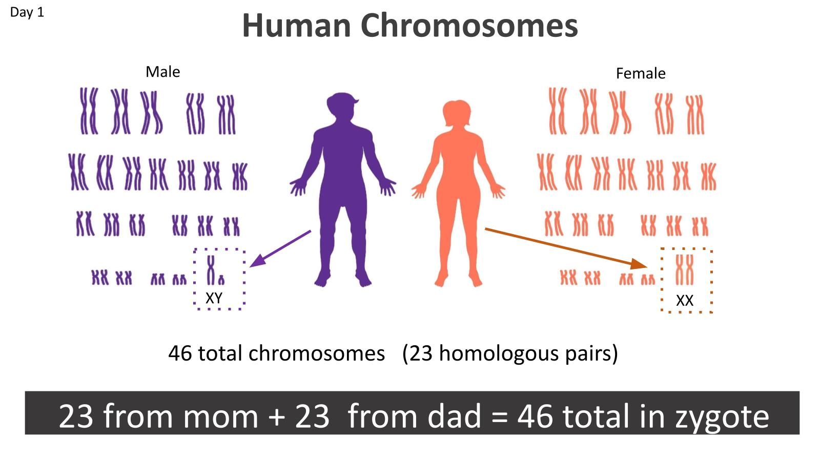
diploid
having 2 sets of chromosomes is called a _. The symbol is 2n.
For humans, 2n = 46
23 chromosomes from father, 23 from mother
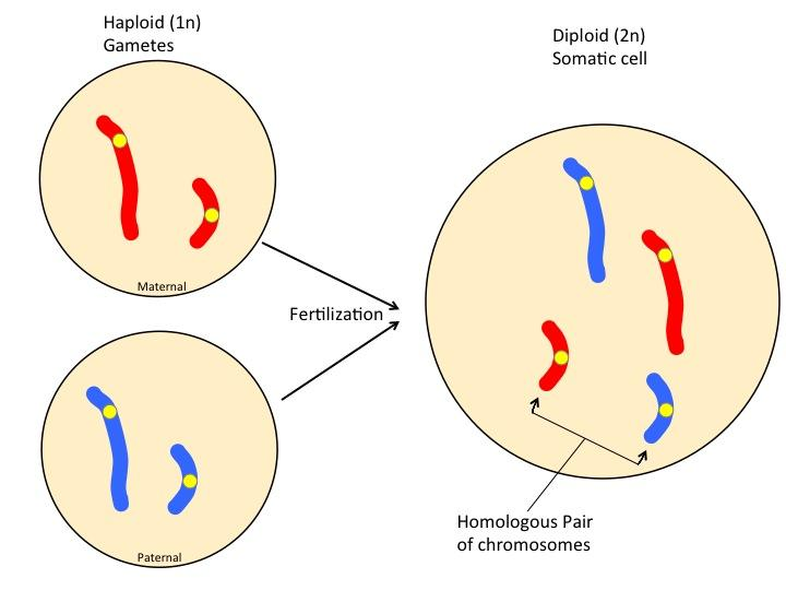
gametes
These are called reproductive cells, such as sperm and egg cells. Unlike somatic cells, _ contain only ONE set of chromosomes.
In humans, sperm cells contain 23 chromosomes, and egg cells contain 23 chromosomes.
haploid
having 1 set of chromosomes is _. The symbol is n.
For humans, n=23
human reproductive cells (sperm and eggs) have only 23 chromosomes


sister chromatids
When a cell prepares to divide, it first needs to copy its DNA, known as DNA replication. After DNA replication, each chromosome has two identical copies attached together. These copies are called _.
Def: one of two identical copies of a replicated chromosome.
When a chromosome is replicated, it produces two identical DNA molecules. Each DNA molecule is packaged into a chromatid. The two chromatids remain attached to one another. Together, they form one duplicated chromosome.
One half of a homologous chromosome, each called _.
Present from the middle of the S phase until anaphase.
DNA, chromosomes
DNA replication doubles the amount of _, but it does NOT immediately double the number of _.
This is because the two sister chromatids are still attached and are counted as ONE chromosome.
cohesin
proteins that hold sister chromatids together after DNA replication.
The attachment of sister chromatids to one another is called sister chromatid _.
This is important because the cell needs to keep the two identical DNA copies together until the correct stage of cell division.

centromere
a specialized region of a chromosome where sister chromatids are most closely associated (joined) and where the kinetochore assembles.
It contains repetitive DNA sequences and proteins that bind to those sequences. In a duplicated chromosome, the _ is the narrow region where the two sister chromatids are joined most closely.

chromosome arms
the portions of a chromatid (chromosome) extending on either side of the centromere. Think of the X shape:
The middle is the centromere.
The sections extending away from the centromere are the _
Each sister chromatid has two.
An unduplicated chromosome also has a centromere and two arms, even though it only has one chromatid.

Chromosome condensation
Before the cell divides, the DNA is replicated. After replication, each chromosome consists of two identical sister chromatids. The DNA then becomes highly condensed.
Def: the process of tightly coiling and folding chromatin into compact chromosomes.
This happens because DNA is extremely long, and the cell needs to move and separate DNA without it becoming tangled. Condensing the DNA makes this much easier.
When chromosomes are highly condensed, they become short and thick enough to be visible under a light microscope. This is why chromosomes are often drawn as X shapes in biology textbooks. Chromosomes don't always look like Xs. They look like Xs when they have been replicated and their sister chromatids are still attached.

Separation of sister chromatids
Eventually, the two sister chromatids separate. Each sister chromatid moves toward an opposite end of the cell. The process:
The cell contains duplicated chromosomes.
Each duplicated chromosome has two identical sister chromatids.
The sister chromatids separate.
One copy moves toward one side of the cell.
The other copy moves toward the opposite side.
Once sister chromatids separate, they are no longer called sister chromatids. They are considered individual chromosomes because each separated chromatid is now an independent chromosome.
The number of chromosomes change. Before sister chromatids separate, 46 chromosomes and 92 chromatids. After sister chromatids separate, there are temporarily 92 individual chromosomes in the dividing cell because the two sister chromatids are now counted as separate chromosomes. However, they are moving toward opposite ends of the cell, so each future daughter cell will receive 46 chromosomes. The separation of sister chromatids is what essentially doubles the chromosome count within the dividing cell.


cytokinesis
After mitosis, the cell still needs to physically separate into two cells. This is called _. division of the cytoplasm. which completes the mitotic phase.
It generally begins during anaphase or telophase.
the process is different in animal cells and plant cells.
At the end of _, the parent cell has become two daughter cells.
Each daughter cell has:
Its own plasma membrane.
Its own nucleus.
A complete set of chromosomes.
Cytosol and organelles.
The daughter cells can now begin their own cell cycles.
meiosis
a specialized type of cell division that produces cells with half the number of chromosomes as the original parent cell. a type of cell division that produces haploid cells used in sexual reproduction (gametes). In humans, _ occurs in special reproductive cells in the:
Ovaries (where eggs are produced)
Testes (where sperm is produced)
In humans: 46 → 23
It allows fertilization to restore the normal chromosome number. _ prevents the doubling of chromosomes per generations by reducing the chromosome number by half. 46 → 23.
Fertilization
the fusion of a sperm cell and an egg cell. Each gamete contains 23 chromosomes. When they combine: 23 + 23 = 46. The result is a fertilized egg (46 chromosomes total)
zygote
a fertilized egg with 46 chromosomes. The _ is diploid because it has two sets of chromosomes.

cell cycle
the life of a cell from the time it is formed by the division of a parent cell until it divides into two daughter cells itself.
everything that happens between one cell division and the next.
2 major stages:
interphase
mitotic phase (M phase)
Interphase
The cell grows, performs its normal functions, copies its DNA, and prepares for division. the period between cell divisions. During this time, the cell:
Grows.
Produces proteins.
Makes new organelles.
Performs its normal functions.
Copies its DNA.
Prepares for cell division.
Includes G1, S, and G2.
about 90% of the cell cycle
mitotic phase
The 2nd major part of the cell cycle, where the cell divides its genetic material and then divides its cytoplasm.
Includes mitosis and cytokinesis.
about 10% of the cell cycle (takes less than 1 hour)
G1 phase (gap 1 phase)
The first major growth period after a cell has been formed. the cell grows and performs its normal functions. During _, the cell:
Increases in size.
Produces proteins.
Produces cytoplasmic organelles.
Performs its normal cellular functions.
Carries out metabolic activities.
The cell needs to grow during _ because when the cell eventually divides into two, each daughter cell needs enough cellular material and organelles to function. The cell needs to build up its resources before splitting.
Takes 5-6 hours (most variable in duration, as some cells divide infrequently or not at all)
Metabolic activity
all the chemical reactions occurring inside a cell that allow it to stay alive and function.
Cytoplasmic organelles
structures inside the cell that perform specific jobs.
Ex: mitochondria, endoplasmic reticulum, Golgi apparatus
S phase (synthesis phase)
where DNA replication occurs. The cell copies its chromosomes so that each daughter cell can receive a complete set
Before _:
46 chromosomes (each with 1 chromatid)
46 DNA molecules.
During _:
Each DNA molecule is copied.
Each chromosome becomes duplicated.
Two identical sister chromatids are produced per chromosome.
After _:
46 duplicated chromosomes (2 sister chromatids each, 92 total)
92 DNA molecules.
_ takes approximately half of the cell cycle (10-12 hours) because copying the entire genome is a very large and complicated process.

G2 phase (gap 2 phase)
The 2nd major growth period after a cell has been formed and DNA replication has occurred.
By this point:
The cell has grown during G1.
The DNA has been replicated during S.
The cell now needs to finish preparing for division.
During _, the cell:
Continues growing.
Produces more proteins.
Produces more organelles.
Completes preparations for cell division.
the final preparation stage before the cell divides (mitosis)
Takes 4–6 hours
G0 phase
Some cells in multicellular organisms divide very infrequently or not at all. These cells enter a phase called _, which is a state in which a cell exits the active cell cycle and carries out its normal functions without actively preparing to divide.
a state outside the active cell cycle in which cells perform their normal functions instead of progressing toward division.
Not every cell in your body needs to constantly divide. Some cells are specialized to perform particular jobs. They may remain in _ for extended periods. Some cells can re-enter the cell cycle under appropriate conditions, while others remain in _ permanently or for most of their lifespan
pancreatic cells
The pancreas is an organ that performs several functions, including producing and secreting digestive enzymes. Some pancreatic cells spend their time carrying out their specialized job rather than repeatedly dividing.
mitosis, cytokinesis
The mitotic phase includes BOTH _ and _
mitosis
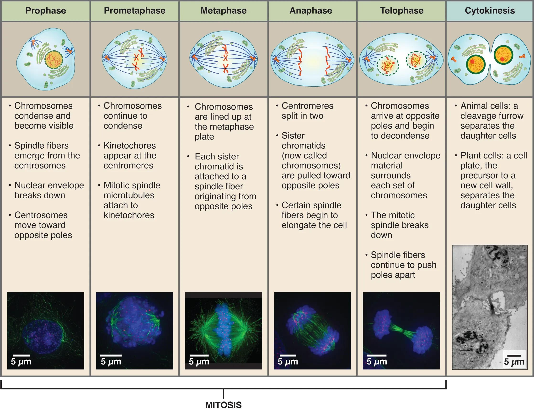
_ is divided in 5 stages:
prophase
prometaphase
metaphase
anaphase
telophase
Learn: “PP MAT”
mitotic spindle
a structure made of microtubules and associated proteins that helps organize and separate (move) chromosomes during mitosis.
Think: a system of cables that attaches to chromosomes and helps move them
As the _ forms, some of the cell's existing cytoskeleton microtubules partially disassemble, providing material for spindle assembly.
begins forming during prophase. While this happens, many of the cell's existing cytoskeletal microtubules partially disassemble. Their tubulin subunits can then be reused to build the _. The cell is reorganizing its microtubules rather than creating all of them from scratch.

microtubules
hollow, tube-shaped structures made from a protein called tubulin. They are part of the cell's cytoskeleton, which helps:
Maintain cell shape.
Organize the inside of the cell.
Move materials.
Help with cell division.
Polymerization
Tubulin subunits are added, making a microtubule longer
Depolymerization
Tubulin subunits are removed, making a microtubule shorter
centrosomes
In animal cells, spindle microtubules begin assembling at structures called _.
regions inside an animal cell that organizes microtubules. helps organize the microtubules that form the mitotic spindle.
During interphase, the centrosome duplicates, producing two _.
These move apart during prophase and prometaphase until they are at opposite ends of the cell. important because it helps establish the two opposite ends, or poles, of the mitotic spindle.
centrioles
Many animal-cell centrosomes contain a pair of _. They are cylindrical structures made of microtubules and are located near the center of the centrosome.
They are NOT essential for cell division. Even without _, the cells were still able to form a mitotic spindle. Also, plant cells do not have _ but can still form mitotic spindles.
a spindle can still form when centrioles are destroyed
Plant cells do not have centrioles, but they can still form mitotic spindles
interphase
During _, an animal cell starts with one centrosome. That centrosome duplicates, producing two centrosomes. These two centrosomes remain near the nucleus until mitosis begins.
The cell needs 2 because the spindle needs two opposite poles. During prophase and prometaphase, the two centrosomes move away from one another toward opposite ends of the cell. Spindle microtubules grow outward from them. By the end of prometaphase, the centrosomes are located at opposite ends of the cell, called the spindle poles.
spindle pores
By the end of prometaphase, the centrosomes are located at opposite ends of the cell. These opposite ends are called the _.
aster
a radial arrangement of short microtubules extending outward from each centrosome.

kinetochore
Each sister chromatid has a structure called a _, which is made of proteins assembled on specific sections of chromosomal DNA at the centromere. Therefore, each duplicated chromosome has 2 _.
The 2 _ on a duplicated chromosome face opposite directions because each _ needs to attach to spindle microtubules coming from a different pole as the chromatids must eventually be pulled toward opposite ends of the cell
It connects the chromosome (the object being moved) to the spindle microtubules (the structures that move it).
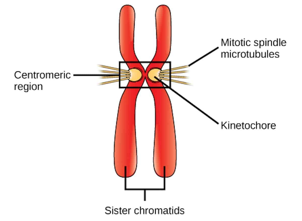
kinetochore microtubules
The spindle microtubules that attach directly to kinetochores (on chromosomes) are called _. Their job is to attach to chromosomes and help move chromosomes. The number of microtubules attached to each kinetochore varies depending on the species.
Yeast cells can have just ONE microtubule attached to each kinetochore.
Some mammalian cells can have approximately 40.
The important concept is that kinetochore microtubules connect the spindle to the chromosomes.

prophase
the first stage of mitosis. Several important events begin happening during this stage.
Chromatins condense into chromosomes: during interphase, DNA exists as relatively loose chromatin. During _, the chromatin condenses into compact chromosomes. Each duplicated chromosome consists of two sister chromatids attached together via cohesins. This makes the chromosomes easier to organize and separate. Occurs because the cell needs to organize its DNA into compact chromosomes so they can be moved and separated more easily
The mitotic spindle begins forming: Microtubules reorganize to form the mitotic spindle. the spindle begins assembling in the cytoplasm.
Centrosomes begin moving towards opposite ends of cell: Remember that centrosomes duplicated during interphase. Now, during prophase, the two centrosomes begin moving toward opposite ends of the cell.
Spindle microtubules begin growing outward from the centrosomes.

Prometaphase
Stage 2 of mitosis. _ comes immediately after prophase. chromosomes begin attaching to the mitotic spindle.
The nuclear envelope breaks down: Because the spindle microtubules need access to the chromosomes. If the nuclear envelope remained intact, the microtubules generally could not reach and attach to the kinetochores in the usual way.
Spindle microtubules attach to kinetochores inside nucleus: The kinetochore microtubules attach to the kinetochores on the chromosomes. Microtubules from opposite spindle poles can attach to them. This arrangement is essential because the cell needs to move the sister chromatids toward opposite ends later.
Chromosomes begin moving toward middle of the cell: When a kinetochore is captured by microtubules from one pole, the chromosome begins moving toward that pole. But when microtubules from the opposite pole attach to the other kinetochore, the chromosome experiences forces in opposite directions (tug of war). The chromosome may move back and forth before settling in the middle of the cell.
Metaphase
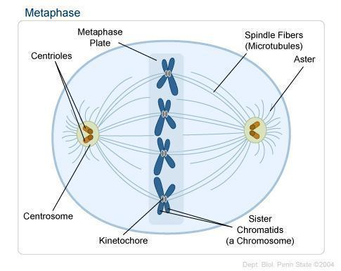
stage 3 of mitosis. _ is when the chromosomes line up in the middle of the cell. Remember that during prometaphase, chromosomes were moving back and forth because of the opposing forces from the two spindle poles. Eventually, chromosomes settle in the middle. The centromeres of all duplicated chromosomes line up along an imaginary plane called the _ plate.
They need to be positioned so that each sister chromatid can be separated toward a different side of the cell
metaphase plate
an imaginary plane located halfway between the two spindle poles. Important because the cell needs to make sure that each daughter cell receives one copy of every chromosome. The chromosomes must be properly attached before they separate.

nonkinetochore microtubules
the cell needs to make sure that each daughter cell receives one copy of every chromosome. The chromosomes must be properly attached before they separate. The other spindle microtubules are called _. They do NOT attach to kinetochores (and therefore don’t attach to chromosomes).
They overlap with microtubules from the opposite spindle poles in the middle of the cell. They help push the spindle poles apart and elongate the cell during anaphase
They interact with motor proteins, which will help push the spindle poles apart later. Meanwhile, the asters have also grown and their microtubules are in contact with the plasma membrane. At this point, the mitotic spindle is complete.

Anaphase
Stage 4 of mitosis. when sister chromatids separate and move apart.
separase cuts cohesins: separase is the enzyme that cuts cohesins, which hold sister chromatids together
sister chromatids become individual chromosomes: once separated, each former chromatid is now considered an individual chromosome
Chromosomes move toward opposite poles via motor proteins
The cell elongates: Nonkinetochore microtubules overlap with microtubules from the opposite spindle poles. Motor proteins attached to the microtubules walk them away from one another using energy from ATP. As the microtubules slide apart, the spindle poles move farther apart, the cell elongates, and the overlapping microtubules can lengthen through the addition of tubulin subunits

Telophase
The final stage of mitosis. During this:
Chromosomes arrive at opposite poles
Nuclear envelopes reform around each group of chromosomes.
Chromosomes begin to decondense, returning to a less-condensed chromatin state
The mitotic spindle disassembles as microtubules depolymerize.
Cytokinesis is occurring or beginning
At the end of _, the cell has two nuclei, one at each end. However, the cell still needs to physically split into two separate cells
Separase
Enzyme that cleaves (cuts) cohesins, allowing sister chromatids to separate
Motor proteins
Proteins that use energy to produce movement
Pac-Man mechanism
Chromosome movement involving kinetochore-associated motors and microtubule shortening
Reeling-in mechanism
Chromosome movement involving motor proteins at spindle poles
ATP
Molecule that supplies energy for cellular activities
Cytokinesis
Division of the cytoplasm
Cleavage
Process by which animal cells divide their cytoplasm
The sequence:
A shallow groove appears near the old metaphase plate.
This groove is called the cleavage furrow.
On the cytoplasmic side of the furrow is a contractile ring made of actin microfilaments associated with myosin proteins.
Actin and myosin interact, causing the ring to contract.
The cleavage furrow deepens.
The parent cell is pinched into two daughter cells.
Each daughter cell receives:
Its own nucleus.
A share of the cytosol.
Organelles.
Other sub-cellular structures.


Cleavage furrow
Groove that forms as an animal cell pinches inward

Contractile ring
Ring made of actin and myosin that contracts during animal-cell cytokinesis
Actin
Protein that forms microfilaments
Myosin
Motor protein that interacts with actin
Vesicle
Small membrane-enclosed sac that transports materials
Golgi apparatus
Organelle that modifies, sorts, and packages molecules
Coalesce
To come together and combine

Cell plate
Plant cells have cell walls, so they cannot divide by simply pinching inward like animal cells. Instead, they form a _.
Structure formed in the middle of a dividing plant cell that develops into a new dividing wall
The sequence:
During telophase, vesicles derived from the Golgi apparatus move along microtubules toward the middle of the cell.
The vesicles coalesce (come together and fuse).
They form a structure called the cell plate.
Cell wall materials carried in the vesicles collect inside the growing cell plate.
The cell plate expands toward the perimeter of the cell.
Its surrounding membrane fuses with the plasma membrane.
A new cell wall forms between the two daughter cells.
Bacterial chromosome
Usually a circular DNA molecule containing most bacterial genes
Nucleoid
Region where bacterial DNA is concentrated
Origin of replication
Specific location where DNA replication begins
Fluorescence microscopy
Technique that uses fluorescent molecules to visualize cellular structures
Evolutionary hypothesis
Proposed explanation for how a biological process evolved over time
The basic idea is that prokaryotes appeared more than a billion years before eukaryotes (binary fission). Scientists hypothesize that mitosis may have evolved from simpler prokaryotic mechanisms of cell reproduction because some proteins involved in bacterial binary fission are related to eukaryotic proteins that function in mitosis. Shows the processes share an evolutionary history
Cell cycle regulation
Cells need to divide at the correct time. Different cell types divide at different rates
Cytoplasmic signals
Experiments showed that signaling molecules in the cytoplasm can stimulate nuclei to enter different stages of the cell cycle.
Cell cycle control system
A group of molecules coordinates and triggers important cell cycle events
This is a cyclically operating set of molecules that:
Triggers important cell cycle events.
Coordinates those events.
Helps determine whether the cell should proceed or stop.
the cell cycle has a built-in sequence, but it can pause when a checkpoint detects a problem.
Checkpoint
A control point where stop and go-ahead signals determine whether a cell can proceed (regulate the cell cycle). G1, G2, and M _s determine whether the cell is allowed to proceed
Animal cells generally have built-in stop signals that prevent the cycle from continuing until appropriate go-ahead signals override them
Checkpoints receive signals from:
Inside the cell: Internal signals report whether important cellular events have been completed correctly
Outside the cell: External signals report whether conditions outside the cell are appropriate
G1 checkpoint (restriction point)
The most important checkpoint for many cells. Determines whether the cell continues through the cell cycle, dividing, or enters G0.
G0
A non-dividing state outside the active cell cycle. Some cells can return to the cycle.
M checkpoint
Ensures all chromosomes are properly attached to spindle microtubules before anaphase. The sequence is:
Chromosomes line up at the metaphase plate.
Spindle microtubules attach to their kinetochores.
The cell checks whether all kinetochores are properly attached.
If even one kinetochore is unattached, anaphase is delayed.
When all chromosomes are properly attached, a regulatory protein complex becomes activated.
This complex triggers molecular events that activate separase.
Separase cleaves cohesins.
Sister chromatids separate, beginning anaphase.
Growth factor
A protein released by certain cells that stimulates other cells to divide.
Different cell types respond to specific _s combinations of _s. They can bind to receptors and activate signal transduction pathways that allow a cell to pass the G1 checkpoint.
PDGF (platelet-derived growth factor)
A growth factor released by platelets that stimulates fibroblast division during wound healing.
An injury occurs.
Platelets release _ near the injured area.
_ binds to receptors on the plasma membranes of fibroblasts.
This activates a signal transduction pathway.
The signal allows fibroblasts to pass the G1 checkpoint.
Fibroblasts divide.
The increased number of fibroblasts helps repair the wound.
Density-dependent inhibition
the phenomenon in which crowded cells stop dividing.
When cells become crowded, they form a single layer covering the inside surface of the flask. Proteins on the surface of one cell bind to their counterparts on neighboring cells. This interaction sends signals to both cells that inhibit cell division. Even if growth factors are present, these signals can prevent the cells from moving forward in the cell cycle.
Anchorage dependence
The requirement that most animal cells be attached to an appropriate surface or structure to divide (ECM)
Cancer cells
Can ignore normal cell cycle controls, continue dividing, and evade apoptosis.
can evade the normal controls that trigger apoptosis
Transformation
the process in which cells in culture acquire the ability to divide indefinitely. These transformed cells begin behaving like cancer cells in terms of cell division.
Apoptosis
Programmed cell death. It is a controlled process that causes a cell to die when appropriate.
ex: a serious mistake might occur during DNA replication. If the mistake cannot be repaired, it may be better for the cell to undergo _ rather than continue dividing and passing along damaged DNA
Malignant tumor
Can spread into other tissues.
Metastasis
the spread of cancer cells from their original location to distant parts of the body. The process:
Cancer cells form an original tumor.
Some cells separate from the original tumor.
They enter blood vessels or lymph vessels.
They travel to other parts of the body.
They may begin dividing at the new location.
A new tumor can form.
cancer cells may:
Lose attachments to neighboring cells.
Lose attachments to the extracellular matrix.
Spread into nearby tissues.
Release signaling molecules that cause blood vessels to grow toward the tumor.
Blood vessels can supply the tumor with oxygen and nutrients.
Taxol
Prevents microtubule depolymerization, freezing the mitotic spindle and stopping progression beyond metaphase
Herceptin
Blocks HER2