HSC 214 Vision
 Call Kai
 Learn
 Practice Test
 Spaced Repetition
 Match
 Flashcards
 Knowt Play
1/37

There's no tags or description
Looks like no tags are added yet.
38 Terms
Optic nerve (CN II)
vision

Optic chiasm
gives binocular vision

Optic tract

Occipital lobe

Superior rectus
elevation, adduction, and medially rotates the eyeball

Inferior rectus
depresses, adducts, and laterally rotates the eyeball

Medial rectus
adducts the eyeball

Lateral rectus
abducts the eyeball

Superior oblique
depresses, abducts, medially rotates eyeball
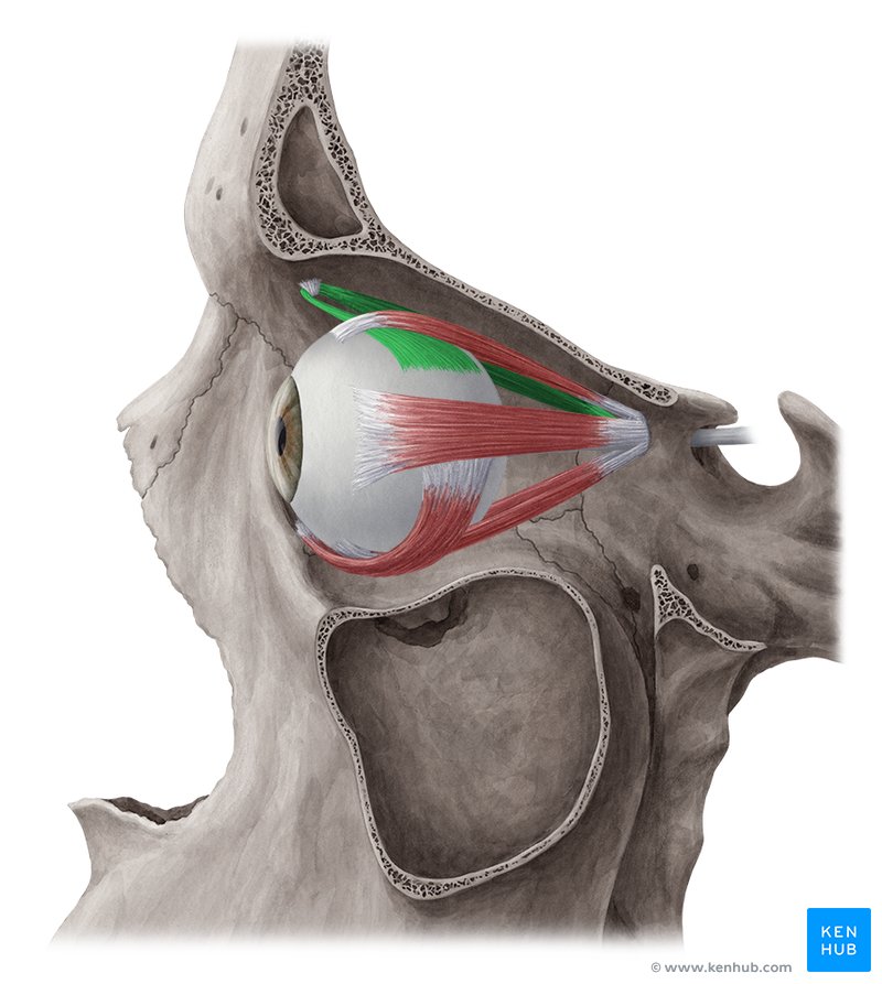
Inferior oblique
elevates, abducts, and laterally rotates eyeball

Eyelids

palpebral conjuctiva
inside of eyelid, pinkish color
ocular conjuctiva
covers the anterior surface of eye (sclera)
Medial Canthus
medial point where eyelids meet
lateral canthus
lateral point where eyelids meet
lacrimal gland ducts
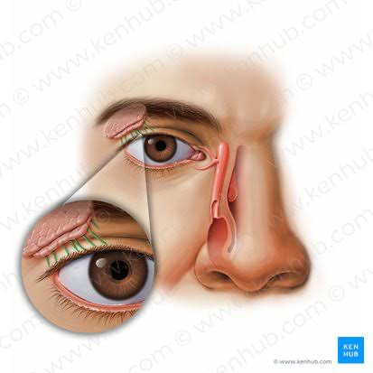
lacrimal gland

lacrimal punctum

Superior lacrimal canal
superior duct drains lacrimal punctum to nasocrimal duct
inferior lacrimal canal
inferior duct drains lacrimal punctum to nasocrimal duct
Nasolacrimal duct

Sclera
the white around the eye
Choroid
layer between sclera and retina
retina
layer under the choroid, contains neural receptors
Cornea

anterior cavity

aqueous humor
fluid that fills the anterior cavity
Anterior chamber of anterior cavity
space between cornea and iris/pupil
Posterior chamber of anterior cavity
space between iris/pupil/ and lens/ciliary body
Iris

Pupil

lens

Ciliary body
attachment point for suspensory ligaments
suspensory ligaments
suspends the lens from the ciliary body
Posterior cavity
space posterior to the lens
Vitreous humor
substance filling the posterior cavity
optic disc
area on the retina where the nerves leaves the eye
Fovea centralis
where cones (color receptors) are concentrated, spot of best vision