chptr 23 digesive system anat&phys 2
 Call Kai
 Learn
 Practice Test
 Spaced Repetition
 Match
 Flashcards
 Knowt Play
1/60

There's no tags or description
Looks like no tags are added yet.
61 Terms
Differentiate between the organs of the alimentary canal (GI tract) and the accessory digestive organs.
alimentary canal: Incudes the mouth, pharynx, esophagus, stomach, small and large intestine
Accessory: Includes teeth, tongue, salivary glands, gall bladder, liver, pancreas
Describe the six major processes of digestive activity.
Indigestion- eating
Propulsion- movement forward through alimentary canal
Mechanical breakdown- breaking down food by increasing surface area
Digestion- catobolizm via enzymes ( chemical breakdown )
Absorption- goes into body or lymph
Defication- elimination of indigestible things
Differentiate between the parietal and visceral peritoneum. What is the function of the peritoneal cavity?
Parietal: lines outside of peritineum. Visceral: covers external surface of most digestive organs
Peritoneal cavity: allows organs to glide easily across one another as they change shape during digestion
What is a mesentery, and what are its major functions?
It is the double layer of peritoneum, two serous membranes fused back-to-back.
Functions: Provides route for blood vessels, lymphatic vessels and nerves, Holds organ in place, Stores fat
Detail the four basic tissue layers (tunics) of the alimentary canal from the lumen outward.
Mucosa: line lumen
Submucosa: underneath mucosa
Muscularis externa: Muscle layer
Serosa: protective layer

What are the specific sub-layers of the mucosa and what does each do?
Epithelium: Secreats mucous to protect organs, lubricate canal, secrete enzymes and hormones
Lamina propria: contains capillaries to nourish epithelium and absorb digested nutrients
Muscularis mucosae: layer of smooth muscle that produce movement for secretion and absorption
Contrast the processes of peristalsis and segmentation.
Peristalsis: occurs from outer muscles, they contract/ relax and move food down/ forward
Segmentation: occurs in inner muscles, they mostly break down food.
Explain the role of the enteric nervous system (ENS). Differentiate between the submucosal plexus and the myenteric plexus.
Enteric Nervous System (ENS): nerve supply of the alimentary canal
Submucosal plexus: controls secretion, absorption and blood flow. Myensentric: controls GI motility
How do short reflexes differ from long reflexes in the GI tract?
Short Reflexes: controls patterns of segmentation and peristalsis in response to stimuli within the GI tract
Long Reflexes: sends information to the CNS via the vagus nerve in response to internal and external stimuli; receives parasympathetic information to enhance GI activity and sympathetic information to inhibit it
Identify the major structures of the oral cavity, including hard and soft palate/uvula.
Picture

Describe the structure of the tongue, including the papillae.
Picture

What are the primary functions of saliva?
Cleanse mouth dissolve food for taste, moisten food to form bolus, begin starch breakdown
Identify the three major extrinsic salivary glands and their general locations, along with their associated duct.
Picture

Differentiate between the two types of secretory cells found in salivary glands
Serious cells: make watery secretion
Mucous cells: make mucous

Describe the composition of saliva.
Mostly water, ph is 6.75-7, electrolytes, has enzymes and metabolic waste, also has proteins.
Identify the different classifications and types of teeth.
Incisors: chisel-shaped for cutting or nipping pieces of food, Canines: fang-like for tearing or piercing. Premolars & Molars: broad crowns with rounded cusps for grinding or crushing
Types of teeth: Deciduous (Primary/Milk/Baby) Teeth- 20 teeth that erupt between ages 6-24 months fall out and replaced by permanent teeth.
Permanent Teeth- 32 teeth that develop beneath the surface.
Wisdom teeth( third molars)- Erupt between ages 17–25 sometimes dont erupt at all
Detail the anatomical structure of a tooth.
Picture

Describe the digestive processes of the mouth.
Ingestion- breakdown through mastication ( chewing )- propulsion ( swallowing )- digestion
Outline the passage of food through the pharynx. How does the epiglottis assist in this process?
Food passes from the mouth to the oropharynx, then to the laryngopharynx. As food moves through the laryngopharynx, the epiglottis closes off the larynx, directing food posteriorly into the esophagus.

Describe the anatomy of the esophagus.
Upper esophageal sphincter (UES): prevents backflow of bolus into pharynx
Lower esophageal sphincter (LES): prevents reflux from the stomach

Detail the four tunics of the esophagus.
Mucosa
Submucosa
Muscularis externa
Adventisha

Detail the steps of the buccal phase and the pharyngeal-esophageal phase of swallowing
Picture

Describe the gross anatomy of the stomach, including its distinct regions, curvatures, omentum, and the function of rugae.
What is the mucosal barrier, and how does it protect the stomach?
Identify the specific secretions produced by the specialized cells in the gastric glands..
Explain the role of HCl and intrinsic factor in the stomach.
How is the muscularis externa of the stomach uniquely modified and why?
Describe the digestive processes of the stomach.
Outline the three phases of gastric secretion regulation.
Identify the four primary lobes of the liver and its major mesenteries.
Right- largest part
Left smaller than the right
Caudate-
Quadrate-
Mesentaries: falciform ligament separates right from left. Lessor omentum anchors liver to lessor curvature of stomach.
What three vessels make up the portal triad at the porta hepatis?
Hepatic artery proper
– Hepatic portal vein
– Common hepatic duct
Describe the microscopic anatomy of a liver lobule, including hepatocytes, portal triad, central veins, and sinusoids.
Trace the flow of oxygenated and deoxygenated/nutrient-rich blood into and through the liver.
Oxygenated blood to Liver:
– Abdominal aorta to Celiac artery To Common hepatic artery to Hepatic artery proper (triad) to Liver sinusoids to Central vein to Hepatic vein to then the IVC.
• Deoxygenated blood to liver (with nutrients from intestines):
– Abdominal aorta to Mesenteric arteries to Intestinal capillaries to Mesenteric veins to Hepatic portal vein (triad) to Liver sinusoids (capillaries) to Central vein to Hepatic vein to then the IVC
Describe the composition of bile. Which components actually aid in digestion?
*Bile salts: fat emulsifier that breaks fat globules into tiny droplets to make them more readily digestible and easily absorbed
– Bile pigments (bilirubin): the yellow waste product of heme group formed in the breakdown of RBC is absorbed by hepatocytes and excreted in bile
Trace the flow of bile through the liver to the gall bladder or duodenum.
What is the function of the gallbladder?
Store bile
What is composition of bile?
What happens if gallstones (cholelithiasis) block the flow of bile?
~ Bilirubin accumulates in the blood, eventually deposits in the skin and sclera, causing a yellow-tint (jaundice)
~ Leads to steatorrhea (fatty stools)
~ Poor absorption of fat
~ Deficiencies fat-soluble vitamins (A, D, E, K)
Describe the digestive processes of the liver and gallbladder.
It’s just used in fat digestion.
Describe the exocrine function of the pancreas. What enzymes are secreted by the acinar cells?
• Exocrine function is to produce enzymes that breakdown types of food and neutralize acidic chyme entering duodenum.
Explain how pancreatic proteases are produced in inactive forms (like trypsinogen) and activated in the duodenum.
Once enzyme is dumped into the duodenum its able to be broken down
Describe the duct systems that unite the liver, gallbladder, and pancreas at the hepatopancreatic ampulla.
Bile duct: delivers bile from the liver to the duodenum
Main pancreatic duct: carries pancreatic juice from the pancreas to the duodenum
accessory pancreatic duct: a smaller duct that empties pancreatic juice directly into the duodenum
Describe the digestive processes of the pancreas.
Digestion: pancreatic enzyme production for protein, carbohydrate, fat, and nucleic acids.
In other words digestion. It’s just digestion.
Identify the three subdivisions of the small intestine. What is the mesentery proper?
Three subdivisions:
1. Duodenum: beginning segment; mostly retroperitoneal; curves around the head of the pancreas
2. Jejunum: middle section
3. Ileum: terminal segment; joins the large intestine at the ileocecal valve.
Mesentery proper hold the intestines in place

List the modifications to the mucosa and submucosa of the small intestines.
Explain how circular folds, villi, and microvilli structurally modify the small intestine to increase surface area.
Circular folds have villi then villi have microvilli. They hope to absorb all nutrients
Describe the five specialized cells found in the intestinal crypts and their specific functions.
1. Enterocytes – lined with microvilli, for nutrient and electrolyte absorption
2. Goblet cells – mucus- secreting cells
3. Enteroendocrine cells – hormone-secreting cells, regulating digestion (cholecystokinin and secretin)
4. Paneth cells – cells that release antimicrobial- agents
5. Stem cells – renew the epithelium

What is the role of Mucosa-Associated Lymphoid Tissue (MALT) and Peyer's Patches in the small intestine?
Protects against microorganisms entering the bloodstream

Describe the digestive processes of the small intestines.
1. Breakdown: mechanical breakdown
2. Propulsion: peristalsis and segmentation
3. Digestion: pancreatic enzymes complete digestion for all classes of foods.
4. ABSORPTION: breakdown products of carbs, proteins, fats, and nucleic acids, plus vitamins, electrolytes, water are absorbed ( absorption is most important )
Identify the major subdivisions of the large intestine.
intestine.
• Five subdivisions:
1. Cecum
2. Appendix
3. Colon (ascending, right colic flexure, transverse, left colic flexure, descending, sigmoid)
4. Rectum
5. Anal canal (internal and external sphincters, anus)
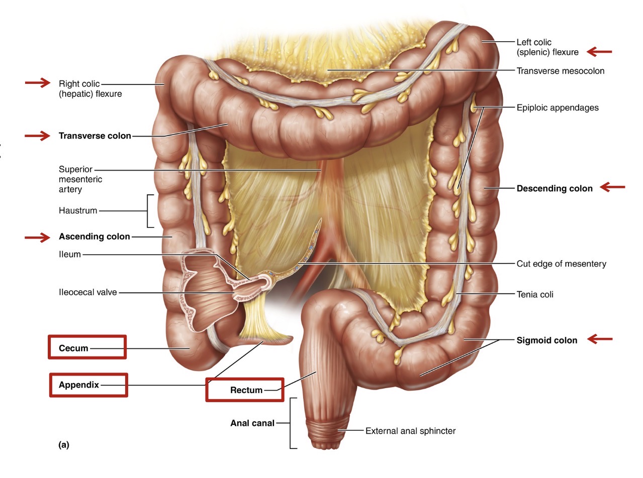
Describe the three unique features of the large intestine.
1. Teniae coli: bands of longitudinal smooth muscle in the muscularis
2. Haustra: pocket-like sacs formed by the muscle tone of the teniae coli
3. Epiploic appendages: small, fat-filled pouches of visceral peritoneu

How do the tunics of the large intestine differ from the small intestine?
4 tunics but the large intestine has modifications.
1. Mucosa - no circular folds, villi, or microvilli;
‒ Thicker mucosa with deeper crypts and abundant goblet cells for mucus secretion, lubricating fecal passage and protecting against irritation and gases released by resident bacteria
2. Submucosa
3. Muscularis externa – modified outer longitudinal layer (teniae coli)
4. Serosa – replaced by adventitia for the ascending and descending colon
Detail the steps of the defecation reflex, including the roles of the internal and external anal sphincters.

Explain the importance of the bacterial microbiota in the gut.
Gut bacteria help to recover energy from otherwise indigestible foods and synthesizing some vitamins
Outline the three processes that regulate movement in the GI.
Enterogastric Reflex: controls pyloric sphincter, prevents water loss and overwhelmed.
Gastroileal Reflex: control iliocecal valve, gets things moving
Gastrocolic Reflex: stimulates haustrral contractions and mass peristalsic waves moving fecal mater forward at one time.
Describe the digestive processes of the large intestines.
1. Propulsion: feces propelled to rectum by mass movements (haustral contractions and mass peristaltic waves))
2. Digestion: tricky…reminding food residues are digested by gut bacteria (not by large intestine tissue)
3. Absorption: absorption of WATER, electrolytes (largely NaCl), and bacterial-produced vitamins
4. Defecation: reflex triggered by rectal distension; eliminates feces from the body
What is hydrolysis, and why is active transport necessary for most nutrient absorption?
hydrolysis, the process of adding a water molecule to each molecular bond to be broken
Summarize carbohydrate digestion: Where does it begin, and what enzymes are involved?
Summarize protein digestion.
Outline the step-by-step process of lipid digestion and absorption, from emulsification by bile salts to absorption by lacteals.
How are nucleic acids digested and absorbed?