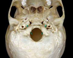
The Skull
 Call Kai
 Learn
 Practice Test
 Spaced Repetition
 Match
 Flashcards
 Knowt Play
1/86

There's no tags or description
Looks like no tags are added yet.
87 Terms
coronal
suture between frontal and parietal bones

squamous (squamosal)
suture between temporal and parietal bones

sagittal
suture between parietal bones
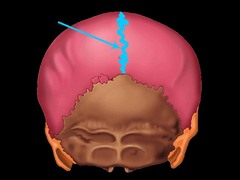
lambdoid
suture between parietal and occipital bones (sutural bones may be present here too)

lacrimal bones
form part of medial wall of the orbit (eye socket)
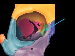
palatine bones
form the posterior portion of the hard palate
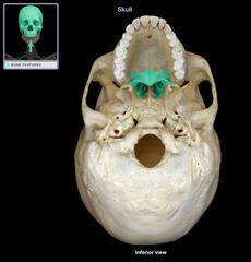
zygomatic bones
form part of the cheekbone and contribute to the rim and lateral wall of the orbit
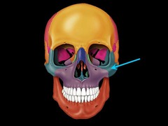
maxillae
Support the upper teeth and form the inferior orbital rim, the lateral margins of the external nares, the upper jaw and most of the hard palate

inferior nasal conchae
create turbulence in air passing through the nasal cavity, and increase the epithelial surface area to warm and humidify inhaled air.

vomer
forms inferior portion of the bony nasal septum

mandible
forms lower jaw

6, 2
there are ____ paired facial bones and _____ single facial bones
frontal bone
forms the forehead

sphenoid bone
forms part of the floor of the cranium, unites the cranial and facial bones, and acts as a cross-brace that strengthens the sides of the skull

ethmoid bone
forms the anteromedial floor of the cranium, the roof of the nasal cavity, and part of nasal septum and medial orbital wall

parietal bones
form part of the superior and lateral surfaces of the cranium

occipital bone (occiput)
contributes to the posterior, lateral, and inferior cranial surfaces

external occipital crest
extends downward from the external occipital protuberance and stabilizes the vertebrae of the neck

temporal bones
form part of the lateral wall of cranium; articulates with mandible and facial bones

mastoid process
attachment for muscles that rotate or extend head

styloid process
attached to ligaments supporting the hyoid bone and tendons of several muscles

temporal lines
ridges marking attachment sites of the temporals muscle

external acoustic meatus
canal beginning on lateral surface of the temporal bone; ends at the tympanic membrane

zygomatic process
articulates with zygomatic bone to form zygomatic arch

mandibular angle
the posterior, inferior corner of the lower jaw

mental protuberance (chin)
attachment site for several facial muscles

alveolar processes
projecting ridges of maxillae and mandible; supports upper and lower teeth
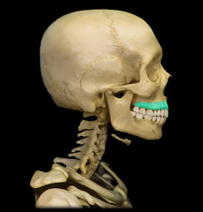
frontal sinuses
hollow spaces front of head

foramen lacerum
jagged slit between sphenoid and petrous portion of temporal bone; contains hyaline cartilage and small arteries

foramen ovale
passage for nerves innervating the jaws

carotid canal
Passage for internal carotid artery.

stylomasoid foramen
posterior to the base of the styloid process; passage for facial nerves

jugular foramen
located between the occipital and temporal bone; passage for jugular vein

foramen magnum
connects cranial cavity with vertebral canal; surrounds connection between brain and spinal cord

occipital condyles
articulation site between skull and C1

nuchal lines
intersect at occipital crest

superior nuchal line

inferior nuchal line

optic canal
passage for optic nerve from eyes to brain

lesser wings
extend horizontally anterior to the sella turcica

greater wings
extend laterally from the body and form part of the cranial floor

hypophyseal fossa
houses the pituitary gland
sella turcica
saddle-shaped enclosure

sphenoidal sinuses
inferior to the sella turcica; hollow spaces on either side of the body

cribriform plate
forms anteromedial cranial floor and nasal cavity roof

crista galli
bony ridge that projects superior to cribriform plate; attachment point for falx cerebri

perpendicular plate
forms superior part of nasal septum

horizontal plate
forms the posterior part of the hard plate

supraorbital notch
passageway for blood vessels to eyebrow, eyelids, and frontal sinuses

paranasal sinuses
lighten the skull bones and amplify the sounds we make as we speak
maxillary sinuses
secrete mucus to flush inferior nasal cavity surfaces; largest sinuses

ramus
begins at the mandibular angle

condyle
articulates with the temporal bone at the temporomandibular joint

mandibular notch
depression that separates the condylar and coronoid processes

coronoid process
insertion point for temporalis muscle

mylohyoid line
marks the insertion of the mylohyoid muscle

mandibular foramen
passageway for blood vessels and nerves supplying the lower teeth

hyoid bone
supports the larynx

nasal bone

ethmoid

zygomatic arch

temporal process

palatine
Name this bone

hard palate
roof of the mouth

anterior cranial fossa
formed by the frontal bone and part of the sphenoid bone

middle cranial fossa

posterior cranial fossa

infratemporal fossa
space on lateral side of skull, below the level of the zygomatic arch and deep (medial) to the ramus of the mandible

orbital portion of frontal bone
#1

orbital portion of sphenoid bone
#2

superior orbital fissure
#3

orbital portion of zygomatic bone
#4

inferior orbital fissure
#5

optic canal
#7

orbital portion of ethmoid bone
#8

lacrimal duct
what passageway is this
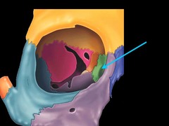
orbital portion of palatine bone
#11

orbital portion of the maxilla
#12

infraorbtial foramen
#13

superciliary arch

orbital margin

frontal crest

basilar process
#8

groove for the transverse sinus

internal occipital protuberance

condyloid canal

hypoglossal canal
what structure are the green things going through